. 2018 Oct 18;8(1):15423.
doi: 10.1038/s41598-018-33641-2.

Drug Screening of Human GBM Spheroids in Brain Cancer Chip

Affiliations

Drug Screening of Human GBM Spheroids in Brain Cancer Chip

Metin Akay et al. Sci Rep. .

Abstract

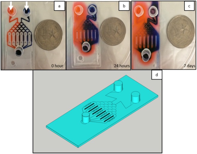
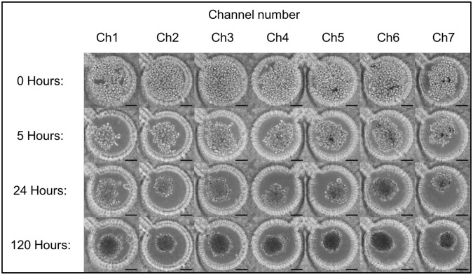
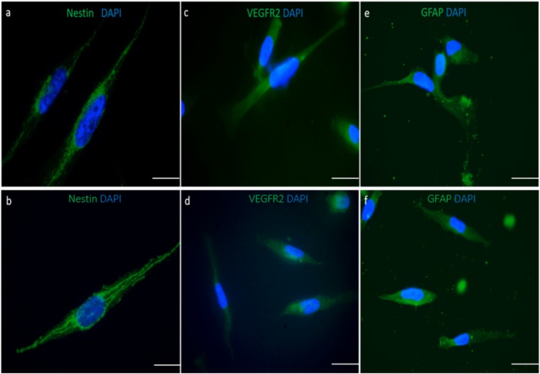
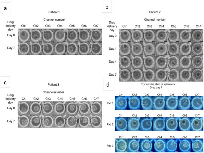
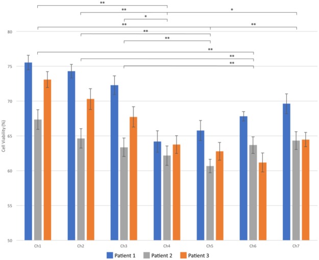
Glioblastoma multiforme (GBM), an extremely invasive and high-grade (grade IV) glioma, is the most common and aggressive form of brain cancer. It has a poor prognosis, with a median overall survival of only 11 months in the general GBM population and 14.6 to 21 months in clinical trial participants with standard GBM therapies, including maximum safe craniotomy, adjuvant radiation, and chemotherapies. Therefore, new approaches for developing effective treatments, such as a tool for assessing tumor cell drug response before drug treatments are administered, are urgently needed to improve patient survival. To address this issue, we developed an improved brain cancer chip with a diffusion prevention mechanism that blocks drugs crossing from one channel to another. In the current study, we demonstrate that the chip has the ability to culture 3D spheroids from patient tumor specimen-derived GBM cells obtained from three GBM patients. Two clinical drugs used to treat GBM, temozolomide (TMZ) and bevacizumab (Avastin, BEV), were applied and a range of relative concentrations was generated by the microfluidic channels in the brain cancer chip. The results showed that TMZ works more effectively when used in combination with BEV compared to TMZ alone. We believe that this low-cost brain cancer chip could be further developed to generate optimal combination of chemotherapy drugs tailored to individual GBM patients.


Conflict of interest statement

Authors J.H., Y.F., N.G.A., Y.A. and M.A. declare no competing financial and/or non-financial interests. Author J.Z. has obtained funding from the following sources: Boston Biomedical Sumitomo Dainippon Pharma Global Oncology; DEKK-TEC, Inc.; Diffusion Pharmaceuticals, LLC; Five Prime Therapeutics, Inc.; Immuno-Cellular Therapeutics, LTD; NRG/RTOG/National Cancer Institute; Novocure, Inc.; Tocagen, Inc. Otherwise, J.Z. declares no other competing financial and/or non-financial interests.

Figures

Figure 1
Figure 1
Eosin red (left) and Chicago blue (right) dyes were loaded into the inlets to characterize the gradient of two solutions generated in the microfluidic channels. The dyes were left in the chip for 24 hours to diffuse into the hydrogel, but was unable to cross the gradient generating gap between channels. Gradient generating gaps are marked with white arrows (a). The layout of the brain cancer chip flows solution loaded into the two inlets, through a gradient generating regions, and into 7 microfluidic channels, each containing a culture array composed of 9–11 microwells 360 µm in diameter (b). The image was taken at day 7 to test the leakage (c). The coins are a US Quarter.
Figure 2
Figure 2
Cells were seeded into the microfluidic chips at 0.5 × 106 cells/mL and captured in the microwells. Images were taken 0, 5, 24, and 120 hours after cell seeding to visualize cell aggregation and spheroid formation. Scale bar is 100 µm.
Figure 3
Figure 3
Characterization of the cells collected from freshly dissociated cells (a,c,e) and from the microfluidic chips (b,d,f). Nestin (a,b), VEGFR2 (c,d), GFAP (e,f) stainings of the cells extracted from fresh primary tumor and cells collected from microfluidic chip, respectively. Cells were seeded on the coverslides, incubated overnight to attach to the coverslides and stained against the antibodies. Nuclei are stained with DAPI. Scale bar is 100 µm.
Figure 4
Figure 4
After 7 days of spheroid culture, TMZ (600 µM, right inlet) and BEV (7.5 µM, left inlet) were applied to the brain cancer chip. Effects of the drug treatment on the cancer spheroids were visualized by the shrinking spheroid volume and the disaggregation of dead cells from the spheroids in patient 1 (a), patient 2 (b), and patient 3 (c). Seven days after drug administration, the cells were briefly rinsed with PBS and loaded with 0.4% trypan blue for semi-quantitative cell viability (d). Quantitative analysis was performed off chip using a trypan blue exclusion. Scale bar is 100 µm.
Figure 5
Figure 5
Cell viabilities in each channel are given for patients 1, 2 and 3. A single factor ANOVA test was used to calculate p-values. *Denotes p ≤ 0.05 and **denotes p ≤ 0.01.

References

    1. Siegel RL, Miller KD, Jemal A. Cancer statistics, 2017. CA: A Cancer Journal for Clinicians. 2017;67(1):7–30. doi: 10.3322/caac.21387. - DOI - PubMed
    1. Huse JT, Holland EC. Targeting brain cancer: advances in the molecular pathology of malignant glioma and medulloblastoma. Nat Rev Cancer. 2010;10:319–331. doi: 10.1038/nrc2818. - DOI - PubMed
    1. Liu S, et al. Gata3 restrains B cell proliferation and cooperates with p18INK4c to repress B cell lymphomagenesis. Oncotarget. 2016;7(39):64007–64020. doi: 10.18632/oncotarget.11746. - DOI - PMC - PubMed
    1. Zhu, P., Du, X. L., Lu, G., & Zhu, J. Survival benefit of glioblastoma patients after FDA approval of temozolomide concomitant with radiation and bevacizumab: A population-based study. Oncotarget., 10.18632/oncotarget.17054 (2017). - PMC - PubMed
    1. Stupp R, et al. Maintenance Therapy with Tumor-Treating Fields Plus Temozolomide vs Temozolomide Alone for Glioblastoma - A Randomized Clinical Trial. JAMA. 2015;314(23):2535–2543. doi: 10.1001/jama.2015.16669. - DOI - PubMed

Publication types

MeSH terms

Substances