

mTOR kinase leads to PTEN-loss-induced cellular senescence by phosphorylating p53
- PMID: 30337688
- PMCID: PMC6755978
- DOI: 10.1038/s41388-018-0521-8
mTOR kinase leads to PTEN-loss-induced cellular senescence by phosphorylating p53
Abstract
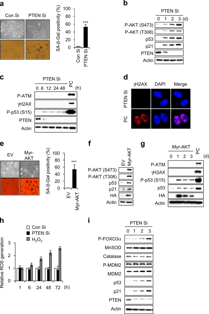
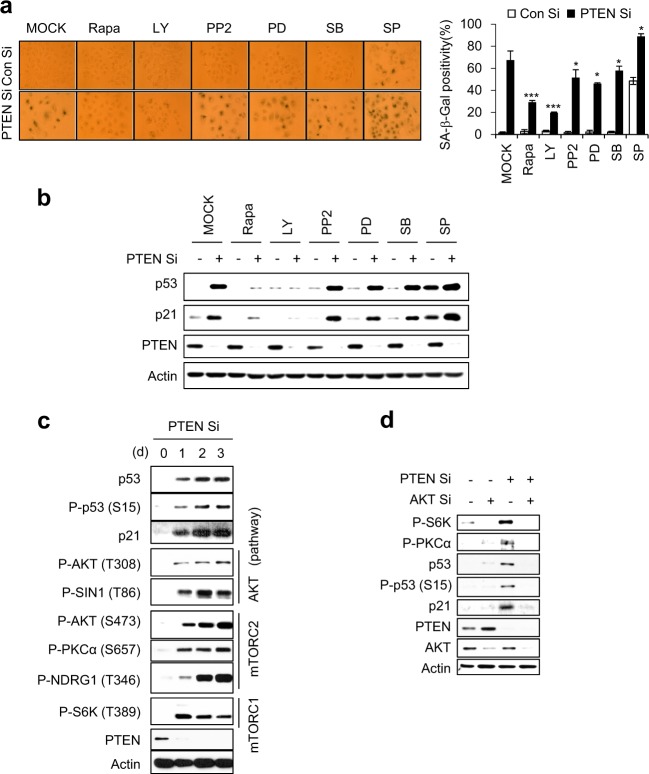
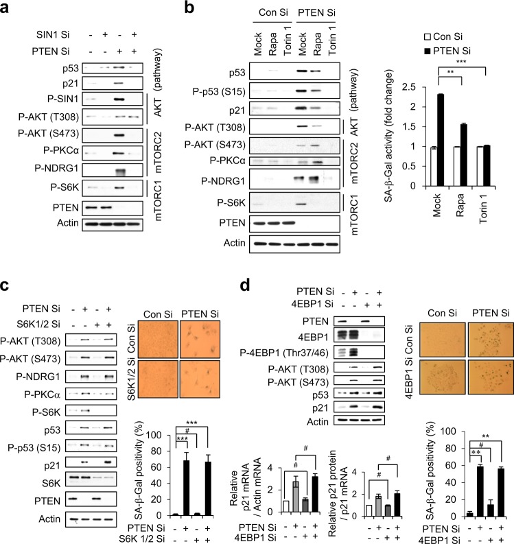
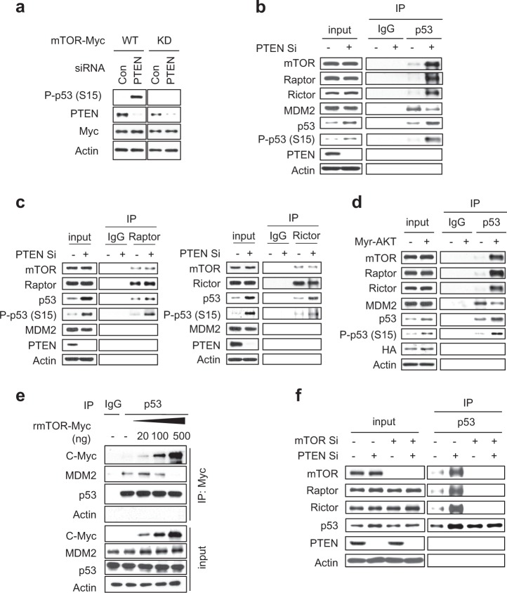
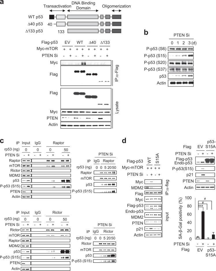
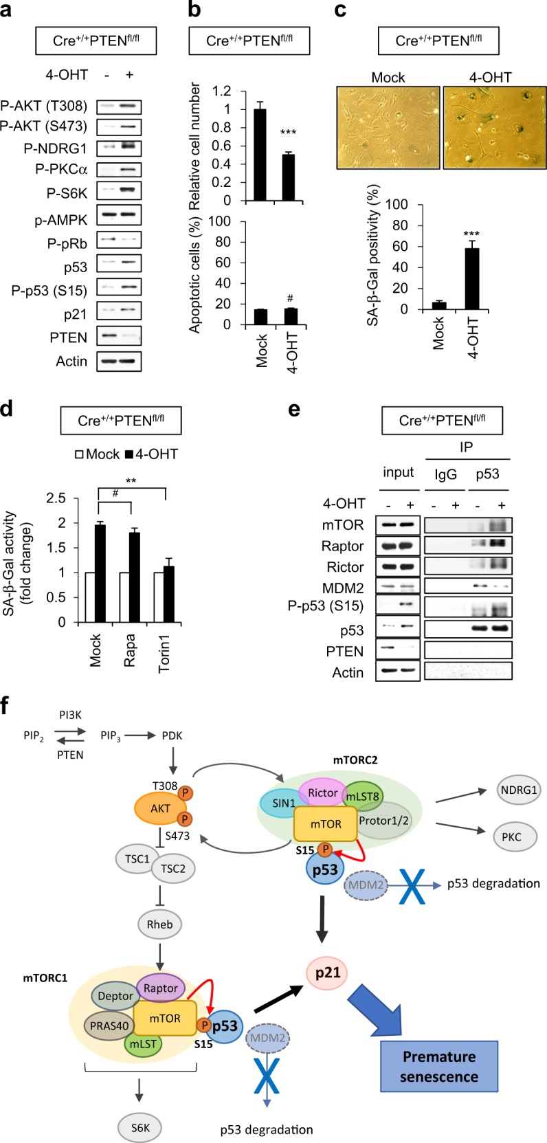
Loss of PTEN, the major negative regulator of the PI3K/AKT pathway induces a cellular senescence as a failsafe mechanism to defend against tumorigenesis, which is called PTEN-loss-induced cellular senescence (PICS). Although many studies have indicated that the mTOR pathway plays a critical role in cellular senescence, the exact functions of mTORC1 and mTORC2 in PICS are not well understood. In this study, we show that mTOR acts as a critical relay molecule downstream of PI3K/AKT and upstream of p53 in PICS. We found that PTEN depletion induces cellular senescence via p53-p21 signaling without triggering DNA damage response. mTOR kinase, a major component of mTORC1 and mTORC2, directly binds p53 and phosphorylates it at serine 15. mTORC1 and mTORC2 compete with MDM2 and increase the stability of p53 to induce cellular senescence via accumulation of the cell cycle inhibitor, p21. In embryonic fibroblasts of PTEN-knockout mice, PTEN deficiency also induces mTORC1 and mTORC2 to bind to p53 instead of MDM2, leading to cellular senescence. These results collectively demonstrate for the first time that mTOR plays a critical role in switching cells from proliferation signaling to senescence signaling via a direct link between the growth-promoting activity of AKT and the growth-suppressing activity of p53.
Conflict of interest statement
The authors declare no conflict of interest.
Figures
Similar articles
-
AKT induces senescence in human cells via mTORC1 and p53 in the absence of DNA damage: implications for targeting mTOR during malignancy.Oncogene. 2012 Apr 12;31(15):1949-62. doi: 10.1038/onc.2011.394. Epub 2011 Sep 12. Oncogene. 2012. PMID: 21909130 Free PMC article.
-
Discrete signaling mechanisms of mTORC1 and mTORC2: Connected yet apart in cellular and molecular aspects.Adv Biol Regul. 2017 May;64:39-48. doi: 10.1016/j.jbior.2016.12.001. Epub 2017 Jan 4. Adv Biol Regul. 2017. PMID: 28189457 Review.
-
Diverse signaling mechanisms of mTOR complexes: mTORC1 and mTORC2 in forming a formidable relationship.Adv Biol Regul. 2019 May;72:51-62. doi: 10.1016/j.jbior.2019.03.003. Epub 2019 Apr 11. Adv Biol Regul. 2019. PMID: 31010692 Review.
-
Fisetin-induced PTEN expression reverses cellular senescence by inhibiting the mTORC2-Akt Ser473 phosphorylation pathway in vascular smooth muscle cells.Exp Gerontol. 2021 Dec;156:111598. doi: 10.1016/j.exger.2021.111598. Epub 2021 Oct 22. Exp Gerontol. 2021. PMID: 34695518
-
Endothelial replicative senescence delayed by the inhibition of MTORC1 signaling involves MicroRNA-107.Int J Biochem Cell Biol. 2018 Aug;101:64-73. doi: 10.1016/j.biocel.2018.05.016. Epub 2018 May 29. Int J Biochem Cell Biol. 2018. PMID: 29857052
Cited by
-
Cystathionine-β-synthase is essential for AKT-induced senescence and suppresses the development of gastric cancers with PI3K/AKT activation.Elife. 2022 Jun 27;11:e71929. doi: 10.7554/eLife.71929. Elife. 2022. PMID: 35758651 Free PMC article.
-
Therapeutic potential of vasculogenic mimicry in urological tumors.Front Oncol. 2023 Sep 21;13:1202656. doi: 10.3389/fonc.2023.1202656. eCollection 2023. Front Oncol. 2023. PMID: 37810976 Free PMC article. Review.
-
Mapping the unicellular transcriptome of the ascending thoracic aorta to changes in mechanosensing and mechanoadaptation during aging.Aging Cell. 2024 Aug;23(8):e14197. doi: 10.1111/acel.14197. Epub 2024 Jun 2. Aging Cell. 2024. PMID: 38825882 Free PMC article.
-
Autophagy suppresses the formation of hepatocyte-derived cancer-initiating ductular progenitor cells in the liver.Sci Adv. 2021 Jun 4;7(23):eabf9141. doi: 10.1126/sciadv.abf9141. Print 2021 Jun. Sci Adv. 2021. PMID: 34088666 Free PMC article.
-
Senescence mechanisms and targets in the heart.Cardiovasc Res. 2022 Mar 25;118(5):1173-1187. doi: 10.1093/cvr/cvab161. Cardiovasc Res. 2022. PMID: 33963378 Free PMC article. Review.
References
Publication types
MeSH terms
Substances
LinkOut - more resources
Full Text Sources
Molecular Biology Databases
Research Materials
Miscellaneous
