. 2018 Oct 11:18:156.
doi: 10.1186/s12935-018-0655-3. eCollection 2018.

R-spodin2 enhances canonical Wnt signaling to maintain the stemness of glioblastoma cells

Affiliations

R-spodin2 enhances canonical Wnt signaling to maintain the stemness of glioblastoma cells

Si Liu et al. Cancer Cell Int. .

Abstract

Background: As newly identified Wnt enhancer, R-spondin gene family members have been linked to various cancers; however, their role in isocitrate dehydrogenase-wildtype subtype of human glioblastoma (GBM) cells remains unknown.

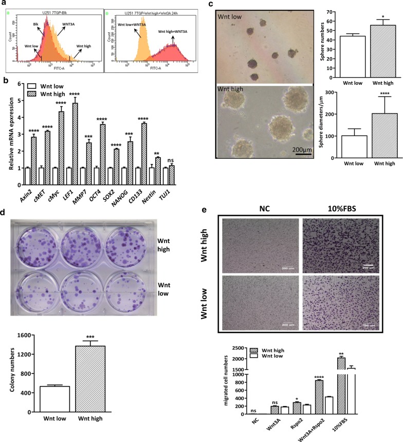
Methods: Human U87 and U251 cell lines were used to perform the experiments. GBM stem-like cells were enriched in stem cell growth media and induced to differentiate using retinoid acid or growth factor deprivation. Wnthigh and Wntlow subpopulations were isolated and evaluated by MTS, sphere formation, transwell migration and xenograft formation assays.

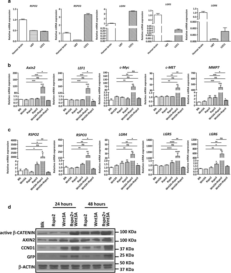
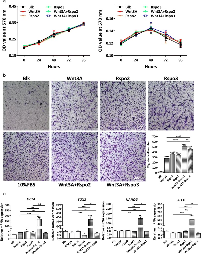
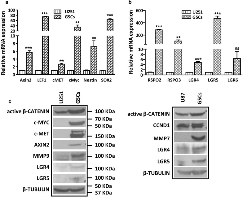
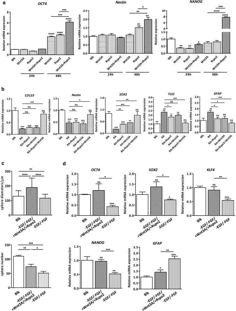
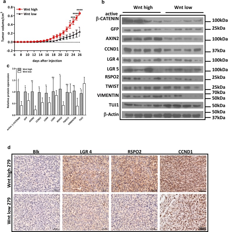
Results: R-spondin 2 but not R-spondin 3 potentiates Wnt/β-catenin signaling in GBM cell lines. While R-spondin 2 does not affect cell growth, it induces the expression of pluripotent stem cell markers in combination with Wnt3A. GBM stem-like cells are endowed with intrinsic high activity of β-catenin signaling, which can be further intensified by R-spondin 2. In addition, R-spondin2 promotes stem cell self-renewal and suppresses retinoid acid- or growth factor deprivation-induced differentiation, indicating R-spondin 2 maintains stem cell traits in GBM. On the other hand, we identify subpopulations of GBM cells that show distinctive responsiveness to Wnt/β-catenin signaling. Interestingly, Wnthigh and Wntlow cells display distinctive biologic properties. Moreover, Wnthigh cell-inoculated xenografts exhibit enhanced tumorigenicity and increased expression levels of R-spondin 2 compared to Wntlow cell-inoculated xenografts.

Conclusion: Our study reveals a novel regulatory mechanisms underlying the over-activation of β-catenin-mediated signaling in the pathogenesis of GBM.

Keywords: Cancer stem cells; Glioblastoma; R-spondin2; Stemness; Wnt.


Figures

Fig. 1
Fig. 1
Expression pattern and potentiation effect of RSPOs in U251 cells. a mRNA expression levels of RSPO2/3 and their receptors LGR4–6 were determined in U87 and U251 cells. U251 cells were pre-treated in serum-free medium for 24 h, then cultured in serum-free medium containing different Wnt ligands (20 ng/ml Wnt3A, Rspo2, Rspo3) for another 24 h. Rspo2 shows prominent potentiation effect on Wnt3A-induced canonical β-catenin downstream targets (b), and RSPO-LGR genes (c). Blk indicates U251 cells cultured in serum-free medium containing 0.1% DMSO. d 7TGP-transduced U251 cells were pre-treated in serum-free medium for 24 h, then cultured in serum-free medium containing different Wnt stimuli for another 24–48 h. Representative Western Blot showing the protein expression levels of canonical β-catenin downstream targets in 7TGP-transduced U251 cells with Wnt ligand treatment
Fig. 2
Fig. 2
Combination of Rspo2 and Wnt3A does not affect cell growth, but elevates the expression levels of stem cell genes in U251 cells. a U87 and U251cells were pre-treated in serum-free medium for 24 h, then cultured in serum-free medium containing different Wnt ligands for another 4 days, and cell growth was determined by MTT assay. Results show neither WNT ligand alone nor in combination has any effect on cell growth. b Transwell migration assay was performed in U251 cells. U251 cells were pre-treated in serum-free medium for 24 h, then 2 × 104 cells were cultured in serum-free medium in chambers, while wells containing different Wnt stimuli were used as chemotaxis source for another 24 h. scale bar represents 200 μm. Quantification analysis of data is expressed as the Mean ± SD from triplicates of two independent experiments. c The mRNA expression levels of stem cell marker OCT4, SOX2, NANOG and KLF4 were determined by real time PCR, results show that the combination of Rspo2 and Wnt3A significantly upregulates stem cell markers
Fig. 3
Fig. 3
GSC-like cells are endowed with upregulated Wnt-Lgr-Rspo axis. a U251 GSCs show dramatic increment of β-catenin targets and neural stem cell markers as determined by real time PCR. b Rspo/Lgr axis is upregulated in U251 GSCs as determined by real time PCR. c the protein expression levels of β-catenin targets and LGRs were upregulated in U251 and U87 GSCs
Fig. 4
Fig. 4
Wnt3A and Rspo2 maintains stemness in GSCs. a Rspo2 and Wnt3A upregulates stem cell marker in U251 GSCs as demonstrated by real time PCR. Blk indicates U251 GSCs cultured in GSC media with 0.1% DMSO. b All-trans retinoic acid (10 µM RA) was used to induce differentiation in U251 GSCs for 24 h with or without Wnt ligands (20 ng/ml). Real-time PCR was used to determine the effect on differentiation. Results show that Rspo2/Wnt3A treatment rescues RA-induced GSC differentiation. Blk indicates U251 GSCs cultured in DMEM with 0.1% DMSO. c GSCs were cultured in GSC media, or GSC media without EGF and FGF, or GSC media without EGF and FGF but with Wnt3A and Rspo2 for 7 days. Results show that lack of EGF/FGF causes reduced sphere formation, which can be compensated by adding Rspo2/Wnt3A. d Real-time PCR shows that Rspo2/Wnt3A treatment abolishes the downregulation of stem cell markers and upregulation of differentiation markers caused by growth factor deprivation. Blk in C and D indicates U251 GSCs cultured in GSC media with 0.1% DMSO
Fig. 5
Fig. 5
Wnthigh and Wntlow sub-populations of U251 cells exhibit different cellular properties in vitro. a Isolation of Wnthigh and Wntlow sub-populations of U251 cells. Left panel, Typical flow cytometry analysis depicting unsorted U251-7TGP cells treated with 20 ng/ml Wnt3A (yellow). For subsequent experiment, the 5% of cells with the highest and lowest GFP levels were sorted. Right panel, shown is the flow cytometric reanalysis of the respective subgroups after several rounds of enrichment. b Real-time PCR demonstrates that Wnthigh cells show higher expression levels of β-catenin targets and stem cell markers. c Sphere formation assay shows that Wnthigh cells have an enhanced sphere formation ability. Scale bar is 200 μm. Quantification analysis of data is expressed as the Mean ± SD from three independent experiments. d Soft agar assay shows Wnthigh cells have a higher anchorage-independent cell growth ability. Quantification analysis of data is expressed as the Mean ± SD from three independent experiments. e Transwell assay shows Wnthigh cells exhibit enhanced cell migratory ability. Quantification analysis of data is expressed as the Mean ± SD from three independent experiments. N. C. indicates vehicle control of Wnthigh and Wntlow cells
Fig. 6
Fig. 6
Wnthigh tumors grow faster and have high RSPO expression. a 1 × 104 Wnthigh or Wntlow cells were inoculated at the flanks of nude mice, and tumor growth curve were recorded. The result shows that Wnthigh cell-injected xenografts (n = 9) grow faster than Wntlow cell-injected xenografts (n = 6). b Protein expression of β-catenin targets, RSPO/LGR, EMT markers and differentiation marker was compared between Wnthigh (n = 4) and Wntlow (n = 4) xenografts. c Statistic analysis of G. d Tumor sections from Wnthigh or Wntlow xenografts were stained by RSPO2, LGR4 and CCND1, where Wnthigh tumors display stronger staining compared to Wntlow tumors. Scale bar is 200 μm

References

    1. Gorlia T, van den Bent MJ, Hegi ME, Mirimanoff RO, Weller M, Cairncross JG, Eisenhauer E, Belanger K, Brandes AA, Allgeier A, et al. Nomograms for predicting survival of patients with newly diagnosed glioblastoma: prognostic factor analysis of EORTC and NCIC trial 26981-22981/CE.3. Lancet Oncol. 2008;9(1):29–38. doi: 10.1016/S1470-2045(07)70384-4. - DOI - PubMed
    1. Huntly BJ, Gilliland DG. Leukaemia stem cells and the evolution of cancer-stem-cell research. Nat Rev Cancer. 2005;5(4):311–321. doi: 10.1038/nrc1592. - DOI - PubMed
    1. Al-Hajj M, Becker MW, Wicha M, Weissman I, Clarke MF. Therapeutic implications of cancer stem cells. Curr Opin Genet Dev. 2004;14(1):43–47. doi: 10.1016/j.gde.2003.11.007. - DOI - PubMed
    1. Bao S, Wu Q, McLendon RE, Hao Y, Shi Q, Hjelmeland AB, Dewhirst MW, Bigner DD, Rich JN. Glioma stem cells promote radioresistance by preferential activation of the DNA damage response. Nature. 2006;444(7120):756–760. doi: 10.1038/nature05236. - DOI - PubMed
    1. Singh SK, Clarke ID, Terasaki M, Bonn VE, Hawkins C, Squire J, Dirks PB. Identification of a cancer stem cell in human brain tumors. Cancer Res. 2003;63(18):5821–5828. - PubMed