. 2018 Sep 25:9:2184.
doi: 10.3389/fimmu.2018.02184. eCollection 2018.

White Spot Syndrome Virus-Induced Shrimp miR-315 Attenuates Prophenoloxidase Activation via PPAE3 Gene Suppression

Affiliations

White Spot Syndrome Virus-Induced Shrimp miR-315 Attenuates Prophenoloxidase Activation via PPAE3 Gene Suppression

Phattarunda Jaree et al. Front Immunol. .

Abstract

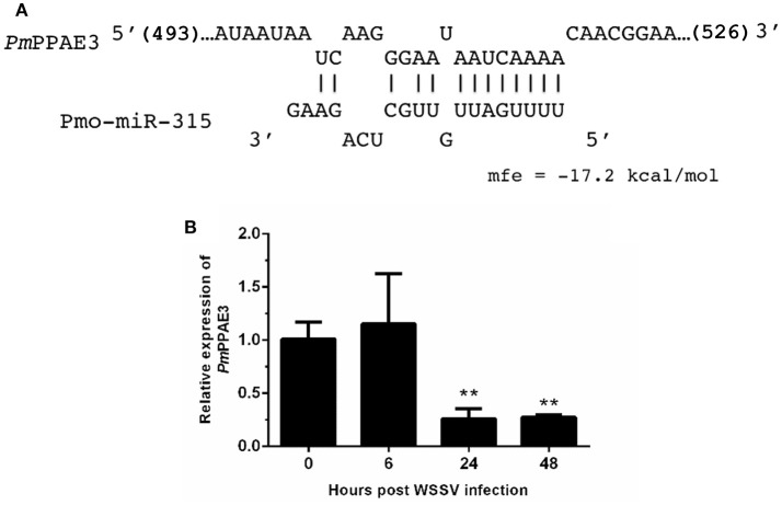
MicroRNAs (miRNAs), the small non-coding RNAs, play a pivotal role in post-transcriptional gene regulation in various cellular processes. However, the miRNA function in shrimp antiviral response is not clearly understood. This research aims to uncover the function of pmo-miR-315, a white spot syndrome virus (WSSV)-responsive miRNAs identified from Penaeus monodon hemocytes during WSSV infection. The expression of the predicted pmo-miR-315 target mRNA, a novel PmPPAE gene called PmPPAE3, was negatively correlated with that of the pmo-miR-315. Furthermore, the luciferase assay indicated that the pmo-miR-315 directly interacted with the target site in PmPPAE3 suggesting the regulatory role of pmo-miR-315 on PmPPAE3 gene expression. Introducing the pmo-miR-315 into the WSSV-infected shrimp caused the reduction of the PmPPAE3 transcript level and, hence, the PO activity activated by the PmPPAE3 whereas the WSSV copy number in the shrimp hemocytes was increased. Taken together, our findings state a crucial role of pmo-miR-315 in attenuating proPO activation via PPAE3 gene suppression and facilitating the WSSV propagation in shrimp WSSV infection.

Keywords: Penaeus monodon; invertebrates; microRNA; prophenoloxidase; viral infection.


Figures

Figure 1
Figure 1
The expression of pmo-miR-315 in WSSV-infected shrimp tissues. Quantitative stem-loop real-time RT-PCR was performed to determine the expression level of pmo-miR-315 in hemocytes, gill, lymphoid organ and stomach at 0, 6, 24, and 48 hpi. Using U6 as an internal control, the relative expression level of miR-315 was calculated. All experiments were performed in triplicate. The * and ** indicate the significant difference at P < 0.05 and P < 0.01, respectively.
Figure 2
Figure 2
The pmo-miR-315 target mRNA, prophenoloxidase-activating enzyme 3 (PmPPAE3). (A) The pmo-miR-315/PmPPAE3 base pairing and the binding energy was predicted using RNA hybrid software. The number in bracket indicates the position of nucleotide of pmo-miR-315 target site on the PmPPAE3 CDS. (B) The relative expression level of PmPPAE3 gene at 0, 6, 24, and 48 h post-WSSV infection was investigated. The EF-1α gene was used as an internal control. All experiments were performed in triplicate. The ** indicates the significant difference at P < 0.05.
Figure 3
Figure 3
The pmo-miR-315/PmPPAE3 interaction by luciferase reporter assay. The target sequence of pmo-miR-315 from PmPPAE3 was amplified and cloned into the pmirGLO vector (pmir-T315). The pmo-miR-315 mimic or scramble pmo-miR-315 were co-transfected with pmir-T315 using Effectene transfection reagent (Qiagen) into HEK293T cells. At 48 h after transfection, the luciferase activity was measured using a Dual-luciferase® reporter assay system (Promega). The data shown is derived from triplicate experiments. The ** indicates significant difference (P < 0.01).
Figure 4
Figure 4
Characterization of PmPPAE3 gene. (A) Phylogenetic analysis of PPAEs from various organisms including crustaceans and insects, e.g., B. mori PO-activating enzyme (BmPAE); M. sexta proPO-activating proteinase 1 (MsPAP1), H. diomphalia proPO-activating factor I (HdPPAFI), G. morsitans (GmPPAE) and D. melanogaster melanization protease1 (DmMP1); shrimp P. monodon proPO-activating enzymes 1, 2 and 3 (PmPPAE1, PmPPAE2, PmPPAE3), shrimp, L. vannamei proPO-activating enzyme (LvPPAE), F. chinensis proPO-activating enzyme (FcPPAE), and crayfish P. leniusculus proPO-activating enzyme (PlPPAE), was performed using ClustaX 2.1 and MEGA7 softwares. (B) Multiple alignment of the deduced amino acid sequences of PmPPAE3 with other crustacean PPAEs was conducted using the Clustal Omega program. The conserved features of PPAEs such as the disulfide bond, the catalytic triad (histidine, aspartic acid, and serine residues), the clip domain, and the serine proteinase domain are shown. (C) The representative result of PmPPAE3 gene tissue distribution analysis by semi-quantitative RT-PCR in the hemocytes (Hc), gill (G), stomach (St), and lymphoid organ (L), is shown. In this analysis, the EF-1α transcript was used as an internal control. All experiments were performed in triplicate. (D) The PmPPAE3 gene knockdown using PmPPAE3-dsRNA was performed to reveal the functional role of PmPPAE in proPO activating system. The effectiveness of the PmPPAE3 gene knockdown is shown. Hemocytes collected from the control groups of 0.85% NaCl and dsGFP injected shrimp and from the experimental shrimp injected with PmPPAE3-dsRNA was analyzed for the expression of PmPPAE3 gene by qRT-PCR using the EF-1α transcript as an internal control. (E) The PO activity was assayed in the PmPPAE3 knockdown shrimp hemocytes. The data are derived from three independently replicated experiments. The * and ** indicate the significant difference at P < 0.05 and P < 0.01, respectively.
Figure 5
Figure 5
The role of pmo-miR-315 in WSSV-infected shrimp hemocytes. In this experiment, either pmo-miR-315 mimic or AMO-miR-315 was injected into WSSV-infected shrimp. Then, several parameters were investigated including (A) pmo-miR-315 and (B) PmPPAE3 gene expression level, (C) PO activity and (D) WSSV copy number. The U6 and EF-1α were used as internal controls for pmo-miR-315 and PmPPAE3 expression, respectively. All experiments were performed in triplicate. The * and ** indicate the significant difference at P < 0.05 and P < 0.01, respectively.
Figure 6
Figure 6
A schematic representation of the pmo-miR-315 role in modulating proPO activity by PmPPE3 suppression during WSSV infection.

References

    1. Bartel DP. MicroRNAs: genomics, biogenesis, mechanism, and function. Cell (2004) 116:281–97. 10.1016/S0092-8674(04)00045-5 - DOI - PubMed
    1. Jopling CL, Yi M, Lancaster AM, Lemon SM, Sarnow P. Modulation of hepatitis C virus RNA abundance by a liver-specific microRNA. Science (2005) 309:1577–81. 10.1126/science.1113329 - DOI - PubMed
    1. Forman JJ, Coller HA. The code within the code: microRNAs target coding regions. Cell Cycle (2010) 9:1533–41. 10.4161/cc.9.8.11202 - DOI - PMC - PubMed
    1. Skalsky RL, Cullen BR. Viruses, microRNAs, and host interactions. Annu Rev Microbiol. (2010) 64:123–41. 10.1146/annurev.micro.112408.134243 - DOI - PMC - PubMed
    1. Huang T, Xu D, Zhang X. Characterization of host microRNAs that respond to DNA virus infection in a crustacean. BMC Genomics (2012) 13:159. 10.1186/1471-2164-13-159 - DOI - PMC - PubMed

Publication types

MeSH terms