. 2018 Oct 10:15:73.
doi: 10.1186/s12986-018-0310-y. eCollection 2018.

Gynura divaricata rich in 3, 5-/4, 5-dicaffeoylquinic acid and chlorogenic acid reduces islet cell apoptosis and improves pancreatic function in type 2 diabetic mice

Affiliations

Gynura divaricata rich in 3, 5-/4, 5-dicaffeoylquinic acid and chlorogenic acid reduces islet cell apoptosis and improves pancreatic function in type 2 diabetic mice

Xiao-Lu Yin et al. Nutr Metab (Lond). .

Abstract

Background: Diabetes mellitus is one of the most common chronic diseases that accompanied by severe complications. Gynura divaricata (GD), a medicinal and edible plant that is usually used for the treatment of diabetes. Therefore, this study investigates the chemical components of GD with hypoglycemic effect and the possible mechanism lowering blood sugar in T2D diabetic mice.

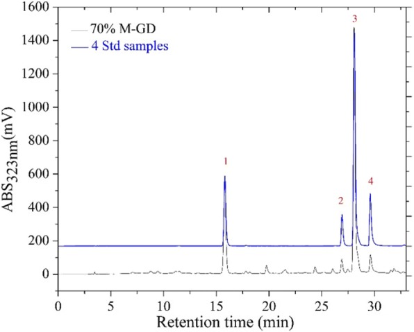
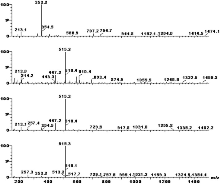
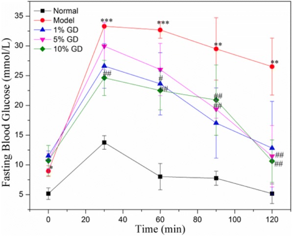
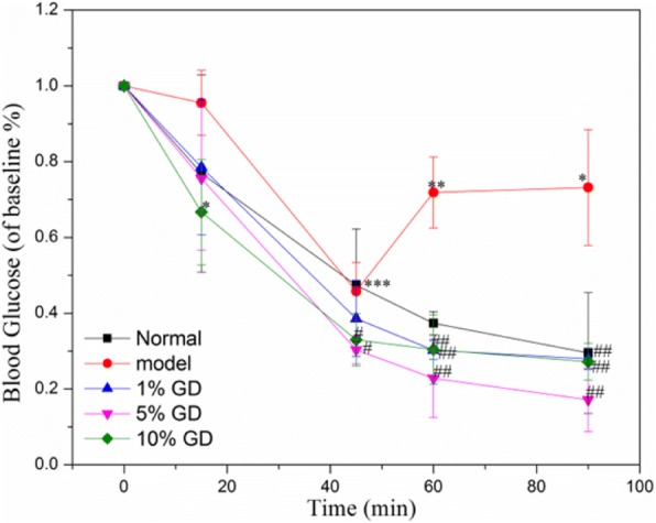
Methods: The methanol extract of GD was analysed by HPLC-DAD. And then mice with type 2 diabetes induced by a high-fat diet in combination with streptozotocin feed the diet containing lyophilized GD powder for 4 weeks. During this period, fasting blood glucose (FBG) levels and body weight were measured.

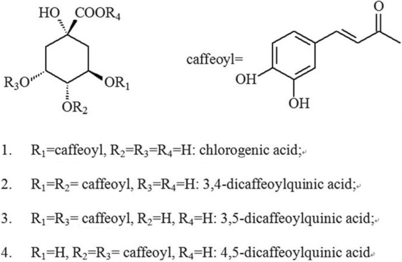
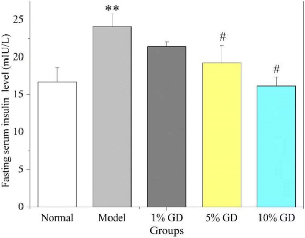
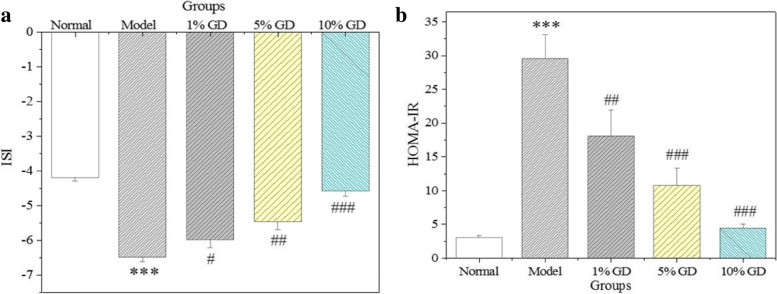
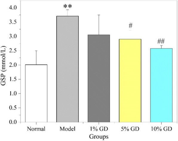
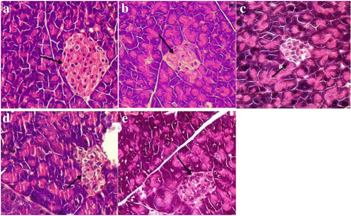
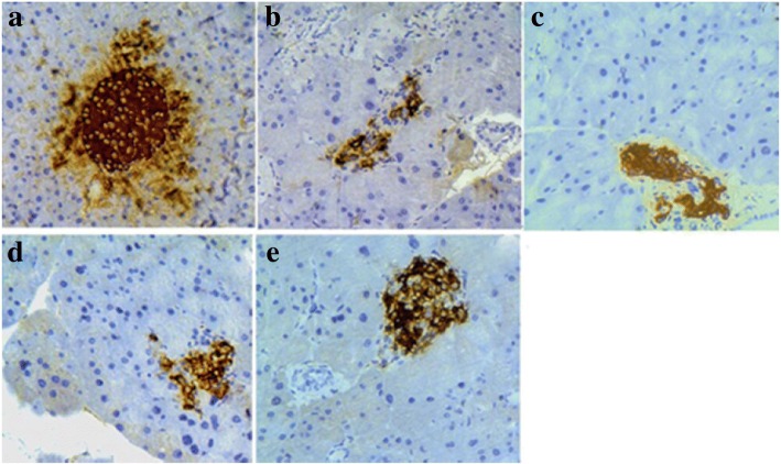
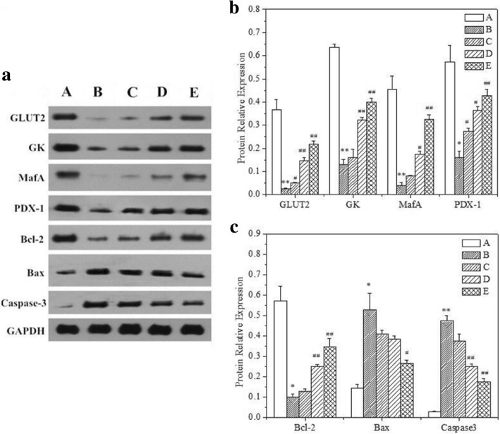
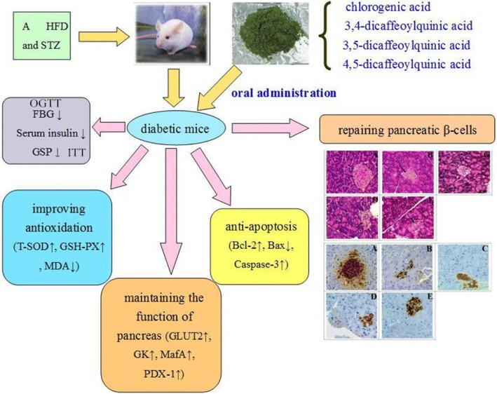
Results: GD was rich in four bioactive components of dicaffeoylquinic acid and chlorogenic acid. These components occupied about 2.37% in the GD powder in which the highest level was 3, 5-dicaffeoylquinic acid. Oral GD significantly reduced FBG, fasting serum insulin, and glycosylated serum protein levels, and enhanced antioxidative activities. HE-staining showed that the pathological damage in pancreatic β-cells was ameliorated. An immunohistochemical assay also showed that GD promoted marked pancreatic β-cell regeneration. GD also caused notable increase in GLUT2, GK, MafA, PDX-1, and Bcl-2 as well as reduction in Bax and caspase-3 expression as shown by western blot analysis.

Conclusions: GD exerts the pronounced hypoglycaemic effect by inhibiting islet cell apoptosis and improving pancreatic function. Therefore, GD might have a potential to improve diabetes.

Keywords: Diabetes mellitus; Fasting blood glucose; Gynura divaricata; Pancreatic β-cell; Serum insulin; Type 2 diabetes.


Conflict of interest statement

All animal experimental protocols used in this study were approved by the Animal Ethics Committee at Soochow University (201504A136).No applicable.The authors declare that they have no competing interests.Springer Nature remains neutral with regard to jurisdictional claims in published maps and institutional affiliations.

Figures

Fig. 1
Fig. 1
HPLC chromatogram of 70% methanol extract of GD and four standards at 323 nm. 70% M-GD, 70% methanol extract of GD lyophilised powder; 4Std, 4 standards including chlorogenic acid (1), 3,4-dicaffeoylquinic acid (2), 3,5-dicaffeoylquinic acid (3) and 4,5-dicaffeoylquinic acid (4)
Fig. 2
Fig. 2
Negative ion mass spectra of four main chemical constituents in 70% methanol extract of GD
Fig. 3
Fig. 3
Structures of chlorogenic acid, 3,4-dicaffeoylquinic acid, 3,5-dicaffeoylquinic acid and 4,5-dicaffeoylquinic acid
Fig. 4
Fig. 4
The effect of different dose of GDP on OGTT in mice
Fig. 5
Fig. 5
The effect of different dose of GDP on ITT in mice
Fig. 6
Fig. 6
Effect of oral GD on the fasting serum insulin levels. The fasting serum insulin levels were determined in mice in the fasting state after 4 weeks of GD treatment. 1% GD, 1% GD-treated diabetic group; 5% GD, 5% GD-treated diabetic group; 10% GD, 10% GD-treated diabetic group. The experimental data are presented as means ± SD. **P < 0.01, versus normal group; #P < 0.05, versus diabetic model group
Fig. 7
Fig. 7
Effects of GD on ISI (a) and HOMA-IR (b). 1% GD, 1% GD-treated diabetic group; 5% GD, 5% GD-treated diabetic group; 10% GD, 10% GD-treated diabetic group. The experimental data are presented as means ± SD. ***P < 0.01, versus normal group; #P < 0.05, ##p < 0.01 and ###p < 0. 001, respectively, versus diabetic model group
Fig. 8
Fig. 8
Effect of GD on GSP. 1% GD, 1% GD-treated diabetic group; 5% GD, 5% GD-treated diabetic group; 10% GD, 10% GD-treated diabetic group. The experimental data are presented as means ± SD. **P < 0.01, versus normal group; #P < 0.05 and ##p < 0.01, respectively, versus diabetic model group
Fig. 9
Fig. 9
Histopathologic examination of pancreas in diabetic mice (HE stain, × 400). a normal group; b diabetic model group; c 1% GD-treated diabetic group; d 5% GD-treated diabetic group; e 10% GD-treated diabetic group. Arrows represent the pancreatic islet with pancreatic β-cells
Fig. 10
Fig. 10
Insulin immunohistochemistry of pancreas in diabetic mice (× 400). a normal group; b diabetic model group; c 1% GD-treated diabetic group; d 5% GD-treated diabetic group; e 10% GD-treated diabetic group
Fig. 11
Fig. 11
Effect of GD on GLUT2, GK, MafA, PDX-1, Bcl-2, Bax, and caspase-3 protein expressions in pancreatic tissues. (A) Western blot analysis of GLUT2, GK, MafA, PDX-1, Bcl-2, Bax, and caspase-3 protein expressions. (B) Quantitative analysis of GLUT2, GK, MafA, and PDX-1 protein expressions. (C) Quantitative analysis of Bcl-2, Bax, and caspase-3 protein expressions. A: normal group; B: diabetic model group; C: 1% GD-treated diabetic group; D: 5% GD-treated diabetic group; E: 10% GD-treated diabetic group. The experimental data are presented as means ± SD. *P < 0.05 and **P < 0.01, respectively, versus normal group; #P < 0.05 and ##P < 0.01, respectively, versus diabetic model group
Fig. 12
Fig. 12
Schematic representation of the protective effect of GD diet in pancreatic tissue of type 2 diabetic mice

Similar articles

Cited by

References

    1. Tandon N, Ali MK, Narayan KV. Pharmacologic prevention of microvascular and macrovascular complications in diabetes mellitus. Am J Cardiovasc Drugs. 2012;12(1):7–22. doi: 10.2165/11594650-000000000-00000. - DOI - PubMed
    1. Blonde L. Benefits and risks for intensive glycemic control in patients with diabetes mellitus. Am J Med Sci. 2012;343(1):17–20. doi: 10.1097/MAJ.0b013e31823ea23e. - DOI - PubMed
    1. Edelman SV. Importance of glucose control. Med Clin N Am. 1998;82(4):665–687. doi: 10.1016/S0025-7125(05)70019-5. - DOI - PubMed
    1. Rosiak M, Grzeszczak S, Kosior DA, Postuła M. Emerging treatments in type 2 diabetes: focus on canagliflozin. Ther Clin Risk Manag. 2014;10:683–689. - PMC - PubMed
    1. Sharma AK, Bharti S, Goyal S, Arora S, Nepal S, Kishore K. Upregulation of pparγ by aegle marmelos, ameliorates insulin resistance and β-cell dysfunction in high fat diet fed-streptozotocin induced type 2 diabetic rats. Phytother Res. 2011;25(10):1457–1465. doi: 10.1002/ptr.3442. - DOI - PubMed