

Evaluation of an Improved Chitosan Scaffold Cross-Linked With Polyvinyl Alcohol and Amine Coupling Through 1-Ethyl-3-(3-Dimethyl Aminopropyl)-Carbodiimide (EDC) and 2 N-Hydroxysuccinimide (NHS) for Corneal Applications
- PMID: 30337966
- PMCID: PMC6182522
- DOI: 10.3889/oamjms.2018.322
Evaluation of an Improved Chitosan Scaffold Cross-Linked With Polyvinyl Alcohol and Amine Coupling Through 1-Ethyl-3-(3-Dimethyl Aminopropyl)-Carbodiimide (EDC) and 2 N-Hydroxysuccinimide (NHS) for Corneal Applications
Abstract
Background: Corneal blindness resulting from various medical conditions affects millions worldwide. The rapid developing tissue engineering field offers design of a scaffold with mechanical properties and transparency similar to that of the natural cornea.
Aim: The present study aimed at to prepare and investigate the properties of PVA/chitosan blended scaffold by further cross-linking with 1-Ethyl-3-(3-dimethyl aminopropyl)-carbodiimide (EDC) and 2 N-Hydroxysuccinimide (NHS) as potential in vitro carrier for human limbal stem cells delivery.
Material and methods: Acetic acid dissolved chitosan was added to PVA solution, uniformly mixed with a homogenizer until the mixture was in a colloidal state, followed by H2SO4 and formaldehyde added and the sample was allowed to cool, subsequently it was poured into a tube and heated in an oven at 60°C for 50 minutes. Finally, samples were soaked in a cross-linking bath with EDC, NHS and NaOH in H2O/EtOH for 24 h consecutively stirred to cross-link the polymeric chains, reduce degradation. After soaking in the bath, the samples were carefully washed with 2% glycine aqueous solution several times to remove the remaining amount of cross-linkers, followed by washed with water to remove residual agents. Later the cross-linked scaffold subjected for various characterization and biological experiments.
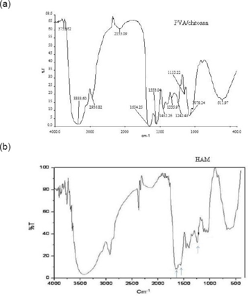
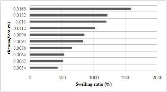
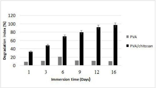
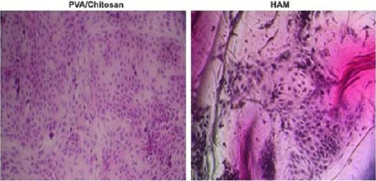
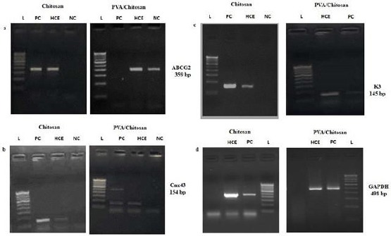
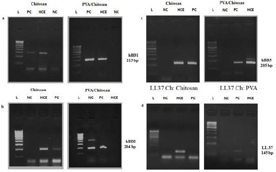
Results: After viscosity measurement, the scaffold was observed by Fourier transform infrared (FT-IR). The water absorbency of PVA/Chitosan was increased 361% by swelling. Compression testing demonstrated that by increasing the amount of chitosan, the strength of the scaffold could be increased to 16×10-1 MPa. Our degradation results revealed by mass loss using equation shows that scaffold degraded gradually imply slow degradation. In vitro tests showed good cell proliferation and growth in the scaffold. Our assay results confirmed that the membrane could increase the cells adhesion and growth on the substrate.
Conclusion: Hence, we strongly believe the use of this improved PVA/chitosan scaffold has potential to cut down the disadvantages of the human amniotic membrane (HAM) for corneal epithelium in ocular surface surgery and greater mechanical strength in future after successful experimentation with clinical trials.
Keywords: Biocompatibility; Chitosan; Corneal cells; PVA; Poly (vinyl alcohol); Tissue engineering.
Figures
Similar articles
-
[Fabrication and properties of a composite chitosan/type II collagen scaffold for tissue engineering cartilage].Zhongguo Xiu Fu Chong Jian Wai Ke Za Zhi. 2005 Apr;19(4):278-82. Zhongguo Xiu Fu Chong Jian Wai Ke Za Zhi. 2005. PMID: 15921318 Chinese.
-
Carbodiimide cross-linked amniotic membranes for cultivation of limbal epithelial cells.Biomaterials. 2010 Sep;31(25):6647-58. doi: 10.1016/j.biomaterials.2010.05.034. Epub 2010 Jun 11. Biomaterials. 2010. PMID: 20541801
-
Fabrication and characterization of spongy denuded amniotic membrane based scaffold for tissue engineering.Cell J. 2015 Winter;16(4):476-87. doi: 10.22074/cellj.2015.493. Epub 2015 Jan 13. Cell J. 2015. PMID: 25685738 Free PMC article.
-
Applications of Human Amniotic Membrane for Tissue Engineering.Membranes (Basel). 2021 May 25;11(6):387. doi: 10.3390/membranes11060387. Membranes (Basel). 2021. PMID: 34070582 Free PMC article. Review.
-
Cross-Linking Agents in Three-Component Materials Dedicated to Biomedical Applications: A Review.Polymers (Basel). 2024 Sep 23;16(18):2679. doi: 10.3390/polym16182679. Polymers (Basel). 2024. PMID: 39339142 Free PMC article. Review.
Cited by
-
Electrospun Nanofibrous Membranes Based on Citric Acid-Functionalized Chitosan Containing rGO-TEPA with Potential Application in Wound Dressings.Polymers (Basel). 2022 Jan 12;14(2):294. doi: 10.3390/polym14020294. Polymers (Basel). 2022. PMID: 35054703 Free PMC article.
-
Chitosan based bioactive materials in tissue engineering applications-A review.Bioact Mater. 2020 Feb 12;5(1):164-183. doi: 10.1016/j.bioactmat.2020.01.012. eCollection 2020 Mar. Bioact Mater. 2020. PMID: 32083230 Free PMC article. Review.
-
Poly(Vinyl Alcohol)-Based Nanofibrous Electrospun Scaffolds for Tissue Engineering Applications.Polymers (Basel). 2019 Dec 18;12(1):7. doi: 10.3390/polym12010007. Polymers (Basel). 2019. PMID: 31861485 Free PMC article. Review.
-
Development of an electrospun poly(ε-caprolactone)/collagen-based human amniotic membrane powder scaffold for culturing retinal pigment epithelial cells.Sci Rep. 2022 Apr 19;12(1):6469. doi: 10.1038/s41598-022-09957-5. Sci Rep. 2022. PMID: 35440610 Free PMC article.
-
The revolutionary role of placental derivatives in biomedical research.Bioact Mater. 2025 Mar 19;49:456-485. doi: 10.1016/j.bioactmat.2025.03.011. eCollection 2025 Jul. Bioact Mater. 2025. PMID: 40177109 Free PMC article. Review.
References
-
- Ambati BK, Nozaki M, Singh N, Takeda A, Jani PD, Suthar T, Albuquerque RJC, Richter E, Sakurai E, Newcomb MT. Corneal avascularity is due to soluble VEGF receptor-1. Nature. 2006;443:993–97. https://doi.org/10.1038/nature05249 PMid:17051153 PMCid:PMC2656128. - PMC - PubMed
-
- World Health Organization. Universal Eye Health. A Global Action Plan 2014-2019. Geneva: WHO; 2013.
-
- VISION 2020. The Right to Sight. [[Last cited on 2017 Jan 19]]. Available from: http://www.iapb.org/vision-2020/
-
- McLaughlin CR, Tsai RJF, Latorre MA, Griffith M. Bioengineered corneas for transplantation and in vitro toxicology. Front Biosci. 2009;14:3326–37. https://doi.org/10.2741/3455. - PubMed
-
- Ruberti JW, Zieske JD. Prelude to corneal tissue engineering-Gaining control of collagen organisation. Prog Retin Eye Res. 2008;27:549–77. https://doi.org/10.1016/j.preteyeres.2008.08.001 PMid:18775789 PMCid:PMC3712123. - PMC - PubMed
LinkOut - more resources
Full Text Sources
Miscellaneous