. 2018 Sep 21;9(74):33896-33911.
doi: 10.18632/oncotarget.26084.

Inhibition of store-operated channels by carboxyamidotriazole sensitizes ovarian carcinoma cells to anti-BclxL strategies through Mcl-1 down-regulation

Affiliations

Inhibition of store-operated channels by carboxyamidotriazole sensitizes ovarian carcinoma cells to anti-BclxL strategies through Mcl-1 down-regulation

Marie-Laure Bonnefond et al. Oncotarget. .

Abstract

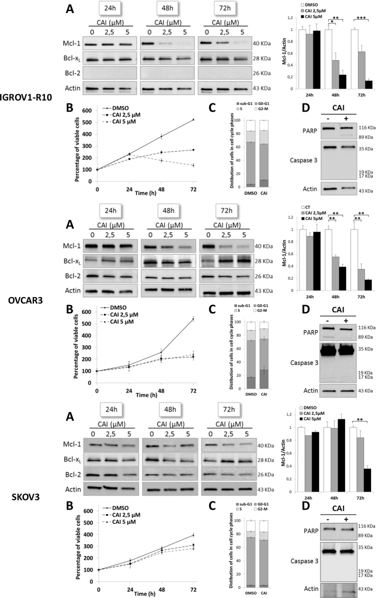
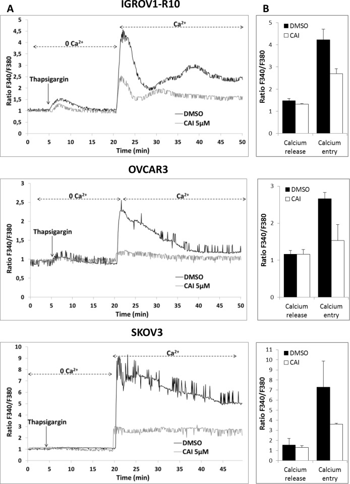
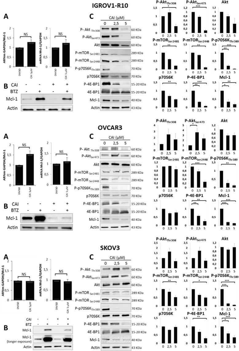
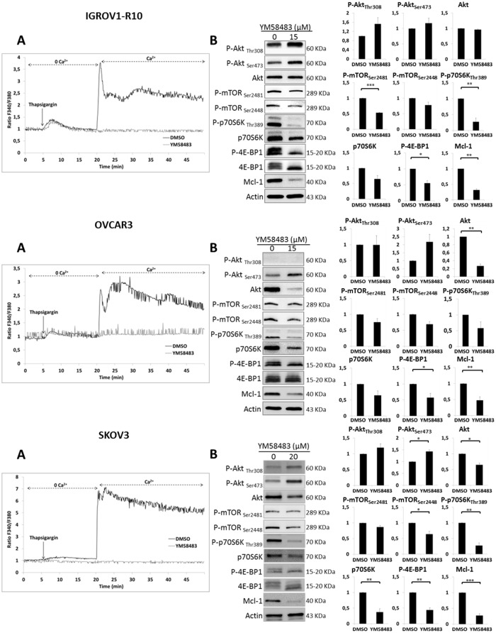
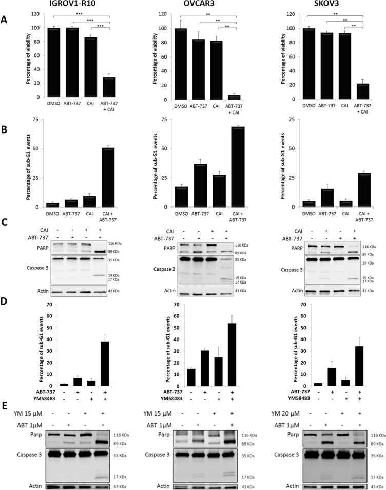
The anti-apoptotic proteins Bcl-xL and Mcl-1 have been identified to play a pivotal role in apoptosis resistance in ovarian cancer and constitute key targets for innovative therapeutic strategies. Although BH3-mimetics (i.e. ABT-737) potently inhibit Bcl-xL activity, targeting Mcl-1 remains a hurdle to the success of these strategies. Calcium signaling is profoundly remodeled during carcinogenesis and was reported to activate the signaling pathway controlling Mcl-1 expression. In this context, we investigated the effect of carboxyamidotriazole (CAI), a calcium channel inhibitor used in clinical trials, on Mcl-1 expression. CAI had an anti-proliferative effect on ovarian carcinoma cell lines and strongly down-regulated Mcl-1 expression. It inhibited store-operated calcium entry (SOCE) and Mcl-1 translation through mTORC1 deactivation. Moreover, it sensitized ovarian carcinoma cells to anti-Bcl-xL strategies as their combination elicited massive apoptosis. Its effect on mTORC1 and Mcl-1 was mimicked by the potent SOCE inhibitor, YM58483, which also triggered apoptosis when combined with ABT-737. As a whole, this study suggests that CAI sensitizes to anti-Bcl-xL strategies via its action on Mcl-1 translation and that modulation of SOCE could extend the therapeutic arsenal for treatment of ovarian carcinoma.

Keywords: ABT-737; MCL-1; Store-operated calcium channels; mTORC1; ovarian cancer.


Conflict of interest statement

CONFLICTS OF INTEREST The authors declare no conflicts of interest.

Figures

Figure 1
Figure 1. CAI inhibits Mcl-1 protein expression and has an anti-proliferative effect on three ovarian cell lines
(A) Expressions of Mcl-1, Bcl-xL and Bcl-2 were assessed by western blot in IGROV1-R10, OVCAR3 and SKOV3. Cells were treated by increasing concentrations of CAI for 24h, 48h and 72h. Mcl-1 protein expression upon CAI treatment in the three cell lines tested was quantified with Image J software. Data are expressed as mean ± SEM of three independent experiments. Statistical differences were analyzed with a Student t-test: *p< 0.05, **p< 0.01, ***p< 0.001 (n=3). (B) Number of viable cells was assessed by blue trypan exclusion. Curves show the percentage of viable cells normalized to the number of viable cells at the beginning of treatment (100%). Results are expressed as mean ± SEM of three independent experiments (n=3). (C) Histograms represent the distribution of cells in cell cycle phases (sub-G1, G0-G1, S and G2-M) induced by 5 μM CAI for 48h (IGROV1-R10 cells) and 72h (for OVCAR3 and SKOV3 cells) and studied by flow cytometry. Results are expressed as mean ± SEM of three independent experiments. (D) The effect of 5μM CAI on PARP and caspase 3 cleavages was assessed by western blot for 24h, 48h and 72h in IGROV1-R10, OVCAR3 and SKOV3 cell lines.
Figure 2
Figure 2. CAI does not block IP3R calcium release but inhibits SOCE
(A) Effect of CAI on IP3R and SOC. Microspectrofluorimetry using Fura-2AM probe was performed in the three carcinoma cell lines IGROV1-R10, OVCAR3 and SKOV3 cells. During exposure to 0Ca2+, depletion of the intracellular stores was triggered by the addition of 2 μM thapsigargin to the bathing medium. Subsequent replenishment of 2 mM Ca2+ to the medium elicited a rise in [Ca2+]i due to Ca2+ influx through open SOC. Black tracings depict the representative changes in [Ca2+]i recorded from DMSO-treated cells and grey tracings depict the representative changes in [Ca2+]i recorded from cells pre-treated for 1h with 5μM CAI. (B) Means ± SEM of the peaks of thapsigargin-induced Ca2+ release and Ca2+ store-operated channel entry recorded from CAI pre-treated cells (black bar) or not (DMSO) (white bar) (n=3).
Figure 3
Figure 3. Mechanisms involved in CAI-induced Mcl-1 decrease
(A) Cells were treated or not (DMSO) with 5 μM CAI for 24h (left panel) and 48h (right panel) for IGROV1-R10 and 48h (left panel) and 72h (right panel) for OVCAR3 and SKOV3. Mcl-1 mRNA expression was assessed by real time quantitative RT-PCR. GAPDH was used as a housekeeping reference gene for normalization. Histograms represent the relative mRNA expression in treated cells normalized to that of control cells. Data are expressed as mean ± SEM of three independent experiments. Statistical differences were analyzed with a Student t-test: NS=non-significant; (B) Ovarian carcinoma cell lines were treated or not (DMSO) with 5 μM CAI for 48h for IGROV1-R10 and 72h for OVCAR3 and SKOV3. One hundred nM bortezomib were added for the last 24h of treatment. Mcl-1 expression was monitored by western blot; (C) Cells were treated with increasing dose of CAI for 48h for IGROV1-R10 and 72h for OVCAR3 and SKOV3. The effect of CAI treatment on the activation of the PI3K/Akt/mTOR pathway was analyzed by studying the protein expression of P-Akt (Ser473 and Thr308) and total Akt, P-mTORC1 (Ser2448) and P-mTORC2 (Ser2448), P-p70S6K (Thr389) and total p70S6K and P-4E-BP1 (Thr70) and total 4E-BP1 by western blot analysis. Protein levels (standardized based on actin) were determined by densitometry scanning with Image J software to generate the values shown in the bar graphs. Results are expressed as mean ± SEM. Statistical differences were analyzed with a Student t-test: *p< 0.05, **p< 0.01, ***p< 0.001 (n=3).
Figure 4
Figure 4. YM48583 blocks SOCE and inhibits mTORC1 targets and Mcl-1
(A) Effect of YM58483 on IP3R and SOC. Microspectrofluorimetry using Fura-2AM probe was performed in the three carcinoma cell lines IGROV1-R10, OVCAR3 and SKOV3 cells. During exposure to 0Ca2+, depletion of the intracellular stores was triggered by the addition of 2 μM thapsigargin to the bathing medium. Subsequent replenishment of 2 mM Ca2+ to the medium elicited a rise in [Ca2+]i due to Ca2+ influx through open store-operated channels. Black tracings depict the representative changes in [Ca2+]i recorded from DMSO treated cells and grey tracings depict the representative changes in [Ca2+]i recorded from cells pre-treated 1h with 15 μM for IGROV1-R10 and OVCAR3 cells or 20 μM for SKOV3 cells (data are representative of three independent experiments). (B) Effect of YM58483 on Akt/mTORC1 pathway. Cells were treated with YM58483 (15 μM for 48h for IGROV1-R10 cells, 15 μM for 72h for OVCAR3 cells and 20 μM for 72h for SKOV3 cells). The effect of YM58483 treatment on the activation of the PI3K/Akt/mTOR pathway was analyzed by studying the protein expression of P-Akt (Ser473 and Thr308) and total Akt, P-mTORC1 (Ser2448) and P-mTORC2 (Ser2448), P-p70S6K (Thr389) and total p70S6K and P-4E-BP1 (Thr70) and total 4E-BP1 by western blot analysis. Protein levels (standardized based on actin) were determined by densitometry scanning with Image J software to generate the values shown in the bar graphs. Results are expressed as mean ± SEM. Statistical differences were analyzed with a Student t-test: *p< 0.05, **p< 0.01, ***p< 0.001 (n=3).
Figure 5
Figure 5. CAI or YM58483 combined with ABT-737 leads to apoptosis in ovarian carcinoma
Cells were treated or not (DMSO) with 5 μM CAI for 48h for IGROV1-R10 or 72h for OVCAR3 and SKOV3, and 1 μM ABT-737 was added 24h before the end of the experiment. (A) Cell viability was assessed by trypan blue exclusion. Data are expressed as mean ± SEM of three independent experiments. Statistical differences were analyzed with a Wilcoxon test: *p< 0.05, **p< 0.01, ***p< 0.001. (B) Sub-G1 events were studied by flow cytometry. Data are expressed as mean ± SEM of three independent experiments. (C) PARP and caspase 3 cleavages were assessed by western blot. (D) YM58483 sensitizes ovarian carcinoma cells to ABT-737. Cells were treated with YM58483 (15 μM for 48h for IGROV1-R10 cells, 15 μM for 72h for OVCAR3 cells and 20 μM for 72h for SKOV3 cells). One micromolar ABT-737 was added for the last 24h hours of treatment. Sub-G1 events were studied by flow cytometry. (E) YM58483 sensitizes ovarian carcinoma cells to ABT-737. Cells were treated with YM58483 (15 μM for 48h for IGROV1-R10 cells, 15 μM for 72h for OVCAR3 cells and 20 μM for 72h for SKOV3 cells). One micromolar ABT-737 was added for the last 24h hours of treatment. PARP and caspase 3 cleavages were assessed by western blot.

Similar articles

Cited by

References

    1. Siegel RL, Miller KD, Jemal A. Cancer Statistics, 2017. CA Cancer J Clin. 2017;67:7–30. - PubMed
    1. Ledermann JA, Raja FA, Fotopoulou C, Gonzalez-Martin A, Colombo N, Sessa C, ESMO Guidelines Working Group Newly diagnosed and relapsed epithelial ovarian carcinoma: ESMO Clinical Practice Guidelines for diagnosis, treatment and follow-up. Ann Oncol. 2013;24(Suppl 6):vi24–32. - PubMed
    1. Hanahan D, Weinberg RA. Hallmarks of cancer: the next generation. Cell. 2011;144:646–74. - PubMed
    1. Juin P, Geneste O, Gautier F, Depil S, Campone M. Decoding and unlocking the BCL-2 dependency of cancer cells. Nat Rev Cancer. 2013;13:455–65. - PubMed
    1. Gloaguen C, Voisin-Chiret AS, Sopkova-de Oliveira Santos J, Fogha J, Gautier F, De Giorgi M, Burzicki G, Perato S, Pétigny-Lechartier C, Simonin-Le Jeune K, Brotin E, Goux D, N'Diaye M, et al. First evidence that oligopyridines, α-helix foldamers, inhibit Mcl-1 and sensitize ovarian carcinoma cells to Bcl-xL-targeting strategies. J Med Chem. 2015;58:1644–68. - PubMed