. 2018 Sep 6;8(10):1691-1702.
doi: 10.1002/2211-5463.12510. eCollection 2018 Oct.

The C-terminal segment of collagenase in Grimontia hollisae binds collagen to enhance collagenolysis

Affiliations

The C-terminal segment of collagenase in Grimontia hollisae binds collagen to enhance collagenolysis

Keisuke Tanaka et al. FEBS Open Bio. .

Abstract

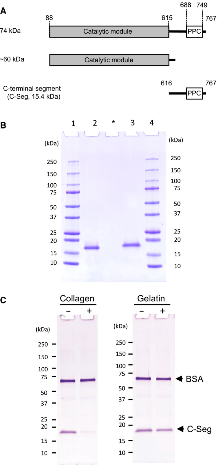
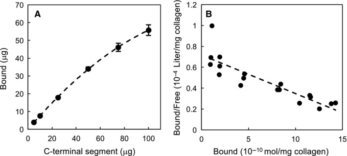
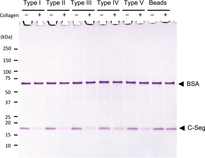
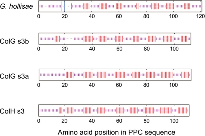
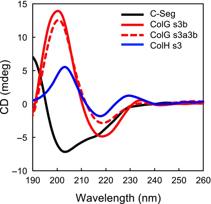
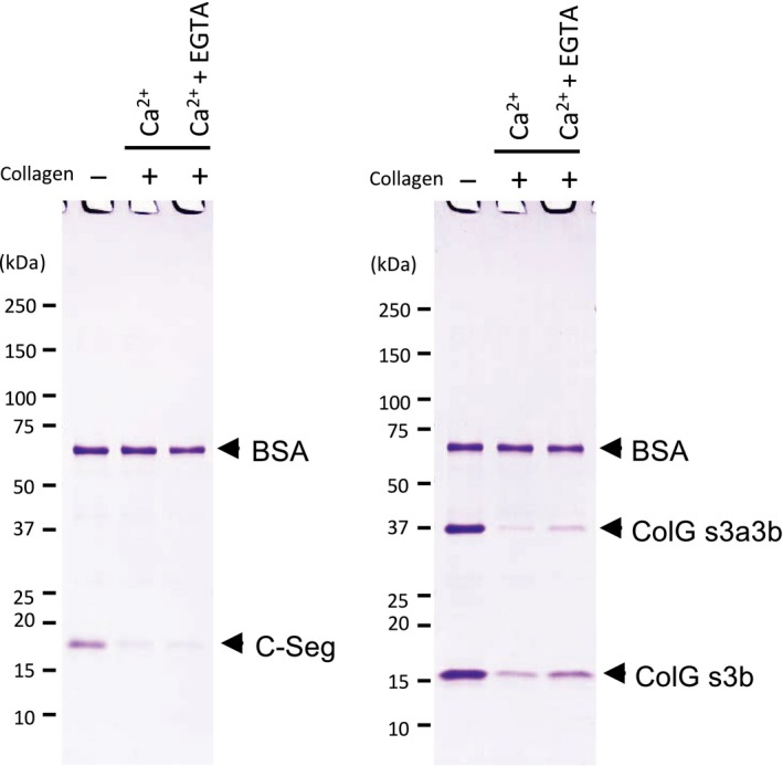
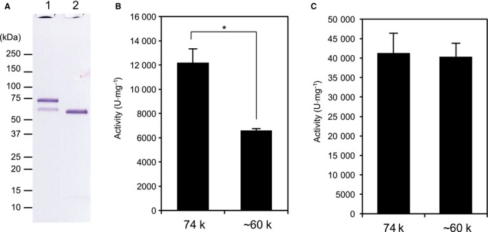
The collagenase secreted by Grimontia hollisae strain 1706B is a 74 kDa protein that consists of two parts: the catalytic module and a C-terminal segment that includes the bacterial pre-peptidase C-terminal domain. Here, we produced a recombinant C-terminal segment protein and examined its ability to bind collagen and other characteristics as compared with collagen-binding domains (CBDs) derived from Hathewaya histolytica (Clostridium histolyticum) collagenases; these CBDs are the only ones thus far identified in bacterial collagenases. We found that the C-terminal segment binds to collagen only when the collagen is in its triple-helical conformation. Moreover, the C-terminal segment and the CBDs from H. histolytica have comparable characteristics, including binding affinity to type I collagen, substrate spectrum, and binding conditions with respect to salt concentration and pH. However, the C-terminal segment has a completely different primary structure from those of the CBDs from H. histolytica. As regards secondary structure, in silico prediction indicates that the C-terminal segment may be homologous to those in CBDs from H. histolytica. Furthermore, we performed collagenase assays using fluorescein isothiocyanate-labeled type I collagen to show that the C-terminal segment positively contributes to the collagenolytic activity of the 74 kDa collagenase from G. hollisae.

Keywords: Grimontia hollisae; PPC domain; bacterial collagenase; metallopeptidase M9 subfamily A; recombinant protein; triple‐helical conformation.


Figures

Figure 1
Figure 1
Graphical representation of C‐terminal segment and its conformation‐specific binding to collagen. (A) Recombinant protein was designed as the C‐terminal segment (aa 616–767) including the PPC domain of G. hollisae collagenase. (B) The C‐terminal segment was purified from Brevibacillus culture medium by affinity chromatography on a Ni column. Two micrograms of purified C‐terminal segment was analyzed by SDS/PAGE using a 4–20% gradient polyacrylamide gel. Lane 1, molecular mass marker; lane 2, C‐terminal segment under non‐reducing conditions; lane 3, C‐terminal segment under reducing conditions; lane 4, molecular mass marker. Asterisk indicates blank lane. (C) Twenty micrograms of C‐terminal segment was incubated in 100 μL of the binding buffer with (+) and without (−) 5 mg of insoluble type I collagen fibers (left panel), or incubated with (+) and without (−) 50 μL of gelatin‐coupled Sepharose beads (right panel). An aliquot (2 μg of protein) of the filtrate was analyzed by SDS/PAGE on a 4–20% gradient gel. Numbers on the left are molecular masses (in kDa) of the markers.
Figure 2
Figure 2
Scatchard analysis of the binding of the C‐terminal segment. Varying concentrations (0.1–2.0 mg·mL−1) of the C‐terminal segment were incubated in 50 μL of the binding buffer with a fixed amount (2.5 mg) of insoluble collagen fibers. After incubation, the filtrate containing unbound proteins was subjected to SDS/PAGE, and the amount of the unbound C‐terminal segment was determined by densitometry of the corresponding band. The results obtained by triplicate assay were analyzed on a Scatchard plot. (A) Saturation binding curve for collagen binding of the C‐terminal segment. (B) Scatchard plot for collagen binding of the C‐terminal segment.
Figure 3
Figure 3
Binding of the C‐terminal segment to various types of collagen. Five micrograms of C‐terminal segment was incubated in 50 μL of the binding buffer with (+) and without (−) various types of collagen coupled‐Sepharose beads (type I, II, III, IV, or V collagen). After incubation, the filtrates were analyzed by SDS/PAGE using a 4–20% gradient polyacrylamide gel. Numbers on the left are molecular masses (in kDa) of the markers.
Figure 4
Figure 4
Consensus secondary structures of the PPC domains from G. hollisae and H. histolytica collagenases. Consensus secondary structures were predicted using a high‐performance method implemented on the NPS@ structure server 11. The predicted structures are represented by colored bars to visualize the schematic architecture: α‐helix, blue; β‐sheet, red; random coil, magenta; unclassified, gray.
Figure 5
Figure 5
CD spectra of the C‐terminal segment and the CBDs from H. histolytica. The CD spectra were measured at 20 °C in 10 mm phosphate, pH 7.5 for the C‐terminal segment (black line), ColG s3b (red solid line), ColG s3a3b (red dashed lines), and ColH s3 (blue line). The concentrations of the C‐terminal segment and the CBDs from H. histolytica were 0.1 mg·mL−1.
Figure 6
Figure 6
Binding of the C‐terminal segment and ColG CBDs with or without calcium ions. Five micrograms of the C‐terminal segment or 5 μg each of ColG CBDs (s3b and s3a3b) in 50 μL of the binding buffer was incubated with (+) and without (−) 2.5 mg of insoluble type I collagen fibers when varying Ca2+ ion concentration (5 mm Ca2+ ion or 5 mm Ca2+ ion with 10 mm EGTA). After incubation, the filtrates were analyzed by SDS/PAGE using a 4–20% gradient polyacrylamide gel. Numbers on the left are molecular masses (in kDa) of the markers.
Figure 7
Figure 7
Collagenolytic and gelatinolytic activities of recombinant 74 kDa and ~ 60 kDa G. hollisae collagenase. (A) Collagenase of 74 kDa and its truncated form consisting of the catalytic module (~ 60 kDa) were purified from Brevibacillus culture medium by DEAE‐Sepharose chromatography. Two micrograms of purified collagenase was analyzed by SDS/PAGE using a 4–20% gradient polyacrylamide gel. Lane 1, 74 kDa collagenase; lane 2, ~ 60 kDa collagenase. Numbers on the left are molecular masses (in kDa) of the markers. (B, C) The collagenolytic and gelatinolytic activities of the recombinant collagenases were determined using FITC‐collagen (B) and FITC‐gelatin (C), respectively. The values represent the average of triplicate trials ± SD. Statistical analyses were performed by Student's t test. *P < 0.01.
Figure 8
Figure 8
Multiple sequence alignment of the PPC domains from G. hollisae collagenase and other M9A collagenases. Multiple sequence alignment of C‐terminal segments from G. hollisae collagenase (NCBI accession number: BAK39964, aa 647–767), Vibrio parahaemolyticus collagenase (NP_797719, aa 698–814), V. alginolyticus collagenase (CAA44501, aa 698–814), V. proteolyticus collagenase (WP_021703968, aa 607–721), V. splendidus collagenase (WP_102548390, aa 613–729), V. cyclitrophicus collagenase (WP_016769033, aa 612–728), V. maritimus collagenase (WP_112460283, aa 608–724), and V. variabilis collagenase (WP_112477837, aa 608–724) was aided by using the clustal omega program 10. Identical residues among the sequences are indicated by asterisks. Cysteine residues are highlighted in red.

References

    1. Hickman FW, Farmer JJ 3rd, Hollis DG, Fanning GR, Steigerwalt AG, Weaver RE and Brenner DJ (1982) Identification of Vibrio hollisae sp. nov. from patients with diarrhea. J Clin Microbiol 15, 395–401. - PMC - PubMed
    1. Thompson FL, Hoste B, Vandemeulebroecke K and Swings J (2003) Reclassification of Vibrio hollisae as Grimontia hollisae gen. nov., comb. nov. Int J Syst Evol Microbiol 53, 1615–1617. - PubMed
    1. Suzuki K (2000) Purification and properties of collagenase from Vibrio hollisae 1706B strain. Hikakukagaku. 45, 272–283.
    1. Teramura N, Tanaka K, Iijima K, Hayashida O, Suzuki K, Hattori S and Irie S (2011) Cloning of a novel collagenase gene from the gram‐negative bacterium Grimontia (Vibrio) hollisae 1706B and its efficient expression in Brevibacillus choshinensis . J Bacteriol 193, 3049–3056. - PMC - PubMed
    1. Lawson PA and Rainey FA (2016) Proposal to restrict the genus Clostridium Prazmowski to Clostridium butyricum and related species. Int J Syst Evol Microbiol 66, 1009–1016. - PubMed