. 2018 Oct 11:25:17.
doi: 10.1186/s40709-018-0089-z. eCollection 2018 Dec.

Combined study on clastogenic, aneugenic and apoptotic properties of doxorubicin in human cells in vitro

Affiliations

Combined study on clastogenic, aneugenic and apoptotic properties of doxorubicin in human cells in vitro

Vasiliki Chondrou et al. J Biol Res (Thessalon). .

Abstract

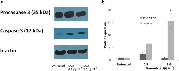
Background: Doxorubicin is a widely used anticancer drug due to its broad spectrum of antitumor activity. Various mechanisms have been proposed for its cytostatic activity, including DNA intercalation, topoisomerase II inhibition, generation of free radicals and apoptosis. The present study aims to further clarify the cytostatic activity of doxorubicin by its specific effect on (a) DNA damage, (b) micronucleation and (c) apoptosis, using a combination of different methods and cell systems such as human lymphocytes and HL-60 human leukemic cells. DNA lesions were analyzed by the alkaline comet assay in combination with formamidopyrimidine (Fpg) and human 8-oxoguanine (hOGG1) repair enzymes. Micronucleation was investigated by the Cytokinesis-Block Micronucleus assay (CBMN) in combination with Fluorescence In Situ Hybridization analysis. Impairment on mitotic apparatus was investigated by double immunofluorescence of β- and γ-tubulin. Apoptotic cell frequency was determined by the CBMN cytome assay. Complementary to the above, caspase-3 level was investigated by Western blot.

Results: It was found that doxorubicin generates DNA breakage induced by oxidative damage in DNA bases, which can be repaired by the Fpg and hOGG1 enzymes. Increased micronucleus frequency was identified mainly through chromosome breakage and, at a lesser extent, through chromosome delay. Analysis of mitotic spindle showed disturbance of chromosome orientation and centrosome duplication and/or separation, leading to aneuploidy. Enhanced frequency of apoptotic leukemic cells was also observed. Caspase-3 seems to be involved in the generation of apoptosis.

Conclusions: The aforementioned findings derived from different treatment schedules, doses and time of exposure on primary versus transformed cells extend our knowledge about doxorubicin genotoxicity and contribute to the better understanding of the mechanisms by which doxorubicin induces genotoxic effects on human cells.

Keywords: Apoptosis-caspase-3; Doxorubicin; Fpg and hOGG1 comet assay; Micronucleation; Mitotic spindle disturbance.


Figures

Fig. 1
Fig. 1
Genetic endpoints studied in different cell types and methodology followed
Fig. 2
Fig. 2
% DNA in tail in HL-60 comets after treatment with various concentrations of doxorubicin. H2O2 (100 μM) was used as positive control. DNA was stained with ethidium bromide. *p ≤ 0.05 in comparison with control (one-way ANOVA). Bars represent standard error
Fig. 3
Fig. 3
% DNA in tail in HL-60 comets after treatment with doxorubicin and incubation of nucleoids with Fpg or hOGG1 endonuclease. Data represent values generated by Fpg (a1) and hOGG1 (a2) incubation, that were calculated by subtracting the value of DNA damage caused in the HL-60 cells that were not treated with the respective enzyme from the DNA damage caused in cells incubated with the enzymes. Subtracted values are pointed out in light grey area marked ‘‘Fpg sites’’ and “hOGG1 sites”. b HL-60 comets, no doxorubicin treatment/no endonuclease incubation (b1); no doxorubicin treatment/Fpg endonuclease incubation (b2); no doxorubicin treatment/hOGG1 endonuclease incubation (b3); doxorubicin treatment at 0.5 μg ml−1/no endonuclease incubation (b4); doxorubicin treatment at 0.5 μg ml−1/Fpg endonuclease incubation (b5); doxorubicin treatment at 0.5 μg ml−1/hOGG1 endonuclease incubation (b6); doxorubicin treatment at 2.0 μg ml−1/no endonuclease incubation (b7); doxorubicin treatment at 2.0 μg ml−1/Fpg endonuclease incubation (b8) and doxorubicin treatment at 2.0 μg ml−1 /hOGG1 endonuclease incubation (b9). DNA was stained with ethidium bromide. * p ≤ 0.05 in comparison with untreated cells (one-way ANOVA). Bars represent standard error
Fig. 4
Fig. 4
a1 Micronucleus frequency in human lymphocytes treated with doxorubicin. a2 Cytokinesis Block Micronucleus Index (CBPI) in human lymphocytes treated with doxorubicin. b FISH analysis with pancentromeric probe. c Lymphocytes with CMN (c1) and C+MN (c2). (CMN: micronucleus without centromeric signal, C+MN: micronucleus exerting centromeric signal. * p ≤ 0.05 in comparison with control (G-test). Vincristine sulfate was used as a positive control. The values represent the average of the two donors. Bars represent standard error
Fig. 5
Fig. 5
a Western blots and b densitometric quantification, showing the caspase-3 level in HL-60 cells after treatment with doxorubicin. * p ≤ 0.05 in comparison with control (G-test). Bars represent standard error
Fig. 6
Fig. 6
a Frequency of abnormal C2C12 metaphases in relation to γ-tubulin signals and chromosome orientation. b Distinct types of metaphase cells: normal metaphase (b1); monopolar metaphase (b2); tripolar metaphase (b3); multipolar metaphase (b4); and bipolar non-congressed metaphases (b5–6). * p ≤ 0.05 in comparison with control (G-test). Demecolcine was used as positive control. Bars represent standard error

Similar articles

Cited by

References

    1. Weiss RB. The anthracyclines: will we ever find a better doxorubicin? Semin Oncol. 1992;19:670–686. - PubMed
    1. Xu X, Persson HL, Richardson DR. Molecular pharmacology of the interaction of anthracyclines with iron. Mol Pharmacol. 2005;68:261–271. - PubMed
    1. Kumaravel TS, Vilhar B, Faux SP, Jha AN. Comet Assay measurements: a perspective. Cell Biol Toxicol. 2006;25:53–64. doi: 10.1007/s10565-007-9043-9. - DOI - PubMed
    1. Liao W, McNutt MA, Zhu WG. The comet assay: a sensitive method for detecting DNA damage in individual cells. Methods. 2009;48:46–53. doi: 10.1016/j.ymeth.2009.02.016. - DOI - PubMed
    1. Piperakis SM. Comet assay: a brief history. Cell Biol Toxicol. 2009;25:1–3. doi: 10.1007/s10565-008-9081-y. - DOI - PubMed