. 2019 Jan 24;25(5):1249-1259.
doi: 10.1002/chem.201803653. Epub 2018 Dec 18.

Electronic Modifications of Fluorescent Cytidine Analogues Control Photophysics and Fluorescent Responses to Base Stacking and Pairing

Affiliations

Electronic Modifications of Fluorescent Cytidine Analogues Control Photophysics and Fluorescent Responses to Base Stacking and Pairing

Kristine L Teppang et al. Chemistry. .

Abstract

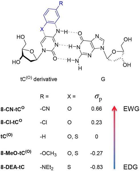
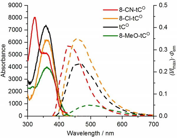
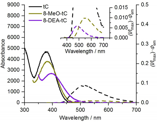
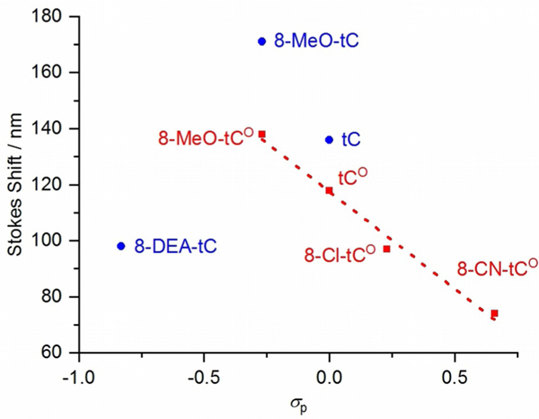
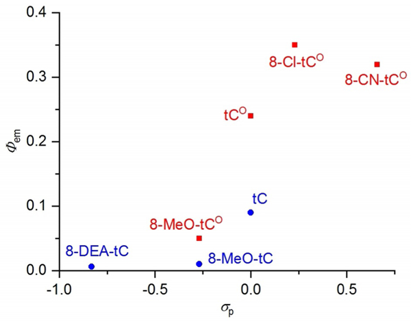
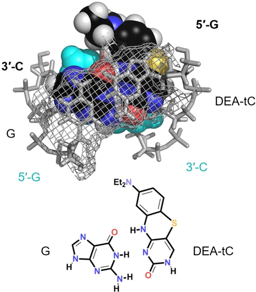
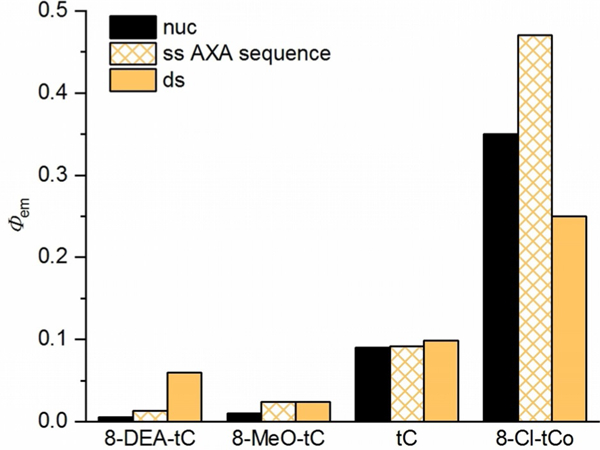
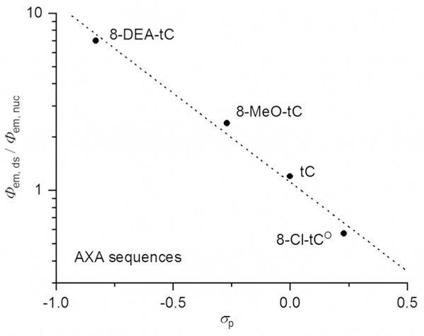
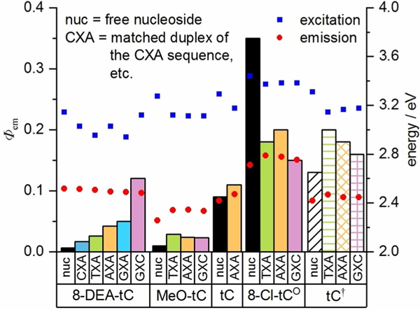
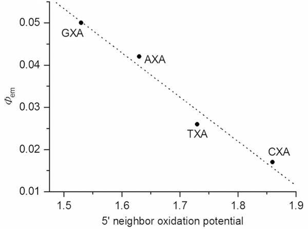
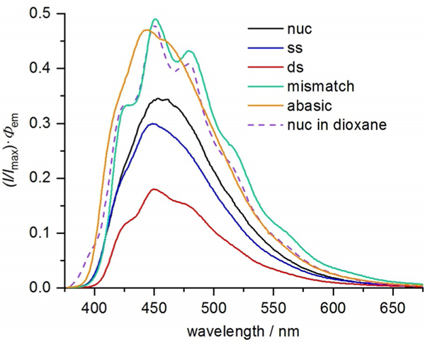
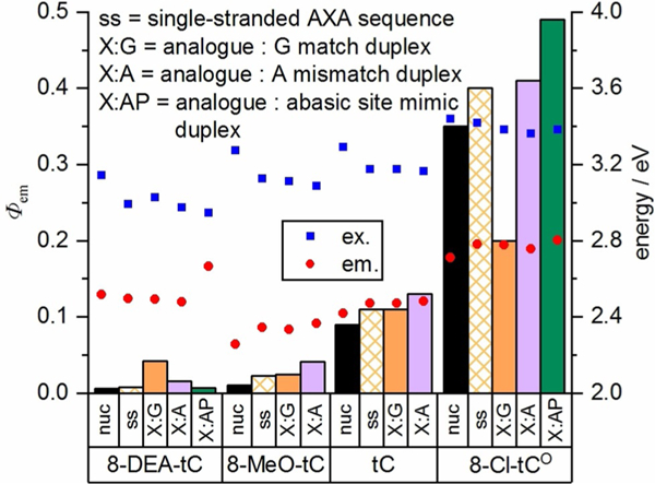
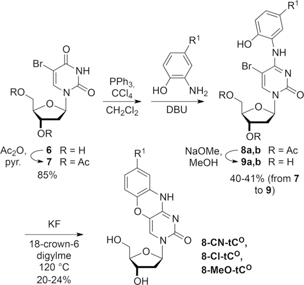
The rational design of fluorescent nucleoside analogues is greatly hampered by the lack of a general method to predict their photophysics, a problem that is especially acute when base pairing and stacking change fluorescence. To better understand these effects, a series of tricyclic cytidine (tC and tCO ) analogues ranging from electron-rich to electron-deficient was designed and synthesized. They were then incorporated into oligonucleotides, and photophysical responses to base pairing and stacking were studied. When inserted into double-stranded DNA oligonucleotides, electron-rich analogues exhibit a fluorescence turn-on effect, in contrast with the electron-deficient compounds, which show diminished fluorescence. The magnitude of these fluorescence changes is correlated with the oxidation potential of nearest neighbor nucleobases. Moreover, matched base pairing enhances fluorescence turn-on for the electron-rich compounds, and it causes a fluorescence decrease for the electron-deficient compounds. For the tCO compounds, the emergence of vibrational fine structure in the fluorescence spectra in response to base pairing and stacking was observed, offering a potential new tool for studying nucleic acid structure and dynamics. These results, supported by DFT calculations, help to rationalize fluorescence changes in the base stack and will be useful for selecting the best fluorescent nucleoside analogues for a desired application.

Keywords: DNA; biophysics; fluorescence; nucleoside analogues; photochemistry.


Conflict of interest statement

Conflict of interest

The authors declare no conflict of interest.

Figures

Figure 1.
Figure 1.
Tricyclic cytidine derivatives used in this study to probe electronic effects on photophysical properties of free nucleosides and the effects of base pairing and stacking. X = O in tCO compounds and X = S in tC compounds.
Figure 2.
Figure 2.
Absorption (solid lines) and corrected emission (dashed lines; normalized at λmax to Φem) spectra of tCO derivatives in 1 × PBS buffer (pH 7.4) recorded at 296 K.
Figure 3.
Figure 3.
Absorption (solid lines) and corrected emission (dashed lines and inset; normalized at λmax to Φem) spectra of tC derivatives in 1 × PBS buffer (pH 7.4) recorded at 296 K.
Figure 4.
Figure 4.
Linear correlation of Stokes shift to Hammett σp for tCO compounds (red) and plot for tC compounds (blue).
Figure 5.
Figure 5.
Correlation of fluorescence quantum yield Φem to Hammett σp for tC (blue) and tCO compounds (red).
Figure 6.
Figure 6.
Normalized fluorescence emission spectra of the 8-Cl-tCO nucleoside in mixtures of 1 × PBS buffer (pH 7.4) and 1,4-dioxane. Vibrational fine structure appears only under nonaqueous conditions.
Figure 7.
Figure 7.
Molecular model (Spartan′08) showing 8-DEA-tC base paired and stacked in idealized B form DNA.
Figure 8.
Figure 8.
Changes in fluorescence quantum yield of four cytidine analogues upon incorporation into single- and double-stranded DNA oligonucleotides, measured in 1 × PBS buffer (pH 7.4). nuc=free nucleoside, ss = single-stranded oligonucleotide with the AXA sequence 5′-CGC-AAX-ATC-G-3′ (Table 2), where X = the fluorescent cytidine analogue, ds = matched, double-stranded oligonucleotide.
Figure 9.
Figure 9.
The fluorescence change upon incorporation of a tC(O) compound into a double-stranded oligonucleotide of the AXA sequence (Table 2), as expressed by Φem,ds/Φem,nuc on a logarithmic scale, is strongly correlated to the Hammett σp for the tC(O) compounds’ substituents.
Figure 10.
Figure 10.
Fluorescence quantum yield of tC(O) derivatives as free nucleosides (black) and in duplex oligonucleotides with varied neighboring bases (full sequences given in Table 2), measured in 1×PBS buffer, pH 7.4 (columns plotted against left y-axis). Excitation and emission energy, derived from λmax,abs and λmax,, respectively are plotted as points against the right y-axis. tC = data from Wilhelmsson for tC-containing duplex oligonucleotides of the same sequences, measured in 50 mM sodium phosphate buffer, pH 7.5.[39]
Figure 11.
Figure 11.
Dependence of the Φem of 8-DEA-tC-containing duplex oligonucleotides (for sequences, see Table 2) on the oxidation potential of the 5′- neighboring nucleobase.[56]
Figure 12.
Figure 12.
Fluorescence quantum yield of tC(O) derivatives as free nucleosides (black) and in duplex oligonucleotides with varied neighboring bases (full sequences given in Table 2), measured in 1 × PBS buffer, pH 7.4. tC = data from Wilhelmsson for tC-containing duplex oligonucleotides of the same sequences, measured in 50 mM sodium phosphate buffer, pH 7.5.[39]
Figure 13.
Figure 13.
Fluorescence emission spectra of 8-Cl-tCO as a free nucleoside (black), in ssDNA (blue; AXA sequence from Table 2), and dsDNA (red; annealed to matched complement), in mismatched dsDNA (green; 8-Cl-tCO:A mismatch), and in dsDNA opposite the 1,2-dideoxy-D-ribose surrogate for an abasic site (orange; 8-Cl-tCO:AP) all in 1 × PBS buffer (pH 7.4), and the emission spectrum of 8-Cl-tCO nucleoside in 1,4-dioxane (purple dashed line).
Figure 14.
Figure 14.
Fluorescence quantum yield of tC(O) derivatives in single-stranded oligonucleotides (AXA sequence; see Table 2), in matched duplexes X:G, with adenosine mismatches X:A, and opposite the abasic site mimic 1,2-dideoxy-D-ribose. Measurements were performed in 1 × PBS buffer, pH 7.4.
Scheme 1.
Scheme 1.
Synthesis of tC derivatives.
Scheme 2.
Scheme 2.
Synthesis of tCO derivatives.

References

    1. Xu W, Chan KM, Kool ET, Nat. Chem. 2017, 9, 1043–1055. - PMC - PubMed
    1. Shin D, Sinkeldam RW, Tor Y, J. Am. Chem. Soc. 2011, 133, 14912–14915. - PMC - PubMed
    1. Rovira AR, Fin A, Tor Y, Chem. Sci. 2017, 8, 2983–2993. - PMC - PubMed
    1. Dumas A, Luedtke NW, J. Am. Chem. Soc. 2010, 132, 18004–18007. - PubMed
    1. Sabale PM, Tanpure AA, Srivatsan SG, Org. Biomol. Chem. 2018, 16, 4141–4150. - PMC - PubMed

LinkOut - more resources