Review
. 2019 Apr;21(4):425-435.
doi: 10.1002/ejhf.1320. Epub 2018 Oct 19.

Treating oxidative stress in heart failure: past, present and future

Affiliations
Review

Treating oxidative stress in heart failure: past, present and future

Atze van der Pol et al. Eur J Heart Fail. 2019 Apr.

Abstract

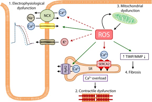
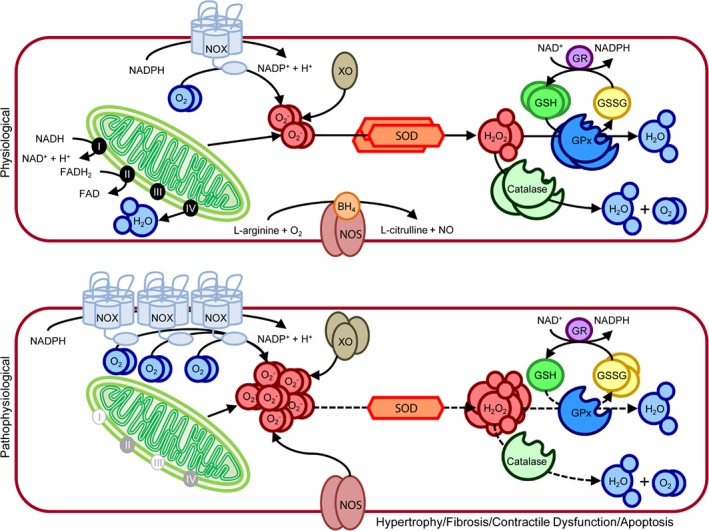
Advances in cardiovascular research have identified oxidative stress as an important pathophysiological pathway in the development and progression of heart failure. Oxidative stress is defined as the imbalance between the production of reactive oxygen species (ROS) and the endogenous antioxidant defence system. Under physiological conditions, small quantities of ROS are produced intracellularly, which function in cell signalling, and can be readily reduced by the antioxidant defence system. However, under pathophysiological conditions, the production of ROS exceeds the buffering capacity of the antioxidant defence system, resulting in cell damage and death. Over the last decades several studies have tried to target oxidative stress with the aim to improve outcome in patients with heart failure, with very limited success. The reasons as to why these studies failed to demonstrate any beneficial effects remain unclear. However, one plausible explanation might be that currently employed strategies, which target oxidative stress by exogenous inhibition of ROS production or supplementation of exogenous antioxidants, are not effective enough, while bolstering the endogenous antioxidant capacity might be a far more potent avenue for therapeutic intervention. In this review, we provide an overview of oxidative stress in the pathophysiology of heart failure and the strategies utilized to date to target this pathway. We provide novel insights into modulation of endogenous antioxidants, which may lead to novel therapeutic strategies to improve outcome in patients with heart failure.

Keywords: Glutathione; Heart failure; Nicotinamide adenine dinucleotide; Oxidative stress; γ-Glutamyl cycle.


Figures

Figure 1
Figure 1
The effects of excessive oxidative stress on the myocardium. As a result of cardiac injury there is a severe accumulation of oxidative stress (reactive oxygen species, ROS), which has several detrimental effects on the myocardium. 1. Cardiomyocyte electrophysiology is severely affected by increased ROS. ROS reverses the function of the Na+/Ca2+ exchanger (NCX), leading to Ca2+ influx and Na+ efflux. ROS also increases the influx of Ca2+ via the L‐type calcium channels. Increased ROS also increases sarcKATP currents, leading to action potential duration shortening, while also reducing KV currents and increasing late sodium currents leading to prolonged action potential durations. 2. Excessive ROS promotes ryanodine receptor 2 (RyR2) activity and inhibits sarcoplasmic reticulum Ca2+‐adenosine triphosphatase 2 (SERCA2) activity, resulting in calcium overload and reduced myofilament calcium sensitivity, eventually leading to contractile dysfunction. 3. The mitochondria react to ischaemic injury by producing increased levels of ROS, however the overabundance of ROS inversely results in further mitochondrial and energy metabolism dysfunction. 4. The increase in ROS is also responsible for increased fibrosis resulting from an increase in tissue inhibitors of metalloproteinases (TIMP) and reduction in matrix metalloproteinase (MMP) expression.
Figure 2
Figure 2
Oxidative stress production and scavenging in cardiomyocytes under physiological and pathophysiological conditions. (Top) Under physiological conditions oxidative stress in the form of reactive oxygen species (ROS) is produced in small quantities by the mitochondrial electron chain, NADPH oxidase (NOX), xanthine oxidase (XO), and nitric oxide synthase (NOS). Mitochondrial respiration converts oxygen to water, resulting in the production of small quantities of superoxide (O2 ) as a by‐product. The process starts with electrons derived from NADH2 and FADH2 moving along the respiratory transport chain through a series of cytochrome‐based complexes (I, III, and IV). These complexes eventually transport electrons to molecular oxygen. The high free energy of the electrons is gradually extracted and converted into adenosine triphosphate. NOX is a multimeric complex composed of a plasma membrane spanning cytochrome b558 (NOX2) and cytosolic components (Rac1, p47phox, p67phox, p40phox). Under physiological conditions this complex is in a resting state, producing minimal O2 , by transferring an electron from NADPH to molecular oxygen. XO, which is a cytoplasmic enzyme that catalyzes the oxidation of hypoxanthine and xanthine to uric acid using molecular oxygen as an electron receptor, produces O2 and hydrogen peroxide (H2O2) in the process. NOS oxidizes the NOS cofactor BH4 utilizing NADPH to generate nitric oxide and L‐citrulline from L‐arginine and oxygen. Superoxide dismutase (SOD) initiates the detoxification of ROS, by scavenging O2 and converting it to H2O2. Both catalase and glutathione peroxidase (GPx) further detoxify the H2O2 to water and oxygen. GPx utilizes two glutathione (GSH) molecules as electron donors in the reduction of H2O2 to water, producing oxidized glutathione (GSSG) in the process. Once GPx oxidizes GSH to GSSG, GSH reductase (GR) can reduce GSSG back to GSH at the expense of NADPH, forming the GSH redox cycle. The ratio of GSH to GSSG largely determines the intracellular redox potential. (Bottom) Under pathophysiological conditions, oxidative stress production is increased as a result of increased NOX and XO expression, coupled to blockage of the mitochondrial electron chain and uncoupling of NOS. Furthermore, the expression and activity (dotted lines) of SOD, catalase, and GPx are reduced. The levels of GSH are also reduced, while the levels of GSSG are increased. This severe increase in oxidative stress eventually leads to hypertrophy, fibrosis, apoptosis, and contractile dysfunction in the myocardium.
Figure 3
Figure 3
Drug therapies targeting endogenous glutathione (GSH) synthesis. GSH is synthesized from cysteine (the rate‐limiting amino acid), glutamate, and glycine by the γ‐glutamyl cycle. GSH is then utilized by GSH peroxidase (GPx) to reduce oxidative stress, and in the process forming oxidized GSH (GSSG). GSSG is then reduced by action of GSH reductase (GR). Improving the γ‐glutamyl cycle's ability to produce GSH has been characterized as a treatment target in heart failure. N‐acetylcysteine (NAC), γ‐glutamylcysteine, and 2‐oxothiazolidine‐4‐carboxylate (OTC, also known as pro‐cysteine) are compounds which have demonstrated the capacity to increase the endogenous production of GSH. OTC is converted to cysteine, by action of 5‐oxoprolinase (OPLAH), to be used for de novo synthesis of GSH. Similarly, NAC is converted to cysteine intracellularly, and used for GSH synthesis. γ‐Glutamylcysteine is utilized by the γ‐glutamyl cycle to form GSH, by addition of glycine. GCL, glutamate cysteine ligase.
Figure 4
Figure 4
Targeting 5‐oxoprolinase (OPLAH) to reduce oxidative stress in heart failure. Following cardiac injury, OPLAH expression is reduced, leading to the accumulation of 5‐oxoproline. 5‐Oxoproline then leads to drastic increase in oxidative stress (reactive oxygen species, ROS). To help reduce the insult of 5‐oxoproline to the injured myocardium, two strategies could be developed: (i) to pharmacologically improve the remaining OPLAH's ability to reduce 5‐oxoproline, or (ii) to increase OPLAH expression by means of gene therapy. ADP, adenosine diphosphate; ATP, adenosine triphosphate; GSH, glutathione.

References

    1. Karimi Galougahi K, Antoniades C, Nicholls SJ, Channon KM, Figtree GA. Redox biomarkers in cardiovascular medicine. Eur Heart J 2015;36:1576–1582. - PubMed
    1. Tsutsui H, Kinugawa S, Matsushima S. Oxidative stress and heart failure. Am J Physiol Heart Circ Physiol 2011;301:H2181‐2190. - PubMed
    1. Ponikowski P, Voors AA, Anker SD, Bueno H, Cleland JG, Coats AJ, Falk V, González‐Juanatey JR, Harjola VP, Jankowska EA, Jessup M, Linde C, Nihoyannopoulos P, Parissis JT, Pieske B, Riley JP, Rosano GM, Ruilope LM, Ruschitzka F, Rutten FH, van der Meer P. 2016. ESC Guidelines for the diagnosis and treatment of acute and chronic heart failure: The Task Force for the diagnosis and treatment of acute and chronic heart failure of the European Society of Cardiology (ESC) . Developed with the special contribution of the Heart Failure Association (HFA) of the ESC. Eur J Heart Fail 2016;18:891–975. - PubMed
    1. Wolfram R, Oguogho A, Palumbo B, Sinzinger H. Enhanced oxidative stress in coronary heart disease and chronic heart failure as indicated by an increased 8‐epi‐PGF 2α. Eur J Heart Fail 2005;7:167–172. - PubMed
    1. Sato Y, Fujiwara H, Takatsu Y. Cardiac troponin and heart failure in the era of high‐sensitivity assays. J Cardiol 2012;60:160–167. - PubMed