Review
. 2019 Apr;94(2):610-628.
doi: 10.1111/brv.12469. Epub 2018 Oct 19.

The enigmatic ATP supply of the endoplasmic reticulum

Affiliations
Review

The enigmatic ATP supply of the endoplasmic reticulum

Maria R Depaoli et al. Biol Rev Camb Philos Soc. 2019 Apr.

Abstract

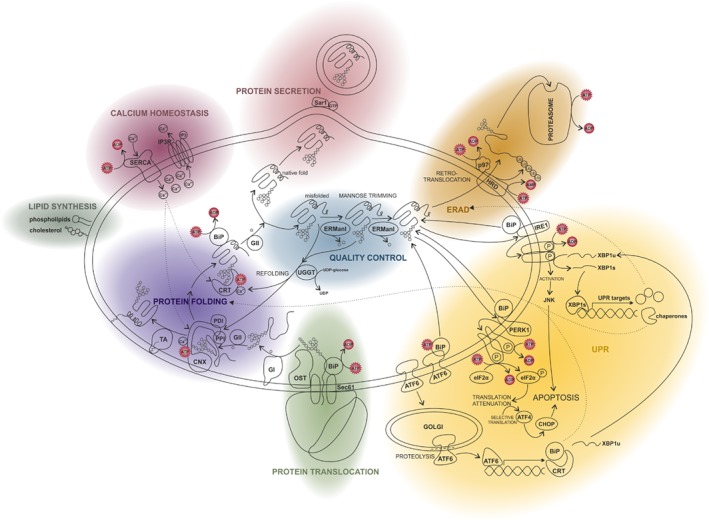
The endoplasmic reticulum (ER) is a functionally and morphologically complex cellular organelle largely responsible for a variety of crucial functions, including protein folding, maturation and degradation. Furthermore, the ER plays an essential role in lipid biosynthesis, dynamic Ca2+ storage, and detoxification. Malfunctions in ER-related processes are responsible for the genesis and progression of many diseases, such as heart failure, cancer, neurodegeneration and metabolic disorders. To fulfill many of its vital functions, the ER relies on a sufficient energy supply in the form of adenosine-5'-triphosphate (ATP), the main cellular energy source. Despite landmark discoveries and clarification of the functional principles of ER-resident proteins and key ER-related processes, the mechanism underlying ER ATP transport remains somewhat enigmatic. Here we summarize ER-related ATP-consuming processes and outline our knowledge about the nature and function of the ER energy supply.

Keywords: ATP; ATP transporter; ER stress; ERAD; endoplasmic reticulum (ER); protein quality control; secretory pathway; unfolded protein response (UPR).


Figures

Figure 1
Figure 1
ATP‐consuming processes in the endoplasmic reticulum (ER). Schematic representation providing an overview of the most important ER‐associated processes and showing where ATP is required. Related functions are highlighted in the same colour. In green we follow protein translocation into the ER. Thereafter proteins are modified and folded (lilac). During protein quality control (blue) the folding state of the protein is monitored. Correctly folded proteins are secreted and transported to the Golgi (red). Unfolded and misfolded proteins are refolded. If all folding attempts fail, proteins undergo ER‐associated protein degradation (ERAD, orange). They are ubiquitinated and transferred into the cytosol via retrotranslocation, where they are degraded by the proteasome. The accumulation of misfolded proteins in the ER may also trigger the unfolded protein response (UPR, yellow). The UPR includes the activation of the kinases IRE1 and PERK1. IRE1 signalling results in the activation of the transcription factor XBP1 and the subsequent expression of UPR targets such as ER chaperones and ERAD factors. PERK1 causes translation inhibition by eIF2α phosphorylation leading to the successive expression of the transcription factors ATF4 and CHOP, which activates apoptosis‐promoting genes. The third important UPR mediator is the transcription factor ATF6 which induces the transcription of its target genes encoding for instance XBP1, BiP or calreticulin. Beside its central functions in protein synthesis, folding and degradation, the ER is also the most important Ca2+ store with an essential contribution to Ca2+ signalling events (magenta). Ca2+ is pumped into the ER by the ATPase SERCA and is primarily released via the IP3 receptor channel. Finally, the ER is mainly responsible for the biosynthesis of phospholipids as well as isoprenoids like cholesterol and steroid hormones (grey). ADP, adenosine diphosphate; ATF4, activating transcription factor 4; ATF6, activating transcription factor 6; ATP, adenosine triphosphate; BiP, binding immunoglobulin protein; CHOP, C/EBP homologous protein; CNX, calnexin; CRT, calreticulin; eIF2α, eukaryotic initiation factor 2α; ERManI, ER mannosidase I; GI, glucosidase I; GII, glucosidase II; GTP, guanosine triphosphate; HRD, ubiquitin‐protein ligase; IP3, inositol trisphosphate; IP3R, inositol triphosphate receptor; IRE1, inositol‐requiring enzyme 1; JNK, c‐Jun N‐terminal kinase; OST, oligosaccharyltransferase; P, indicates phosphorylation; PDI, protein disulfide isomerase; PERK1, proline‐rich receptor‐like protein kinase 1; PPI, prolyl peptidyl isomerase; Sar1, COPII‐associated small GTPase; Sec61, ER membrane protein translocator (translocon); SERCA, sarco/endoplasmic reticulum Ca2+‐ATPase; TA, GPI transamidase; UGGT, UDP‐glucose glycoprotein glucosyltransferase; UPR, unfolded protein response; XBP1, x‐box binding protein 1.
Figure 2
Figure 2
Methods for the investigation of endoplasmic reticulum (ER) ATP dynamics. (A) ATP transport into ER‐derived vesicles or reconstituted liposomes containing ER proteins can be measured using an in vitro approach. Proteoliposomes are incubated with radioactively labeled ATP and then applied to a gel filtration column. Free ATP binds to the matrix, while proteoliposomes and incorporated ATP are eluted in the void volume. Proteoliposomes are then lysed to determine the amount of imported ATP via high‐performance liquid chromatography (HPLC) or liquid scintillation counting. (B) Alterations of ER ATP levels can be measured using genetically encoded fluorescent ATP sensors targeted to the ER. The ERAT4.01 probe for example is a Förster/fluorescence resonance energy transfer (FRET)‐based sensor consisting of two fluorescent proteins – an enhanced cyan fluorescent protein (CFP) and the yellow fluorescent protein (YFP) variant citrine – flanking the ATP binding ϵ‐subunit of the FoF1‐ATP synthase from Bacillus subtilis (Imamura et al., 2009; Vishnu et al., 2014). The N‐terminal fusion of the calreticulin signal sequence and the C‐terminal addition of the ER retention signal with the amino acid sequence KDEL (lysine–aspartic acid–glutamic acid–leucine) allow the efficient targeting of the probe to the ER. Binding of ATP to the ϵ‐subunit causes a conformational change of the sensor, which increases the FRET signal intensity. (C) Alternatively, ER ATP levels can be determined using an ER‐targeted heterologously expressed firefly luciferase, which catalyses the reaction of oxyluciferin to luciferin, resulting in light emission. Since this reaction is ATP dependent, the intensity of the emitted light is proportional to the ATP concentration.
Figure 3
Figure 3
Possible ATP transport routes into the endoplasmic reticulum (ER). It is not known how ATP is transported into the ER, but several possible import pathways are shown. Experiments with rat liver microsomes provided some evidence for a specific ATP transporter and an ATP/ADP antiport mechanism. ATP may also be transported non‐specifically through anion channels. The ER membrane may also be ‘leaky’ for ATP, allowing its passive transport driven by a concentration gradient. One such source of leakiness may be translocon channels, which are widely distributed across the membrane of the rough ER; ATP could enter the ER concomitantly with the inserted peptide or by a translocon‐associated mechanism. Since ATP is mainly produced in mitochondria it seems reasonable that it is directly transported from there into the ER via membrane contact sites (mitochondria‐associated ER membranes; MAMs). Finally, the dynamics of the membrane structures evoked by membrane trafficking via vesicles might also afford the opportunity for ATP uptake.

References

    1. Abele, R. & Tampé, R. (2011). The TAP translocation machinery in adaptive immunity and viral escape mechanisms. Essays in Biochemistry 50, 249–264. - PubMed
    1. Ainscow, E. K. & Rutter, G. A. (2002). Glucose‐stimulated oscillations in free cytosolic ATP concentration imaged in single islet β‐cells evidence for a Ca2+‐dependent mechanism. Diabetes 51, S162–S170. - PubMed
    1. Alder, N. N. , Shen, Y. , Brodsky, J. L. , Hendershot, L. M. & Johnson, A. E. (2005). The molecular mechanisms underlying BiP‐mediated gating of the Sec61 translocon of the endoplasmic reticulum. The Journal of Cell Biology 168, 389–399. - PMC - PubMed
    1. Allan, B. B. , Moyer, B. D. & Balch, W. E. (2000). Rab1 recruitment of p115 into a cis‐SNARE complex: programming budding COPII vesicles for fusion. Science (New York, N.Y.) 289, 444–448. - PubMed
    1. Amin‐Wetzel, N. , Saunders, R. A. , Kamphuis, M. J. , Rato, C. , Preissler, S. , Harding, H. P. & Ron, D. (2017). A J‐protein co‐chaperone recruits BiP to monomerize IRE1 and repress the unfolded protein response. Cell 171, 1625–1637.e13. - PMC - PubMed

Publication types

MeSH terms

Substances