

DICER1 Syndrome: Characterization of the Ocular Phenotype in a Family-Based Cohort Study
- PMID: 30339877
- PMCID: PMC6348055
- DOI: 10.1016/j.ophtha.2018.09.038
DICER1 Syndrome: Characterization of the Ocular Phenotype in a Family-Based Cohort Study
Abstract
Purpose: To characterize the ocular phenotype of DICER1 syndrome.
Design: Prospective, single-center, case-control study.
Participants: One hundred three patients with an identified germline pathogenic DICER1 variant (DICER1 carriers) and 69 family control participants underwent clinical and ophthalmic examination at the National Institutes of Health between 2011 and 2016.
Methods: All participants were evaluated with a comprehensive ophthalmic examination, including best-corrected visual acuity, slit-lamp biomicroscopy, and a dilated fundus examination. A subset of patients returned for a more detailed evaluation including spectral-domain OCT, color fundus photography, fundus autofluorescence imaging, visual field testing, full-field electroretinography, and genetic testing for inherited retinal degenerative diseases.
Main outcome measures: Visual acuity and examination findings.
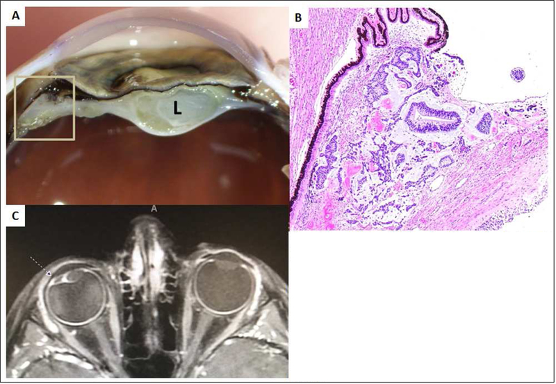
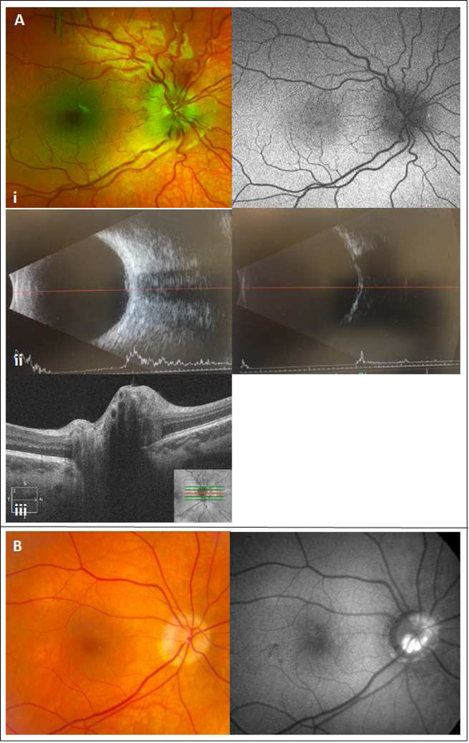
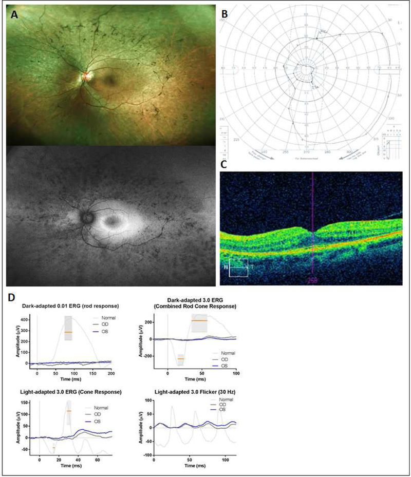
Results: Most DICER1 carriers (97%) maintained a visual acuity of 20/40 or better in both eyes. Twenty-three DICER1 carriers (22%) showed ocular abnormalities compared with 4 family controls (6%; P = 0.005). These abnormalities included retinal pigment abnormalities (n = 6 [5.8%]), increased cup-to-disc ratio (n = 5 [4.9%]), optic nerve abnormalities (n = 2 [1.9%]), epiretinal membrane (n = 2 [1.9%]), and drusen (n = 2 [1.9%]). Overall, we observed a significant difference (P = 0.03) in the rate of retinal abnormalities in DICER1 carriers (n = 11 [11%]) versus controls (n = 1 [1.5%]). One patient demonstrated an unexpected diagnosis of retinitis pigmentosa with a novel variant of unknown significance in PRPF31, and 1 showed optic nerve elevation in the setting of increased intracranial pressure (ICP) of unclear cause. Three patients (3%) demonstrated DICER1-related ciliary body medulloepithelioma (CBME), 2 of which were identified during routine examination, a higher rate than that reported previously.
Conclusions: Ophthalmologists should be aware of the ophthalmic manifestations of DICER1 syndrome, and individuals and families should be counseled on the potential signs and symptoms. We recommend that children with a germline pathogenic variant in DICER1, especially those younger than 10 years, undergo annual dilated ophthalmic examination, looking for evidence of CBME, signs of increased ICP, and perhaps changes in the retinal pigment epithelium.
Published by Elsevier Inc.
Conflict of interest statement
Figures
References
-
- Foulkes WD, Priest JR, Duchaine TF. DICER1: mutations, microRNAs and mechanisms. Nature Reviews Cancer. 2014;14(10):662. - PubMed
-
- Schultz KAP, Rednam SP, Kamihara J, et al. PTEN, DICER1, FH, and Their Associated Tumor Susceptibility Syndromes: Clinical Features, Genetics, and Surveillance Recommendations in Childhood. Clinical Cancer Research. 2017;23(12):e76–e82. - PubMed
Publication types
MeSH terms
Substances
Grants and funding
LinkOut - more resources
Full Text Sources
Medical
