

Comment
doi: 10.1016/j.cell.2018.10.011.
Flipping ATP to AMPlify Kinase Functions
Affiliations
- PMID: 30340038
- PMCID: PMC6421561
- DOI: 10.1016/j.cell.2018.10.011
Item in Clipboard
Comment
Flipping ATP to AMPlify Kinase Functions
Cell.
.
Abstract
Understanding protein kinase family members that lack key catalytic residues-or pseudokinases-is a major challenge in cell signaling. In this issue of Cell, Sreelatha et al. (2018) describe how one pseudokinase transfers adenosine monophosphate (AMP) rather than phosphate to protein substrates, revealing unexpected catalytic diversity for the kinase fold.
Copyright © 2018 Elsevier Inc. All rights reserved.
Conflict of interest statement
DECLARATION OF INTERESTS
The authors declare no competing interests.
Figures
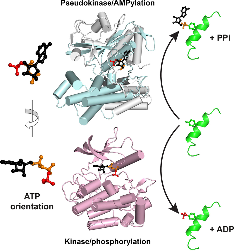
The structure of SelO (upper), from PDB entry 6EAC, is show in pale cyan
and grey, with the kinase fold colored cyan and the SelO-specific components
grey. In the lower panel, the structure of the insulin receptor kinase in its
active conformation (PDB entry 1IR3) is shown in pale magenta. Bound ATP is
shown for each protein, with the γ-phosphate colored red, the α-
and β-phosphates orange, and the rest of the molecule black. As depicted
with the magnified molecules at left, the ATP orientation is flipped by
~180° about a vertical axis between the two proteins. As a result,
whereas the γ-phosphate (red) faces ‘out’ of the kinase
active site to be appended to substrate (right), AMP instead faces out of the
SelO active site for substrate AMPylation (top right).
Comment on
-
Protein AMPylation by an Evolutionarily Conserved Pseudokinase.Cell. 2018 Oct 18;175(3):809-821.e19. doi: 10.1016/j.cell.2018.08.046. Epub 2018 Sep 27. Cell. 2018. PMID: 30270044 Free PMC article.
References
-
- Garcia-Pino A, Zenkin N, and Loris R (2014). The many faces of Fic: structural and functional aspects of Fic enzymes. Trends Biochem. Sci 39, 121–129. - PubMed
-
- Min X, Lee BH, Cobb MH, and Goldsmith EJ (2004). Crystal structure of the kinase domain of WNK1, a kinase that causes a hereditary form of hypertension. Structure 12, 1303–1311. - PubMed
Publication types
MeSH terms
Substances
Grants and funding
LinkOut - more resources
Full Text Sources
