. 2018 Oct 18;175(3):679-694.e22.
doi: 10.1016/j.cell.2018.09.004.

Dysregulated Microbial Fermentation of Soluble Fiber Induces Cholestatic Liver Cancer

Affiliations

Dysregulated Microbial Fermentation of Soluble Fiber Induces Cholestatic Liver Cancer

Vishal Singh et al. Cell. .

Abstract

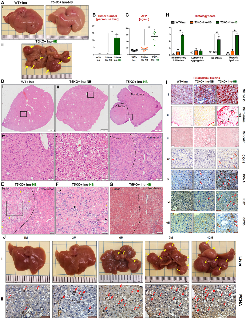
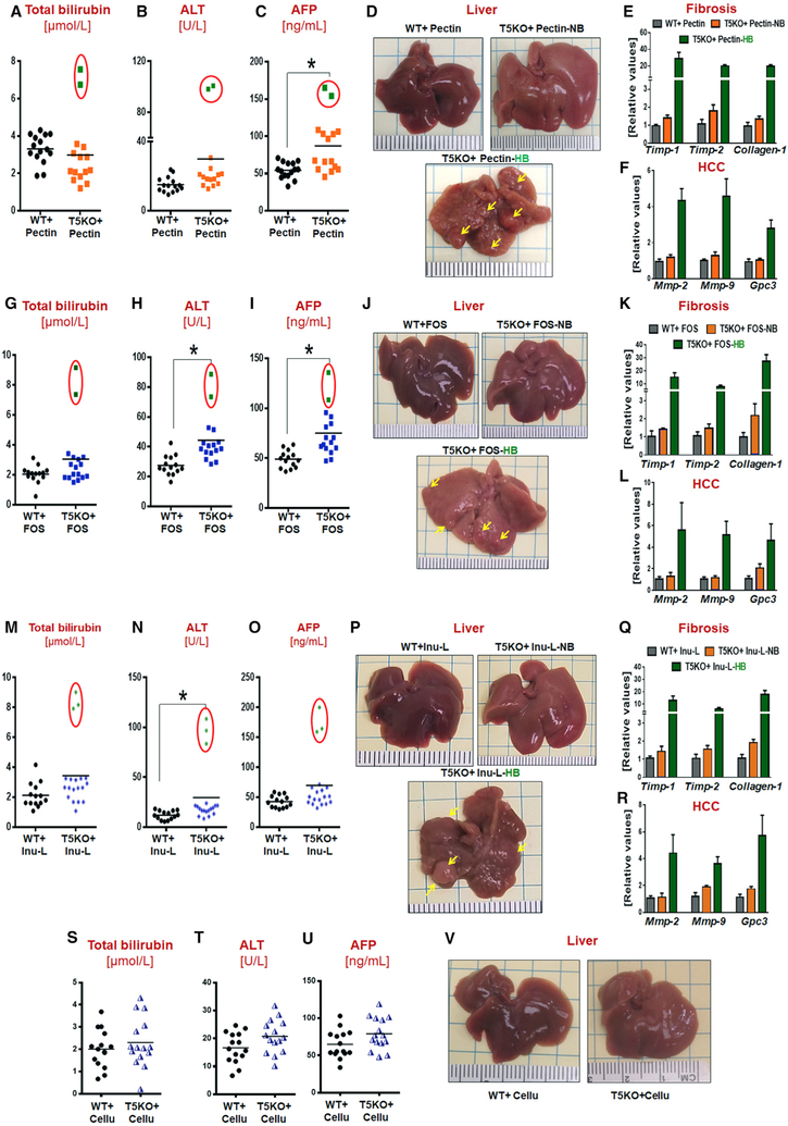
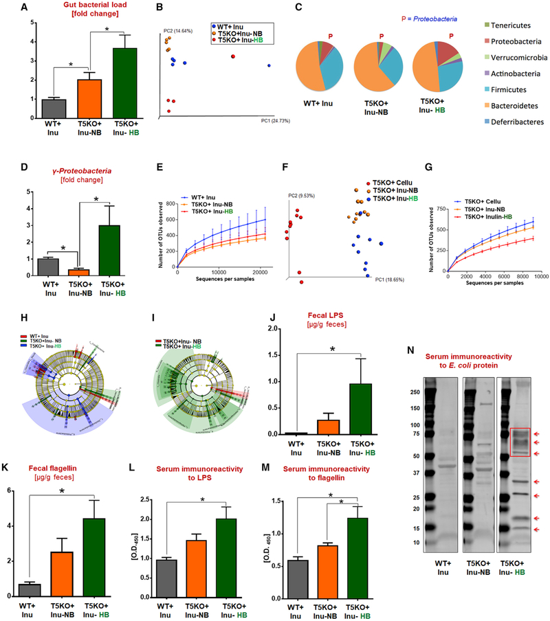
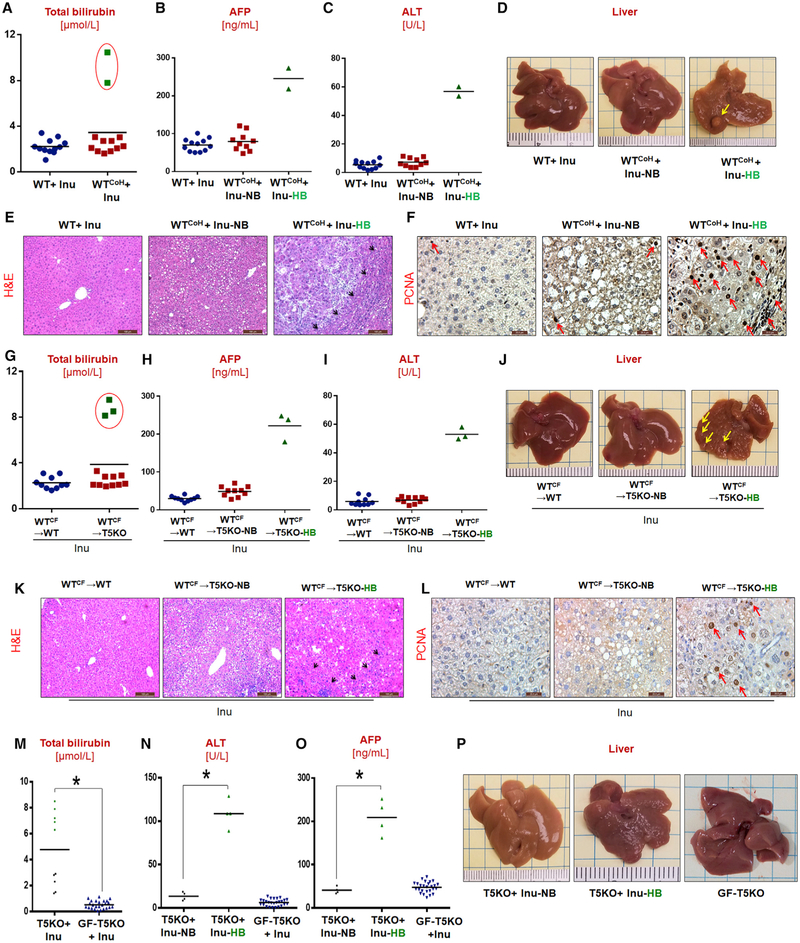
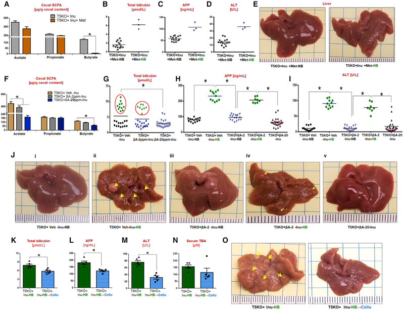
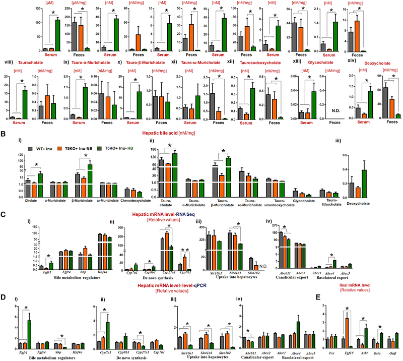
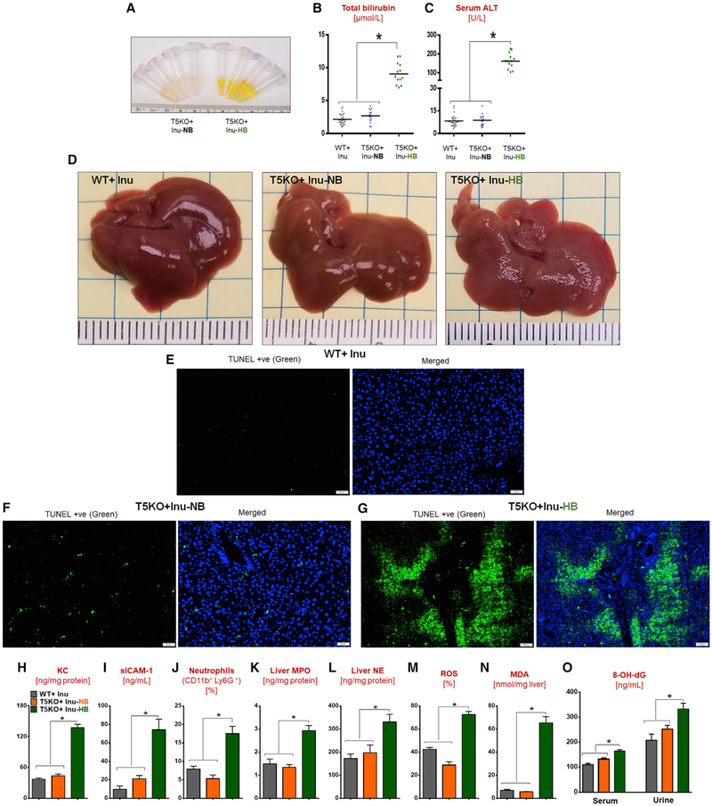
Dietary soluble fibers are fermented by gut bacteria into short-chain fatty acids (SCFA), which are considered broadly health-promoting. Accordingly, consumption of such fibers ameliorates metabolic syndrome. However, incorporating soluble fiber inulin, but not insoluble fiber, into a compositionally defined diet, induced icteric hepatocellular carcinoma (HCC). Such HCC was microbiota-dependent and observed in multiple strains of dysbiotic mice but not in germ-free nor antibiotics-treated mice. Furthermore, consumption of an inulin-enriched high-fat diet induced both dysbiosis and HCC in wild-type (WT) mice. Inulin-induced HCC progressed via early onset of cholestasis, hepatocyte death, followed by neutrophilic inflammation in liver. Pharmacologic inhibition of fermentation or depletion of fermenting bacteria markedly reduced intestinal SCFA and prevented HCC. Intervening with cholestyramine to prevent reabsorption of bile acids also conferred protection against such HCC. Thus, its benefits notwithstanding, enrichment of foods with fermentable fiber should be approached with great caution as it may increase risk of HCC.

Keywords: bile acids.


Figures

Figure 1.
Figure 1.. Dietary Soluble Fiber Inulin-Induced Icteric Hepatocellular Carcinoma in Innate Immune-Deficient Mice
(A) Macroscopic liver images (yellow arrows indicate multinodular HCC in T5KO + Inu-HB, HCC incidence: ~40%). (B) Average number of liver tumors. (C) Serum α-fetoprotein (AFP). (D) (i-vi) H&E stained liver sections (scale bar, 1 mm). The boxed regions in (i-iii) are magnified (iv-vi, scale bar, 100 μm). Dotted line demarcates the tumor and non-tumor region. (E and F) Presence of (E) trabecular pattern (black square), mitotic figures (yellow arrow), and necrotic hepatocyte (black arrow, scale bar, 100 μm) and (F) bile ducts (black arrow, scale bar, 50 μm). (G) Masson’s trichrome staining (scale bar, 100 μm). (H) Liver histology score. (I) Staining for (i) lipid (scale bar, 20 μm), (ii) collagen (scale bar, 20 μm), (iii) reticulin (black line, scale bar, 100 μm), (iv) cytokeratin 19 (scale bar, 50 μm,) proliferating cellular nuclear antigen (PCNA, scale bar, 20 μm), (vi) Ki67 (scale bar, 20 μm), and (vii) glypican (scale bar, 20 μm). (J) (i) Gross appearance of T5KO + Inu-HB livers at indicated time points. (ii) PCNA staining (scale bar, 50 μm). The data are representative of 4 independent experiments and from 3 different facilities. See also Figures S1, S2, and S3, Table S2, and Video S1.
Figure 2.
Figure 2.. Soluble Fibers Pectin (PCD) and Fructooligosaccharides, but Not Insoluble Fiber Cellulose (CCD), Induced HCC in T5KO Mice
(A-F) PCD-fed mice: (A) serum total bilirubin, (B) ALT, (C) AFP, (D) gross liver (~14% HCC incidence), and hepatic mRNA level of (E) fibrosis and (F) HCC markers. (G-L) Fructooligosaccharides (FOS)-fed mice: (G) total bilirubin, (H) ALT, (I) AFP, (J) gross liver (~13% HCC incidence), and (K) hepatic fibrosis and (L) HCC markers. (M-R) Low dose ICD-fed group: (M) total bilirubin, (N) ALT, (O) AFP, (P) gross liver (~16% HCC incidence), and (Q) hepatic fibrosis and (R) HCC markers. (S-V) CCD-fed group: (S) total bilirubin, (T) ALT, (U) AFP, and (V) gross liver (0% HCC incidence). Red circles represent mice with high serum bilirubin that developed HCC on 6 months feeding of indicated diets. Each dot represents the data from one mouse.
Figure 3.
Figure 3.. T5KO Mice with Soluble Fiber-Induced HCC Displayed Profound Alterations in Gut Microbiota Composition and Systemic Dissemination of Microbial Products
(A) Total fecal bacterial load. (B-G) Fecal microbiota composition was analyzed by 16S rRNA sequencing: (B) principal coordinate analysis (PCoA) plot, (C) abundance of major bacterial phyla (%), (D) γ-proteobacteria, (E) rarefaction curves of OTUs observed at various sequencing depths, (F) PCoA plot, and (G) rarefaction curves of OTUs. (H and I) Taxonomic cladogram from LEfSe analysis. Taxa specifically increased were colored as follows: (H) red, WT-Inu; green, T5KO + Inu-NB; blue, T5KO + Inu-HB; (I) red, T5KO + Inu-NB; green, T5KO + Inu-HB. (J and K) Quantification of fecal (J) LPS and (K) flagellin. (L and M) Serum immunoreactivity to (L) LPS and (M) flagellin. (N) Immunoblot of serum immunoreactivityto E. coli proteins (arrows showing new bands/increased intensity in T5KO-inu-HB mice). The data are representative of 2 independent experiments. See also Figure S4.
Figure 4.
Figure 4.. ICD-Induced HCC Is Microbiota-Dependent
(A-F) Co-housed group (n = 12): serum (A) total bilirubin, (B) AFP, (C) and ALT, and (D) gross liver (~17% HCC incidence). (E) H&E (scale bar, 100 μm) and (F) PCNA (scale bar, 20μm) -stained liver sections. (G-L) Cross-fostered group: serum (G) total bilirubin, (H) AFP, (I) ALT, and (J) gross liver (~16% HCC incidence). (K) H&E (scale bar, 100 μm) and (L) PCNA (scale bar, 20 μm)-stained liver sections. (M-O) Germ-free (GF) T5KO group: (M) total bilirubin, (N) ALT, and (O) AFP. (P) Gross liver (HCC incidence: conventional T5KO: ~45%; germ-free T5KO: 0%). Yellow arrows indicate tumor nodule. Red arrows point the PCNA-positive cells.
Figure 5.
Figure 5.. Targeting Butyrate Producers, Bacterial Fermentation, and Dietary Interventions Prevent HCC in ICD-Fed T5KO Mice
(A-E) Metronidazole treatment group: (A) cecal SCFA, (B) total bilirubin, (C) AFP, (D) ALT, and (E) gross liver. (F-J) β-acid treatment group: (F) cecal SCFA, (G) total bilirubin, (H) AFP, (I) ALT, and (J) gross liver (arrows indicate tumor nodules). HCC incidence: vehicle, ~39%; 2 ppm β-acid, ~10%; 20 ppm β-acid, 0%. (K-O) ICD → CCD switch group: (K) total bilirubin, (L) AFP, (M) ALT, (N) TBA, and (O) gross liver (arrows indicate tumor nodules).
Figure 6.
Figure 6.. Dysregulated Bile Acid Metabolism in ICD-Fed Hyperbilirubinemic T5KO Mice
(A) Bile acids in serum and feces: (i and ii) total bile acids, (iii-vii) unconjugated primary bile acids, (viii-xiii) conjugated primary bile acids, and (xiv) secondary bile acids. (B) Bile acids in liver. (C and D) Relative expression of genes encoding for bile acid synthesis, metabolism, and transport via (C) RNA-seq and (D) qRT-PCR in liver and terminal ileum. See also Figure S6.
Figure 7.
Figure 7.. Short-Term ICD Feeding Induces Hyperbilirubinemia, Apoptosis, and Neutrophil Infiltration in the Liver of T5KO Mice
(A–D) One-month ICD fed group: (A) serum appearance, (B) total bilirubin, (C) ALT, and (D) gross liver. (E–G) TUNEL-stained liver sections: (E) WT, (F) T5KO-NB, and (G) T5KO-HB. Positive cells in green, DAPI in blue. Scale bars, 50 μm (E and F); 100 μm (G). (H) Liver KC. (I) Serum sICAM-1. (J-O) Hepatic (J) neutrophils (K) MPO (L) NE (M) ROS (N) MDA (O) 8-hydroxy-2’-guanosine (8-OH-dG) in serum and urine. See also Figures S5 and S7

Comment in

References

    1. Arpaia N, Campbell C, Fan X, Dikiy S, van derVeeken J, deRoos P, Liu H, Cross JR, Pfeffer K, Coffer PJ, and Rudensky AY (2013). Metabolites produced by commensal bacteria promote peripheral regulatory T-cell generation. Nature 504, 451–455. - PMC - PubMed
    1. Arteel GE (2013). Build a better mouse model, and the world will beat a path to your door. Hepatology 58, 1526–1528. - PMC - PubMed
    1. Asimakopoulou A, Borkham-Kamphorst E, Tacke F, and Weiskirchen R (2016). Lipocalin-2 (NGAL/LCN2), a “help-me” signal in organ inflammation. Hepatology 63, 669–671. - PubMed
    1. Bailey RL, Gahche JJ, Lentino CV, Dwyer JT, Engel JS, Thomas PR, Betz JM, Sempos CT, and Picciano MF (2011). Dietary supplement use in the United States, 2003–2006. J. Nutr 141, 261–266. - PMC - PubMed
    1. Belcheva A, Irrazabal T, Robertson SJ, Streutker C, Maughan H, Rubino S, Moriyama EH, Copeland JK, Surendra A, Kumar S, et al. (2014). Gut microbial metabolism drives transformation of MSH2-deficient colon epithelial cells. Cell 158, 288–299. - PubMed

Publication types