. 2018 Dec 7:13:334-346.
doi: 10.1016/j.omtn.2018.09.016. Epub 2018 Sep 27.

Aptamer-miR-34c Conjugate Affects Cell Proliferation of Non-Small-Cell Lung Cancer Cells

Affiliations

Aptamer-miR-34c Conjugate Affects Cell Proliferation of Non-Small-Cell Lung Cancer Cells

Valentina Russo et al. Mol Ther Nucleic Acids. .

Abstract

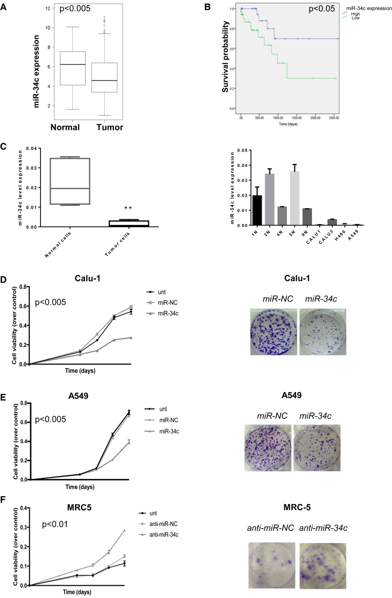
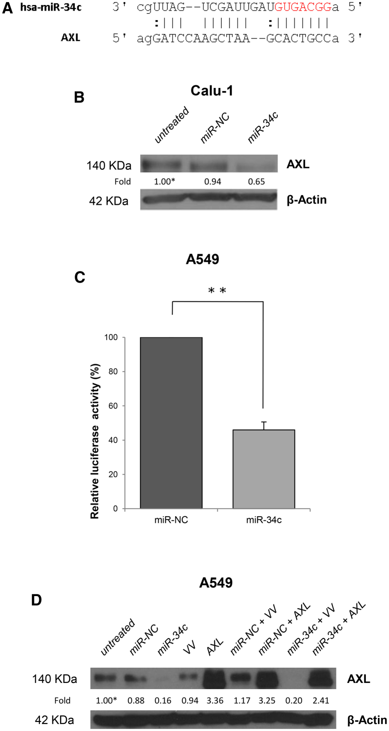
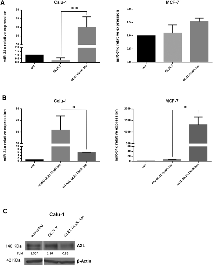
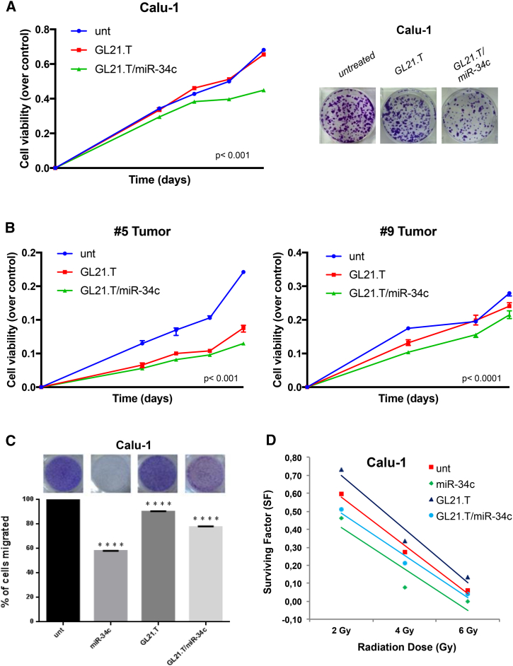
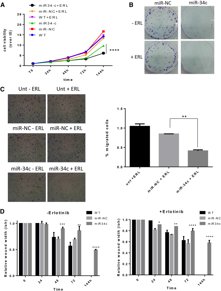
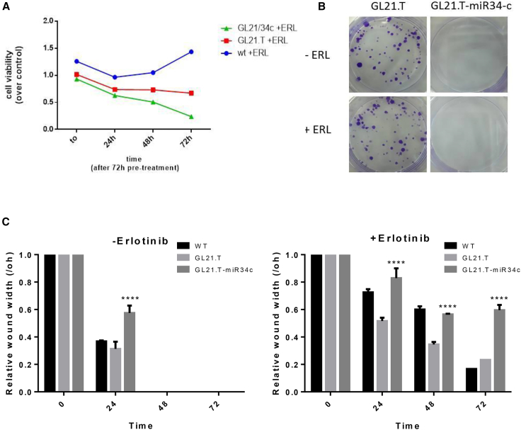
MicroRNAs (miRNAs) are key regulators of different human processes that represent a new promising class of cancer therapeutics or therapeutic targets. Indeed, in several tumor types, including non-small-cell lung carcinoma (NSCLC), the deregulated expression of specific miRNAs has been implicated in cell malignancy. As expression levels of the oncosuppressor miR-34c-3p are decreased in NSCLC compared to normal lung, we show that reintroduction of miR-34c-3p reduces NSCLC cell survival in vitro. Further, in order to deliver the miR-34c-based therapeutic selectively to tumor cells, we took advantage of a reported nucleic acid aptamer (GL21.T) that binds and inhibits the AXL transmembrane receptor and is rapidly internalized in the target cells. By applying methods successfully used in our laboratory, we conjugated miR-34c to the GL21.T aptamer as targeting moiety for the selective delivery to AXL-expressing NSCLC cells. We demonstrate that miR-34c-3p and the GL21.T/miR-34c chimera affect NSCLC cell proliferation and are able to overcome acquired RTK-inhibitor resistance by targeting AXL receptor. Thus, the GL21.T/miR-34c chimera exerts dual inhibition of AXL at functional and transcriptional levels and represents a novel therapeutic tool for the treatment of NSCLC.

Keywords: NSCLC; aptamer; lung cancer; miRNA; therapeutics.


Figures

Figure 1
Figure 1
miR-34c in Human NSCLC Tissues (A) Significant increase of miR-34c expression was identified in normal lung versus adenocarcinoma tissues collected from the TCGA database (t test, p < 0,005). (B) Kaplan-Meier survival analysis for TCGA NSCLC patients with high and low miR-34c expression. The survival data were compared using the log rank test (p < 0.05). (C) Expression of miR-34c-3p in several tumor cell lines (Calu-1, A549, Calu-3, and H460) was lower compared to normal primary lung cell lines (1N, 2N, 4N, 5N, and 9N). MiR-34c-3p expression was assessed by real-time PCR. The transcript level was normalized over RNU6B expression, used as an internal reference. Bar graphs indicate mean value ± SD and the p value is calculated by using Student’s t test, **p < 0.01. (D) Calu-1 and (E) A549 cells were transfected with control miR (miR-NC) or miR-34c-3p, and cell proliferation was analyzed by MTS assay 3, 4, 5, and 6 days after transfection (left). Bar graphs indicate mean value ± SD and the p value is calculated by using Student’s t test, p < 0.005 compared to the non-transfected cells (Unt); Calu-1 and A549 cells were transfected with miR-NC or miR-34c-3p. Overexpression of miR-34c-3p significantly inhibited colony formation (right). (F) MTS assay determined cell proliferation in MRC-5 cells following downregulation of miR-34c-3p (left). Bar graphs indicate mean value ± SD and the p value is calculated by using Student’s t test, p < 0.01, compared to non-transfected cells (Unt); colony-formation assay determined the effect of downregulated miR-34c-3p on colony-forming ability in MRC-5 cell lines (right). Downregulation of miR-34c-3p in normal lung cells promotes cell proliferation and allows colony formation.
Figure 2
Figure 2
miR-34c Targets AXL-3′ UTR and Regulates AXL Expression (A) The predicted miR-34c-3p binding sites on the 3′ UTR of AXL mRNA (predicted by the RNA HYBRID program). (B) AXL expression was analyzed in Calu-1 cells, untreated or transfected with miR-NC or miR-34c-3p for 72 hr, by western blot analysis. β-actin was used as internal control. (C) A549 cells were transiently transfected with AXL-3′ UTR in the presence of miR-34c-3p or miR-NC. Luciferase activity was evaluated 48 hr after transfection. Bar graphs indicate mean value ± SD and the p value is calculated by using Student’s t test, **p < 0.01. (D) Western blot analysis of AXL protein expression in A549 cells co-transfected with vector control (VV) or AXL plasmid lacking the 3′ UTR region (AXL) and miR-34c-3p or miR-NC. β-actin was used as internal control.
Figure 3
Figure 3
Synergistic Effects of the GL21.T-miR-34c Chimera (A) Calu-1 and MCF-7 cells were treated with GL21.T or GL21.T/miR-34c (400 nM). After 72 hr, miR-34c was quantified by RT-PCR. Bar graphs indicate mean value ± SD and the p value is calculated by using Student’s t test, **p < 0.01. (B) Calu-1 cells were transfected with si-AXL or siRNA control (si-NC) for 48 hr; MCF-7 cells were transfected with AXL or control vector (VV) for 24 hr and treated with GL21.T/miR-34c for 72 hr. miR-34c levels were quantified by RT-PCR. Bar graphs indicate mean value ± SD and the p value is calculated by using Student’s t test, *p < 0.05. (C) Calu-1 cells were treated as indicated, and AXL protein expression was analyzed by western blot after 72 hr. β-actin was used as internal control.
Figure 4
Figure 4
Role of GL21.T/miR-34c on NSCLC Cell Proliferation (A) Calu-1 cells were treated with GL21.T/miR-34c and GL21.T aptamer alone. Cell proliferation was analyzed by MTS assay 3, 4, 5, and 6 days upon transfection (left). p < 0.0001, compared to the non-transfected cells (Unt). Cells were treated with GL21.T/miR-34c and GL21.T aptamer alone and colony-formation assay was performed (right). (B) #5T (AXL+) or #9T (AXL) cells were treated with GL21.T/miR-34c or GL21.T aptamer alone. Cell viability was analyzed by MTS assay. Bar graphs indicate mean value ± SD and the p value is calculated by using Student’s t test, p < 0.001 and < 0.0001, respectively, compared to the non-transfected cells (Unt). (C) Calu-1 cells were transfected with miR-34c-3p or treated with GL21.T aptamer or GL21.T/miR-34c conjugate. The cell migration capability was analyzed by transwell migration assay using 10% FBS as migration inducer. Migrated cells were stained with crystal violet and photographed. ****p < 0.0001, compared to the non-transfected cells (Unt). (D) Effect of GL21.T/miR-34c conjugate on long-term survival of Calu-1 cells after irradiation at different doses.
Figure 5
Figure 5
The Role of miR-34c on Erlotinib Resistance (A) ER3 cells were transfected with miR-34c-3p or miR-NC. After 72 hr, transfected ER3 cells were seeded in 96-wells and treated or not with erlotinib. Cell proliferation was measured by the MTT assay 1, 2, 3, and 6 days after transfection. (B) ER3 cells were transfected with miR-34c-3p or miR-NC. After 72 hr, transfected ER3 cells were treated or not with erlotinib. Overexpression of miR-34c-3p restores sensitivity to the drug by reducing cell proliferation and colony formation. (C) Cell migration assay of ER3 cells upon miR-34c-3p and erlotinib treatment. Cells transfected with scrambled miRNA or miR-34c-3p were harvested after 72 hr and cultured on a membrane (8.0 μm pore size) inserted in the wells of a 24-well plate. Percentage of migrated cells was evaluated by eluting crystal violet solution with 1% SDS and reading the absorbance at 570 nm. Data were obtained from three independent experiments and bar graphs indicate mean value ± SD and the p value is calculated by using Student’s t test, *p < 0.05; **p < 0.01; ***p < 0.001 (over control). (D) Wound-healing assays. ER3 cells were transfected with miR-34c-3p for 72 hr and then seeded into 6-well plates at 80%–90% confluence and treated for 72 hr with GL21.T/miR-34c molecule. Bar graphs indicate mean value ± SD and the p value is calculated by using two-way ANOVA. *p < 0.05; **p < 0.01; ***p < 0.001; ****p < 0.0001 (over control).
Figure 6
Figure 6
GL21.T/miR-34c Affects Erlotinib Resistance (A) ER3 cells were treated with 400 nM of GL21.T aptamer or GL21.T/miR34c chimera for 72 hr and treated with erlotinib. Cell proliferation was measured by the MTT assay. (B) ER3 cells were treated for 72 hr with 400 nM GL21.T or GL21.T/miR34c, renewing treatment after 72 hr and treated or not with erlotinib. Long-term proliferation was determined by colony-formation assay. (C) A wound of approximately 1 mm in width was scratched with a 20-μL pipette tip. Wound closure was monitored at the indicated time intervals and imaged with phase contrast microscopy on an inverted microscope (Olympus 1 × 51 using a 5 × phase contrast objective). Bar graphs indicate mean value ± SD, and p is calculated by using two-way ANOVA. ****p < 0.0001 (over control).

References

    1. Spira A., Ettinger D.S. Multidisciplinary management of lung cancer. N. Engl. J. Med. 2004;350:379–392. - PubMed
    1. Yang P. Epidemiology of lung cancer prognosis: quantity and quality of life. Methods Mol. Biol. 2009;471:469–486. - PMC - PubMed
    1. Sequist L.V., Martins R.G., Spigel D., Grunberg S.M., Spira A., Jänne P.A., Joshi V.A., McCollum D., Evans T.L., Muzikansky A. First-line gefitinib in patients with advanced non-small-cell lung cancer harboring somatic EGFR mutations. J. Clin. Oncol. 2008;26:2442–2449. - PubMed
    1. Zhang Z., Lee J.C., Lin L., Olivas V., Au V., LaFramboise T., Abdel-Rahman M., Wang X., Levine A.D., Rho J.K. Activation of the AXL kinase causes resistance to EGFR-targeted therapy in lung cancer. Nat. Genet. 2012;44:852–860. - PMC - PubMed
    1. Bartel D.P. MicroRNAs: genomics, biogenesis, mechanism, and function. Cell. 2004;116:281–297. - PubMed