. 2019 Feb 6;27(2):365-379.
doi: 10.1016/j.ymthe.2018.09.018. Epub 2018 Sep 27.

Promoter Methylation-Regulated miR-145-5p Inhibits Laryngeal Squamous Cell Carcinoma Progression by Targeting FSCN1

Affiliations

Promoter Methylation-Regulated miR-145-5p Inhibits Laryngeal Squamous Cell Carcinoma Progression by Targeting FSCN1

Wei Gao et al. Mol Ther. .

Abstract

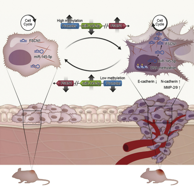
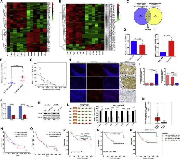
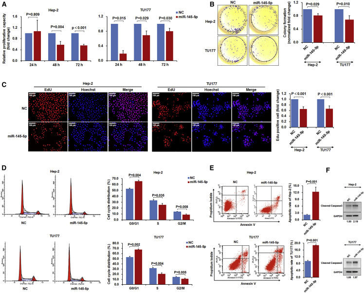
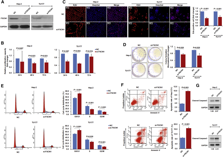
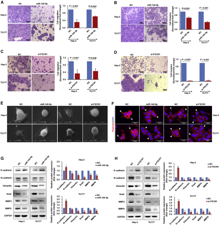
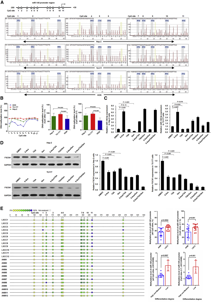
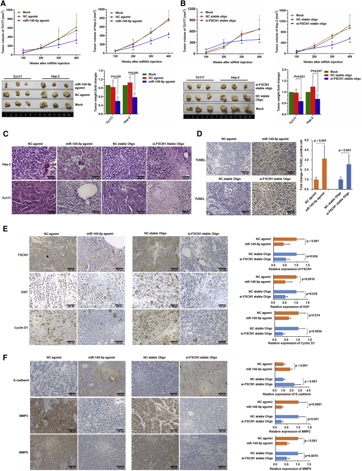
Laryngeal squamous cell carcinoma (LSCC) is a common form of head and neck cancer with poor prognosis. However, the mechanism underlying the pathogenesis of LSCC remains unclear. Here, we demonstrated increased expression of fascin actin-bundling protein 1 (FSCN1) and decreased expression of microRNA-145-5p (miR-145-5p) in a clinical cohort of LSCC. Luciferase assay revealed that miR-145-5p is a negative regulator of FSCN1. Importantly, low miR-145-5p expression was correlated with TNM (tumor, node, metastasis) status and metastasis. Moreover, cases with low miR-145-5p/high FSCN1 expression showed poor prognosis, and these characteristics together served as independent prognostic indicators of survival. Gain- and loss-of-function studies showed that miR-145-5p overexpression or FSCN1 knockdown inhibited LSCC migration, invasion, and growth by suppressing the epithelial-mesenchymal transition along with inducing cell-cycle arrest and apoptosis. Additionally, hypermethylation of the miR-145-5p promoter suggested that repression of miR-145-5p arises through epigenetic inactivation. LSCC tumor growth in vivo could be inhibited by using miR-145-5p agomir or FSCN1 small interfering RNA (siRNA), which highlights the potential for clinical translation. Collectively, our findings indicate that miR-145-5p plays critical roles in inhibiting the progression of LSCC by suppressing FSCN1. Both miR-145-5p and FSCN1 are important potential prognostic markers and therapeutic targets for LSCC.

Keywords: FSCN1; epithelial-mesenchymal transition; laryngeal squamous cell carcinoma; miR-145-5p; promoter methylation.


Figures

None
Graphical abstract
Figure 1
Figure 1
miR-145-5p Is a Negative Regulator of FSCN1 in LSCC and Is Associated with Prognosis of LSCC Patients Heatmap of (A) mRNAs and (B) miRNAs most increased and decreased in expression in six LSCC tissues as compared with six matched non-tumor tissues analyzed by Arraystar Chip. (C) Screening of miRNA target FSCN1 in LSCC. Intersection of bioinformatics-predicted miRNAs targeting FSCN1 (181) and miRNAs with decreased expression in LSCC miRNA profiling (6) showing miR-145-5p as a potential negative regulator of FSCN1 in LSCC. (D) miR-145-5p was downregulated in LSCC. qPCR of miR-145-5p level in 40 pairs of fresh LSCC tissue and corresponding ANM tissue. (E) FSCN1 was upregulated in LSCC. qPCR of FSCN1 level in 40 pairs of fresh LSCC tissue and corresponding ANM tissue. (F) Relative miR-145-5p expression in LSCC tissue with high and low FSCN1 expression. (G) Pearson correlation analysis of miR-145-5p and FSCN1 mRNA levels in LSCC tissue. (H) Expression of miR-145-5p and FSCN1 in LSCC tissue determined by fluorescence in situ hybridization and immunohistochemical staining. In immunohistochemical staining, black arrows indicate tumor areas, and black stars indicate interstitial tissues. (I) qPCR analysis of RNA levels of miR-145-5p and FSCN1 in LSCC cells and human oral keratinocyte HOK cells. (J) qPCR analysis of FSCN1 expression in miR-145-5p mimic-transfected Hep-2 and TU177 cells. (K) FSCN1 protein level in miR-145-5p-transfected Hep-2 and TU177 cells examined by western blot analysis. (L) miR-145-5p binds to the 3′ UTR of FSCN1 to inhibit its expression. HEK293T cells were co-transfected with luciferase reporter constructs containing wild-type (WT) or mutated FSCN1 3′ UTRs and miR-145-5p mimic, and relative luciferase activity was measured and normalized to negative control (NC) mimic-transfected cells. Red circle indicates mutation of miR-145-5p binding site. siCHECK2 indicates cells transfected with the empty vector psiCHECK-2, which does not contain the 3′ UTR region of FSCN1. (M) Differential expression analysis of FSCN1 mRNA of head and neck squamous cell carcinoma (HNSCC) patients in the TCGA cohort (tumor group, 519; normal group, 44; *p < 0.01). (N and O) Kaplan-Meier survival curves of HNSCC patients with different FSCN1 (N) and miR-145-5p (O) level in the TCGA cohort via Oncolnc (http://www.oncolnc.org). Upregulated FSCN1 was significantly correlated with poor outcome for patients with HNSCC (log rank test, p = 0.00587), and miR-145-5p level was positively correlated with survival time of HNSCC (log rank test, p = 0.718). (P–R) Kaplan-Meier survival analysis of 188 LSCC patients by expression of miR-145-5p (P), FSCN1 (Q), and a combination of miR-145-5p and FSCN1 (R). p value was determined by log rank test. Error bars represent SD of the indicated clinical samples or three independent assays.
Figure 2
Figure 2
miR-145-5p Inhibits LSCC Cell Proliferation, Colony Formation, and Induces Cell-Cycle Arrest and Apoptosis Hep-2 and TU177 cells were transfected with miR-145-5p mimic or negative control (NC) mimic for 48 hr. Cell proliferation was measured by cell counting kit-8 analysis (A), colony formation assay (B), and EdU staining (C); cell cycle (D) and apoptosis (E) were measured by flow cytometry. (F) Hep-2 and TU177 cells were transfected with miR-145-5p mimic or NC mimic for 48 hr. Protein level of cleaved caspase-3 was determined by immunoblotting. Data are mean ± SD of three independent experiments. Error bars represent SD of three independent assays.
Figure 3
Figure 3
FSCN1 Knockdown Inhibits LSCC Cell Proliferation and Induces Cell-Cycle Arrest and Apoptosis Hep-2 and TU177 cells were transfected with siRNA targeting FSCN1 (si-FSCN1) or negative control (NC) for 48 hr, and FSCN1 levels were determined by western blot assay (A). Assays of cell proliferation (B), EdU staining (C), colony formation (D), cell cycle (E), and apoptosis (F) after indicated treatments. (G) Hep-2 and TU177 cells were transfected with si-FSCN1 or NC for 48 hr. Protein level of cleaved caspase-3 was determined by immunoblotting. Data are mean ± SD of three independent experiments. Error bars represent SD of three independent assays.
Figure 4
Figure 4
miR-145-5p Overexpression or FSCN1 Knockdown Inhibits LSCC Cell Migration, Invasion, and Endothelial-to-Mesenchymal Transition Hep-2 and TU177 cells were transfected with miR-145-5p mimic or si-FSCN1 for 48 hr. (A and C) Cell migration of miR-145-5p overexpressed (A) and FSCN1 knockdown (C) cells were measured by Transwell assays. (B and D) Cell invasion of miR-145-5p overexpressed (B) and FSCN1 knockdown (D) cells were measured by Transwell assays. (E) Surface structure of cells was observed by scanning electron microscopy. (F) F-actin was stained with rhodamine phalloidin, and nuclei were stained with DAPI. As indicated by white arrows, miR-145-5p mimic or si-FSCN1 treatment decreased cell lamellipodial and filopodial structures as compared with the NC. (G and H) Expression of EMT markers E-cadherin, N-cadherin, Vimentin, Snail, MMP2, and MMP9 in miR-145-5p overexpressed (G) and FSCN1 knockdown (H) cells were determined by western blot assays. Representative images and data from three independent experiments. Data are mean ± SD of three independent experiments. Error bars represent SD of three independent assays. *p < 0.05, **p < 0.01.
Figure 5
Figure 5
Hypermethylation of miR-145-5p Promoter Suppresses miR-145-5p Expression in LSCC Cells (A and B) Pyrosequencing analysis of miR-145-5p promoter methylation status in LSCC cell lines Hep-2 and TU177 and normal control HOK cells. The methylation level in Hep-2 and TU177 cells was normalized to that in HOK cells. Pyrosequencing map (A) and statistical results (B) of methylation levels of Hep-2, TU177 and HOK cells. (C) Hep-2 and TU177 cells were treated with 5-Aza, trichostatin A (TSA), and genistein (Gen) as indicated; the expression of miR-145-5p was determined by qPCR. (D) Hep-2 and TU177 cells were treated with 5-Aza, TSA, and Gen as indicated; FSCN1 protein level was measured by western blot assay. Data are mean ± SD of three independent experiments. (E) Methylation analysis of miR-145-5p in clinical LSCC and paired ANM tissues. DNA purified from LSCC and paired ANM tissues by laser capture microdissection was treated with bisulfite and subjected to MassARRAY methylation analysis. Error bars represent SD of the indicated clinical samples or three independent assays.
Figure 6
Figure 6
miR-145-5p Overexpression or FSCN1 Knockdown Inhibits LSCC Xenograft Tumor Formation in Mice Nude mice were subcutaneously injected with Hep-2 and TU177 cells. After tumor formation, miR-145-5p agomir, NC agomir, si-FSCN1 stable oligonucleotides, and corresponding negative oligonucleotides were subcutaneously injected into tumor sites. Tumor volume and weight after injection of miR-145-5p agomir (A) or si-FSCN1 (B) were plotted. Data are mean ± SD. *p < 0.05. (C) Representative H&E staining images of Hep-2 and TU177 xenograft tumors. (D) TUNEL assay of apoptotic cells in xenograft tumors. (E) Representative immunohistochemical staining of FSCN1 and proliferation markers Ki67 and CyclinD1 in Hep-2 and TU177 xenograft tumors. (F) Representative immunohistochemical staining of EMT markers E-cadherin, MMP2, and MMP9 in xenograft tumors. Data are mean ± SD of three independent experiments. Error bars represent SD of three independent experiments.
Figure 7
Figure 7
Proposed Regulatory Model for miR-145-5p/FSCN1 Axis in LSCC Progression Expression of miR-145-5p was silenced by hypermethylation of its promoter in LSCC. miR-145-5p inhibits malignant phenotypes of LSCC by directly targeting FSCN1 to inhibit the EMT and cytoskeletal organization. Downregulation of miR-145-5p upregulates FSCN1 in LSCC, thus promoting tumorigenesis and progression.

References

    1. Cancer Genome Atlas Network Comprehensive genomic characterization of head and neck squamous cell carcinomas. Nature. 2015;517:576–582. - PMC - PubMed
    1. Gao W., Zhang C., Feng Y., Chen G., Wen S., Huangfu H., Wang B. Fascin-1, ezrin and paxillin contribute to the malignant progression and are predictors of clinical prognosis in laryngeal squamous cell carcinoma. PLoS ONE. 2012;7:e50710. - PMC - PubMed
    1. Liu H.C., Chen G.G., Vlantis A.C., Tong M.C., van Hasselt C.A. Chemotherapy for laryngeal cancer—an apoptotic approach. Curr. Drug Targets. 2008;9:878–886. - PubMed
    1. Jenckel F., Knecht R. State of the art in the treatment of laryngeal cancer. Anticancer Res. 2013;33:4701–4710. - PubMed
    1. Lin C., Zhang S., Wang Y., Wang Y., Nice E., Guo C., Zhang E., Yu L., Li M., Liu C. Functional role of a novel long noncoding RNA TTN-AS1 in esophageal squamous cell carcinoma progression and metastasis. Clin. Cancer Res. 2018;24:486–498. - PubMed

Publication types

MeSH terms