. 2018 Nov 2;7(11):bio033001.
doi: 10.1242/bio.033001.

Drosophila Ror is a nervous system-specific co-receptor for Wnt ligands

Affiliations

Drosophila Ror is a nervous system-specific co-receptor for Wnt ligands

Caroline Ripp et al. Biol Open. .

Abstract

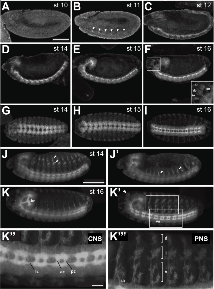
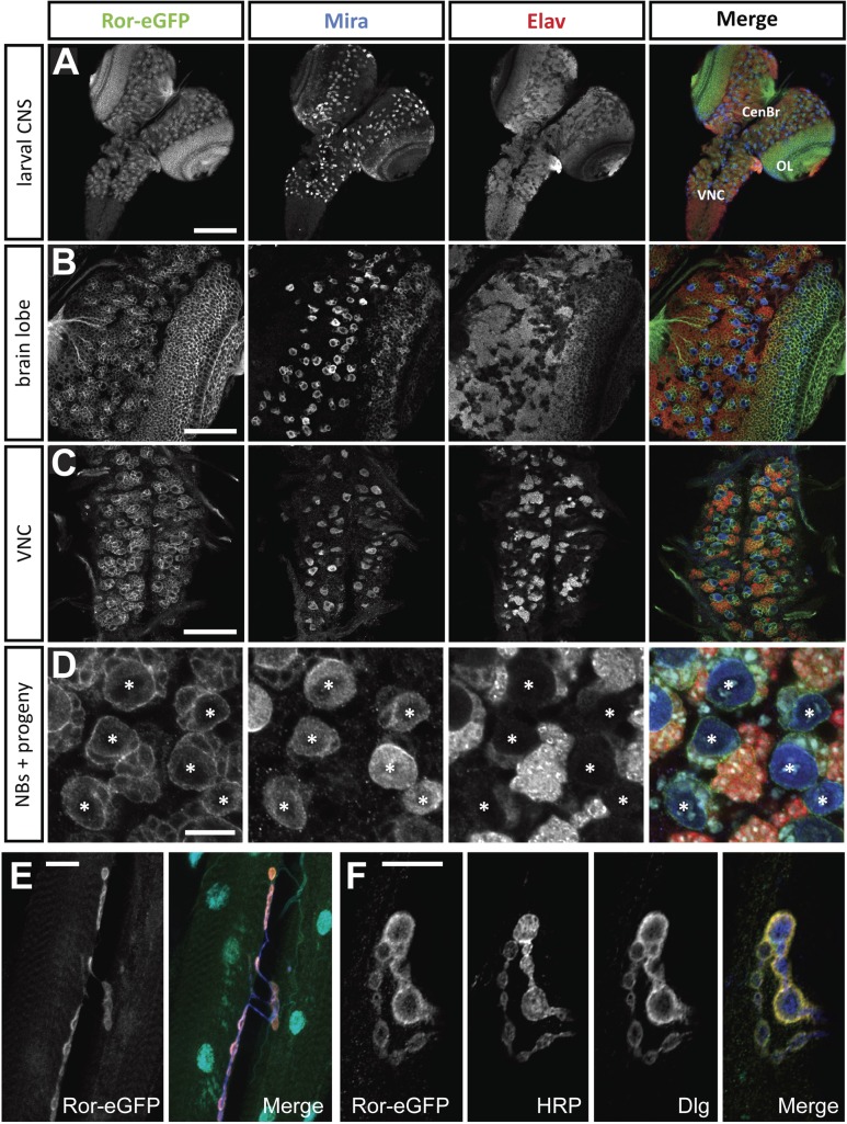
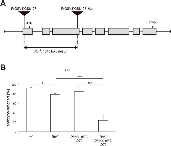
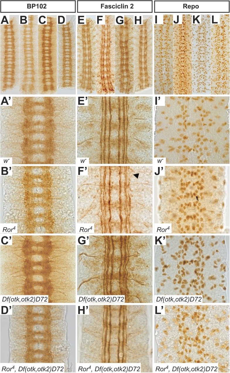
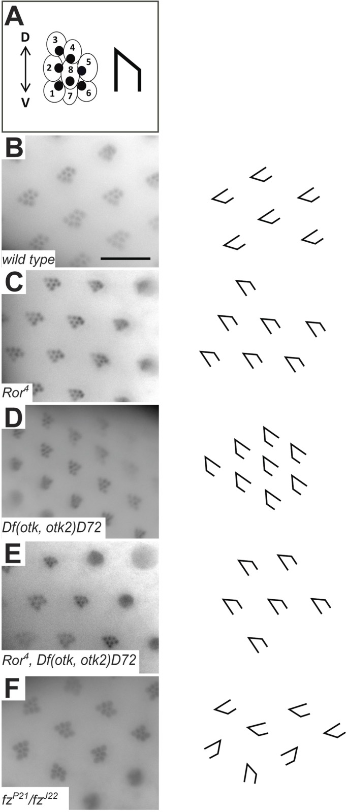
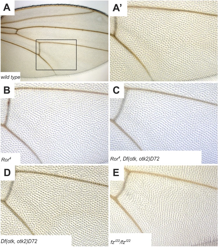
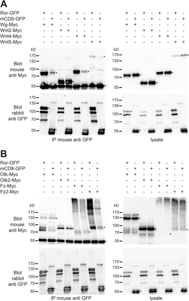
Wnt ligands are secreted glycoproteins that control many developmental processes and are crucial for homeostasis of numerous tissues in the adult organism. Signal transduction of Wnts involves the binding of Wnts to receptor complexes at the surface of target cells. These receptor complexes are commonly formed between a member of the Frizzled family of seven-pass transmembrane proteins and a co-receptor, which is usually a single-pass transmembrane protein. Among these co-receptors are several with structural homology to receptor tyrosine kinases, including Ror, PTK7, Ryk and MUSK. In vertebrates, Ror-2 and PTK7 are important regulators of planar cell polarity (PCP). By contrast, PCP phenotypes were not reported for mutations in off-track (otk) and off-track2 (otk2), encoding the Drosophila orthologs of PTK7. Here we show that Drosophila Ror is expressed in the nervous system and localizes to the plasma membrane of perikarya and neurites. A null allele of Ror is homozygous viable and fertile, does not display PCP phenotypes and interacts genetically with mutations in otk and otk2 We show that Ror binds specifically to Wingless (Wg), Wnt4 and Wnt5 and also to Frizzled2 (Fz2) and Otk. Our findings establish Drosophila Ror as a Wnt co-receptor expressed in the nervous system.

Keywords: Nervous system development; Planar cell polarity; Ror; Wnt signaling.


Conflict of interest statement

Competing interestsThe authors declare no competing or financial interests.

Figures

Fig. 1.
Fig. 1.
Expression of a Ror-eGFP fusion protein under control of the endogenous Ror promoter in Drosophila embryos. (A-F) Lateral views of stage 10-12 and 14-16 embryos. (G-I) Stage 14-16 embryos viewed from the ventral side, anterior to the left. (J-K‴) Light sheet fluorescence microscopy images of Ror-eGFP embryos. The images show maximum intensity projections of stacks taken from whole embryos. (J,J′) and (K,K′) show the same embryo, respectively, scanned from both sides. (J,J′) At stage 14 Ror-eGFP expression is strong in the embryonic CNS and already visible in the developing PNS. (K,K′) At stage 16 Ror-eGFP is expressed throughout the entire nervous system. (K″) Enlarged view of the CNS seen in (K′). (K‴) Enlarged view of the PNS seen in (K′). Areas shown at higher magnification in (K″) and (K‴) are indicated by boxes in (K′). Scale bars for A-K′: 100 µm; K″,K‴: 20 µm. bo, bolwig's organ; do, dorsal organ; to, terminal organ; lpo, lateropharyngeal organ; br, embryonic brain; vn, ventral nerve cord; lc, longitudinal connectives; ac, anterior commissures; pc, posterior commissures; sa, sensory axon; d, dorsal cluster; l, lateral cluster; v, ventral cluster. In (A-F) and (J-K′) anterior is to the left and dorsal up. Arrowheads in (B) point to the first cell clusters showing Ror-eGFP expression during embryonic development. Arrowheads in (J, J′ and K′) point to sensilla of the PNS.
Fig. 2.
Fig. 2.
Ror-eGFP expression in the central nervous system and body wall muscles of third instar larvae. (A) Overview of the larval central nervous system. Ror-eGFP is expressed in neuroblasts (NBs) and their neuronal progeny. (B) Larval brain lobe. (C) Larval ventral nerve cord. (D) Magnification of a section of a larval brain lobe including NBs, neurons and most likely also GMCs. In (D) the merged image additionally contains Hoechst staining (cyan) labeling the DNA. (E) Neuromuscular junctions (NMJs) of third instar larval body wall muscles. (F) High magnification image of NMJ boutons. The merged images in (E) and (F) show Ror-eGFP in green, HRP in blue, Dlg in red and DAPI in cyan. Scale bars: A, 100 µm; B,C, 50 µm; E, 20 µm; D,F, 10 µm. CenBr, central brain; OL, optic lobe; VNC, ventral nerve cord; *, NB.
Fig. 3.
Fig. 3.
Generation of the null allele Ror4. (A) The exon-intron structure of the Ror locus including the positions of the ATG and stop codons is shown. The P-element P{GSV3}GS8107 was first mobilized using P-transposase and re-integrated into the third exon of the Ror coding region (P{GSV3}GS8107-Hop). Then the region between the two P-elements was excised in a second transposase-mediated step. (B) Embryonic viability assay. Data were obtained by repeating each experiment at least three times. The error bars represent the standard deviation of the mean. *P<0.05; ***P<0.001 (independent samples Student's t-test).
Fig. 4.
Fig. 4.
Phenotypic analysis of the CNS in mutants for Ror, otk and otk2. (A-D′) Axon tracts of the CNS are visualized using the BP102 antibody in w (A), Ror4 (B), Df(otk,otk2)D72 (C) and Ror4, Df(otk,otk2)D72 (D) embryos. All mutant embryos resemble the wild type. (E-H′) Fasciclin II labels the axons of a subset of neurons within the CNS. In Ror4 mutant embryos (F,F′) occasional disruptions in the lateral fascicle are visible (arrowhead). In otk, otk2 double mutants (G,G′) and Ror, otk, otk2 triple mutants (H,H′) all fascicles look intact. (I-L′) Glial cells were visualized with the anti-Repo antibody. The pattern is not disturbed in any of the investigated mutants. Images (A′-L′) are higher magnifications of regions of the embryos shown in images (A-L). All images show three abdominal segments of late stage embryos. Anterior is up.
Fig. 5.
Fig. 5.
Planar cell polarity in Ror4 mutant eyes is not disturbed. (A) Schematic representation of a Drosophila ommatidium. Each ommatidium is formed by eight photoreceptor cells. In a cross section only seven photoreceptor cells are visible, because the R7 and the R8 cell are positioned on top of each other. The visible cells resemble an arrowhead. (B) Wild-type ommatidia. (C) Ror4 mutant eye. (D) Otk and otk2 double mutant Df(otk,otk2)D72. (E) Triple mutant Ror4,Df(otk,otk2)D72. (F) fzJ22/fzP2 eye as positive control. The polarity of ommatidia is disturbed, all arrow-like shapes point in different directions. Scale bar: 20 µm.
Fig. 6.
Fig. 6.
Planar cell polarity in Ror mutant wings is normal. (A) Overview of a wild-type Drosophila wing. (A′) Magnification of the boxed region in (A). (B) Ror4 mutant wing. (C) Triple mutant Ror4, Df(otk,otk2)D72. (D) otk, otk2 double mutant Df(otk,otk2)D72. (E) fzJ22/fzJ22 wing as positive control showing disturbed orientation of wing hairs.
Fig. 7.
Fig. 7.
Ror-GFP binds to Myc-tagged Wnts and Wnt receptors. (A) The indicated constructs for Ror-GFP, CD8-GFP as negative control and Myc-tagged Wnts were transfected into Drosophila S2R+ cells. Co-immunoprecipitation from cell lysates was performed using mouse anti-GFP antibody followed by western blotting using mouse anti-Myc and rabbit anti-GFP antibodies. In the blot probed with anti-Myc antibody the denatured heavy chain of the immunoglobulin used in the IP is visible (marked with °). Bands representing the respective full-length proteins are marked with asterisks (*). (B) The indicated constructs for Ror-GFP, CD8-GFP as negative control and Myc-tagged Wnt receptors were co-transfected into Drosophila S2R+ cells. Co-immunoprecipitation from cell lysates was performed as above. IP, immunoprecipitation; Blot, western blot. Protein sizes are indicated in kilo Dalton (kD).
Fig. 8.
Fig. 8.
The morphology of the ventral nerve cord in Ror-Myc overexpressing embryos is normal. (A-B′) Axon tracts of the CNS visualized using the BP102 antibody in wild-type (A,A′) and Ror-Myc overexpressing embryos (B,B′). The CNS of embryos overexpressing Ror resembles the wild type. (C-D′) Longitudinal axon tracts visualized with Fasciclin II. In da::Gal4/UAS::Ror-Myc embryos (D,D′) all three fascicles are intact and resemble the wild type (C,C′). (E-F′) Glial cells visualized with the anti-Repo antibody. The pattern in da::Gal4/UAS::Ror-Myc embryos (F,F′) resembles the wild type (E,E′). Images (A′-F′) are higher magnifications of the preparations shown in images (A-F). All images show three abdominal segments of late stage embryos; anterior is up. (G,H) Confocal images of the CNS of a third instar da::Gal4/UAS::Ror-Myc larva. Expression patterns of the indicated proteins are normal and resemble the wild type (cf. Fig. 2, Fig. S2). (G) Overview of a brain hemisphere. (H) Higher magnification of the larval brain lobe. Scale bars: G, 50 µm; H, 20 µm.

Similar articles

Cited by

References

    1. Adler P. N., Krasnow R. E. and Liu J. (1997). Tissue polarity points from cells that have higher Frizzled levels towards cells that have lower Frizzled levels. Curr. Biol. 7, 940-949. 10.1016/S0960-9822(06)00413-1 - DOI - PubMed
    1. Angers S. and Moon R. T. (2009). Proximal events in Wnt signal transduction. Nat. Rev. Mol. Cell Biol. 10, 468-477. 10.1038/nrm2717 - DOI - PubMed
    1. Axelrod J. D. and McNeill H. (2002). Coupling planar cell polarity signaling to morphogenesis. ScientificWorldJournal 2, 434-454. 10.1100/tsw.2002.105 - DOI - PMC - PubMed
    1. Bai Y., Tan X., Zhang H., Liu C., Zhao B., Li Y., Lu L., Liu Y. and Zhou J. (2014). Ror2 receptor mediates Wnt11 ligand signaling and affects convergence and extension movements in zebrafish. J. Biol. Chem. 289, 20664-20676. 10.1074/jbc.M114.586099 - DOI - PMC - PubMed
    1. Berger H., Wodarz A. and Borchers A. (2017). PTK7 faces the Wnt in development and disease. Front. Cell Dev. Biol. 5, 31 10.3389/fcell.2017.00031 - DOI - PMC - PubMed