. 2018 Nov;28(11):1611-1620.
doi: 10.1101/gr.231696.117. Epub 2018 Oct 19.

Aberrant PRDM9 expression impacts the pan-cancer genomic landscape

Affiliations

Aberrant PRDM9 expression impacts the pan-cancer genomic landscape

Armande Ang Houle et al. Genome Res. 2018 Nov.

Abstract

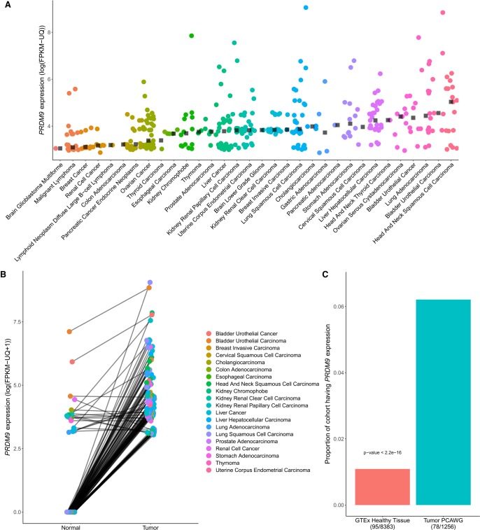
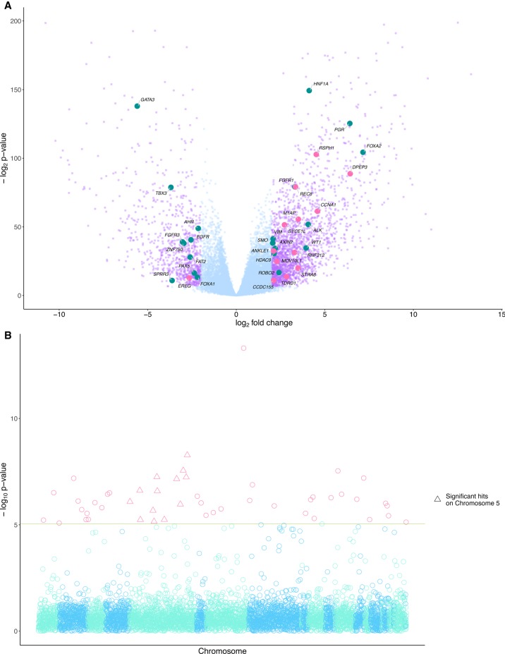
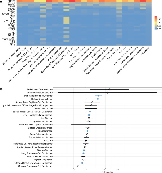
The binding of PRDM9 to chromatin is a key step in the induction of DNA double-strand breaks associated with meiotic recombination hotspots; it is normally expressed solely in germ cells. We interrogated 1879 cancer samples in 39 different cancer types and found that PRDM9 is unexpectedly expressed in 20% of these tumors even after stringent gene homology correction. The expression levels of PRDM9 in tumors are significantly higher than those found in healthy neighboring tissues and in healthy nongerm tissue databases. Recurrently mutated regions located within 5 Mb of the PRDM9 loci, as well as differentially expressed genes in meiotic pathways, correlate with PRDM9 expression. In samples with aberrant PRDM9 expression, structural variant breakpoints frequently neighbor the DNA motif recognized by PRDM9, and there is an enrichment of structural variants at sites of known meiotic PRDM9 activity. This study is the first to provide evidence of an association between aberrant expression of the meiosis-specific gene PRDM9 with genomic instability in cancer.


Figures

Figure 1.
Figure 1.
Characterization of aberrant PRDM9 expression across cancer types. (A) A total of 365 samples expressing PRDM9 above 10 FPKM-UQ across 39 cancer types from the PCAWG and TCGA data sets (n = 1879), after a correction to account for the high homology between PRDM9 and PRDM7 (Methods; see Supplemental Fig. S2). Black squares represent the median of PRDM9 expression above a threshold of 10 FPKM-UQ within each cancer type. (B) PRDM9 expression in 128 tumor samples expressing PRDM9 above a threshold of 10 FPKM-UQ and their matching normal samples, across cancer types within the TCGA and the PCAWG cohorts (n = 813 pairs). Data points are colored according to cancer type, and lines connect tumor and normal samples originating from the same patient. Cancer samples have higher PRDM9 expression than their matching healthy tissues (one-sided Wilcoxon signed-rank test: P < 2.2 × 10⁻¹⁶), a result consistent within each cancer type where at least three sample pairs were expressing PRDM9 (Fisher's method with one-sided Wilcoxon signed-rank test: P = 2.28 × 10⁻²⁰) (Supplemental Table S1). (C) Proportion of samples expressing PRDM9 in the cancer cohorts (PCAWG and TCGA) compared with the proportion of samples expressing PRDM9 in the GTEx cohort, excluding testes. The proportion of samples expressing PRDM9 in cancer samples is significantly higher than that of healthy tissues.
Figure 2.
Figure 2.
Differentially expressed genes and recurrently mutated regions are associated with PRDM9 expression in cancer. (A) A total of 3114 genes were differentially expressed (in purple) above a threshold of 2 log2 fold change and a P-value adjusted for multiple testing using the Benjamini-Hochberg procedure (FDR < 0.05) between groups of cancers partitioned on PRDM9 expression. Of these genes, 2224 were overexpressed in cancers expressing PRDM9, and 890 were underexpressed. Twenty-two known cancer driver genes (Gonzalez-Perez et al. 2013) were differentially expressed in tumors expressing PRDM9 (in teal), and 13 had functions related to meiosis (in pink). (B) P-values from the associations between PRDM9 expression levels and recurrently mutated regions. The red line shows P < 0.05, with a Bonferroni correction for multiple testing for the number of tested regions (n = 1,507,106). Forty-nine recurrently mutated regions were significantly associated with aberrant PRDM9 expression across cancers. Highlighted with pink triangles are loci significantly associated with aberrant expression located on Chromosome 5, where the PRDM9 locus is located.
Figure 3.
Figure 3.
Structural variant (SV) breakpoints are enriched at sites of PRDM9 binding and activity. (A) Proportion of SV breakpoint sequences (SVBSs) significantly matching the motifs recognized by proteins in the JASPAR database, in cancer samples expressing PRDM9. We focused on sequences located within 100 bp of SV breakpoints, which is the mean distance separating DSBs and PRDM9 binding sites in meiosis (Baker et al. 2014). Each row shows the proportion of SVBSs matching the given motif per cancer type. The far-right column shows the proportion of SVBSs matching each motif across all cancer types. Supplemental Figure S7 shows the robustness of these results to significance thresholds used (Methods). (B) Enrichment for SV breakpoints at sites of highly recombining regions (HRRs) in samples expressing PRDM9, as shown by odds ratios >1 for each cancer type. Significant cancer types are shown in blue, as determined using Fisher's exact test (P < 0.05 with Bonferroni correction for the number of cancer types tested). Brain glioblastoma multiforme, kidney chromophobe, head and neck squamous cell carcinoma, liver hepatocellular carcinoma, and breast cancer samples all exhibited significant associations between PRDM9 expression and the colocalization of SV breakpoints and meiotic recombination hotspots. Ovarian cancer, lung squamous cell carcinoma, and uterine corpus endometrial carcinoma showed odds ratios <1, indicating significant enrichment for SV breakpoints in non-HRRs in samples expressing PRDM9.

References

    1. Alexandrov LB, Nik-Zainal S, Wedge DC, Aparicio SAJR, Behjati S, Biankin AV, Bignell GR, Bolli N, Borg A, Børresen-Dale A-L, et al. 2013. Signatures of mutational processes in human cancer. Nature 500: 415–421. - PMC - PubMed
    1. Altemose N, Noor N, Bitoun E, Tumian A, Imbeault M, Chapman JR, Radu Aricescu A, Myers SR. 2017. A map of human PRDM9 binding provides evidence for novel behaviors of PRDM9 and other zinc-finger proteins in meiosis. eLife 6: e28383. - PMC - PubMed
    1. Alves I, Ang Houle A, Hussin JG, Awadalla P. 2017. The impact of recombination on human mutation load and disease. Philos Trans R Soc Lond B Biol Sci 372: 20160465. - PMC - PubMed
    1. Anders S, Pyl PT, Huber W. 2015. HTSeq: a Python framework to work with high-throughput sequencing data. Bioinformatics 31: 166–169. - PMC - PubMed
    1. Andersen SL, Sekelsky J. 2010. Meiotic versus mitotic recombination: two different routes for double-strand break repair. The different functions of meiotic versus mitotic DSB repair are reflected in different pathway usage and different outcomes. Bioessays 32: 1058–1066. - PMC - PubMed

Publication types

MeSH terms

Substances

LinkOut - more resources