. 2018 Dec 28;293(52):20085-20098.
doi: 10.1074/jbc.RA118.004889. Epub 2018 Oct 19.

ATP7A and ATP7B copper transporters have distinct functions in the regulation of neuronal dopamine-β-hydroxylase

Affiliations

ATP7A and ATP7B copper transporters have distinct functions in the regulation of neuronal dopamine-β-hydroxylase

Katharina Schmidt et al. J Biol Chem. .

Abstract

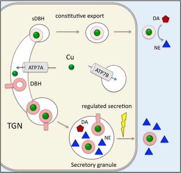
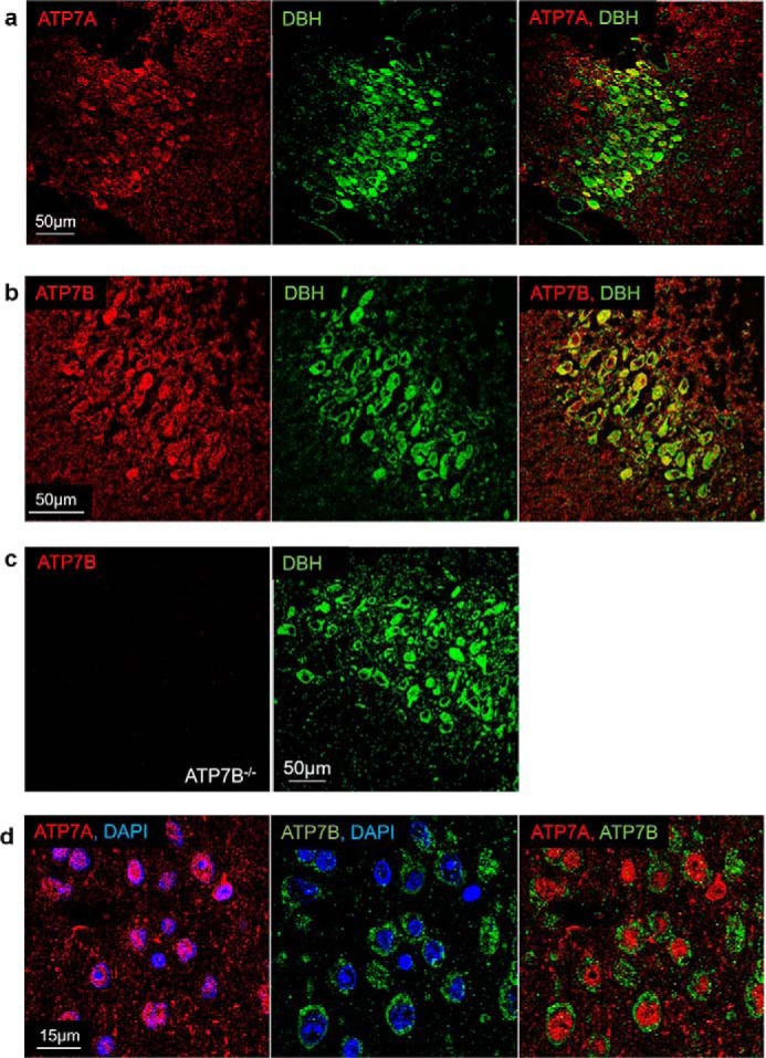
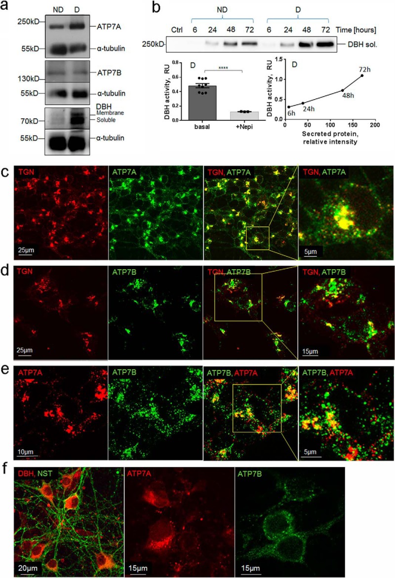
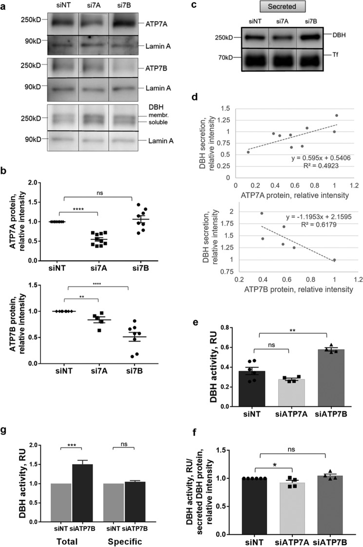
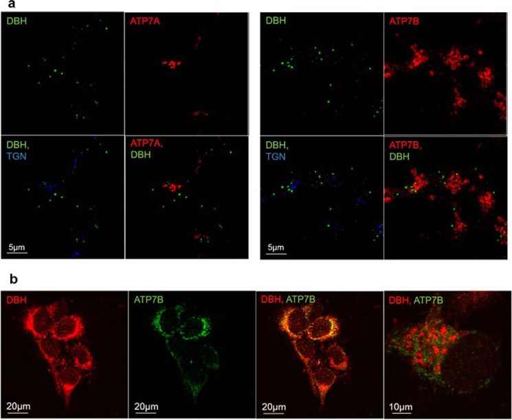
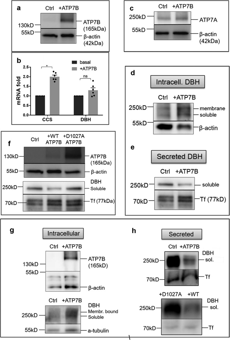
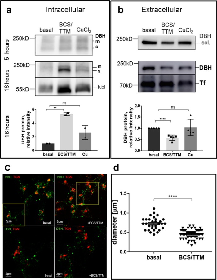
The copper (Cu) transporters ATPase copper-transporting alpha (ATP7A) and ATPase copper-transporting beta (ATP7B) are essential for the normal function of the mammalian central nervous system. Inactivation of ATP7A or ATP7B causes the severe neurological disorders, Menkes disease and Wilson disease, respectively. In both diseases, Cu imbalance is associated with abnormal levels of the catecholamine-type neurotransmitters dopamine and norepinephrine. Dopamine is converted to norepinephrine by dopamine-β-hydroxylase (DBH), which acquires its essential Cu cofactor from ATP7A. However, the role of ATP7B in catecholamine homeostasis is unclear. Here, using immunostaining of mouse brain sections and cultured cells, we show that DBH-containing neurons express both ATP7A and ATP7B. The two transporters are located in distinct cellular compartments and oppositely regulate the export of soluble DBH from cultured neuronal cells under resting conditions. Down-regulation of ATP7A, overexpression of ATP7B, and pharmacological Cu depletion increased DBH retention in cells. In contrast, ATP7B inactivation elevated extracellular DBH. Proteolytic processing and the specific activity of exported DBH were not affected by changes in ATP7B levels. These results establish distinct regulatory roles for ATP7A and ATP7B in neuronal cells and explain, in part, the lack of functional compensation between these two transporters in human disorders of Cu imbalance.

Keywords: ATP7A; ATP7B; Menkes disease; SH-SY5Y cells; Wilson's disease; cellular regulation; constitutive secretion; copper transport; dopamine-β-hydroxylase; intracellular trafficking; locus coeruleus; noradrenergic; vesicles.


Conflict of interest statement

The authors declare that they have no conflicts of interest with the contents of this article

Figures

Figure 1.
Figure 1.
Cartoon illustrating the intracellular distribution of ATP7A, ATP7B, and DBH. ATP7A is located in the TGN and transports copper (green balls) into the lumen of this compartment. ATP7B is in vesicles where it sequesters Cu from the cytosol and regulates the amount of cytosolic Cu available to ATP7A. DBH receives its Cu cofactor as it matures within the secretory pathway (most likely from ATP7A). The fraction of DBH is then proteolytically processed. Soluble DBH is sorted to two main destinations. Some soluble DBH and a noncleaved membrane-bound DBH are sorted to secretory granules. In secretory granules, DBH converts DA into NE, which is secreted together with the soluble DBH into the extracellular space in response to extracellular stimuli (regulated secretion). Another fraction of soluble DBH is modified within the secretory pathway and sorted to vesicles, which undergo constitutive trafficking to the plasma membrane. The vesicles fuse with the membrane and export active soluble DBH in the absence of neuronal stimulation (constitutive export).
Figure 2.
Figure 2.
Copper is enriched in the mouse locus coeruleus. a, the diagram of a mouse brain (a sagittal view); the line shows the location where coronal sections were taken. The red dot represents the LC. b, representative tiled confocal image of the coronal section immunostained for DBH. Regions with high expression of DBH indicate LC (circles). c, XFM images for metals. The map of phosphate, which has stable levels in cells, is used as a control. The enrichment of Cu in the LC region is evident from the lighter color. Other metals (potassium (K), phosphorus (P), and zinc (Zn)) were not elevated in the LC and are shown to illustrate the specificity of Cu enrichment. d, consecutive brain sections were used to verify that high Cu levels occur in the LC; one section was imaged using XFM and the adjacent section was immunostained for DBH. Left, XFM image shows high Cu fluorescence (indicated by a light color) in ventricles and in the LC. Right, immunostaining of DBH confirms high Cu in the LC. e, left, a schematic illustrating the areas (in turquoise) that were used for comparison of Cu levels: LC (circled) and the region outside of LC (the pons). Right, Cu/Zn ratio in the LC and pons regions (1.01 ± 0.08 and 0.33 ± 0.01, respectively; n = 3; p = 0.001).
Figure 3.
Figure 3.
Expression and localization of ATP7A and ATP7B in neurons of LC. a and b, co-immunostaining of ATP7A (a) or ATP7B (b) shown in red with DBH (in green) illustrates the presence of ATP7A and ATP7B in neurons expressing DBH (yellow). c, specificity of anti-ATP7B antibody is confirmed by the lack of staining in sections from Atp7b−/− mice, the LC in these sections is identified by DBH expression, as elsewhere. d, high-magnification image of DBH-expressing neurons shows distinct intracellular localization of ATP7A (red) and ATP7B (green). Left, ATP7A is clustered asymmetrically in the vicinity of the nuclei, identified by DAPI staining (blue). Middle, ATP7B is vesicular and distributed throughout the cytosol. Right, co-localization of ATP7A and ATP7B shows no overlap between the patterns.
Figure 4.
Figure 4.
SH-SY5Y as a model system to study the relationship between Cu and DBH. a, representative Western blotting demonstrates expression of ATP7A, ATP7B, and DBH in nondifferentiated (ND) and differentiated (D) SH-SY5Y cells. Staining with α-tubulin is used as a loading control. b, top, constitutive secretion of DBH is time-dependent and is higher in differentiated cells. Bottom left, extracellular DBH is active. The specificity of the assay was confirmed using the DBH inhibitor nepicastat (Nepi), which inhibited 75% of the signal (0.48 ± 0.03, n = 9, versus 0.12 ± 0.01, n = 3; p < 0.001). Bottom right, the amount of DBH protein and DBH activity in the extracellular medium increases over time. c, in differentiated SH-SY5Y cells, ATP7A (green) is targeted mostly to the TGN (red). Co-localization of ATP7A and TGN (yellow) also shows a smaller fraction of ATP7A in vesicles. d, in basal conditions, ATP7B (green) is targeted mostly to vesicles with a smaller fraction overlapping with the TGN (red). e, co-localization of ATP7A (red) and ATP7B (green) shows a partial overlap in the TGN (yellow) and no overlap in vesicles. ATP7A is localized to small vesicles that are distinct from ATP7B-positive large vesicles. f, left, primary cortical neurons visualized by immunostaining for neurospecific β3-tubulin (green) express DBH (red). Middle and right, ATP7A (red) and ATP7B (green) are present in cortical primary neurons and show distinct localization pattern. ATP7A staining is perinuclear and asymmetric, whereas ATP7B is distributed more uniformly in vesicles. In panels c–e, the regions in boxes are enlarged at the adjacent panel.
Figure 5.
Figure 5.
Cu-ATPases play opposite roles in DBH secretion and activity. a, Western blotting illustrating protein levels following siRNA-mediated down-regulation of ATP7A (si7A) or ATP7B (si7B). A nontargeting siRNA (siNT) was used as a control. b, densitometry data. Top, ATP7A was decreased on average by 45% (0.55 ± 0.05, n = 10; p < 0.001) following transfection with si7A, whereas si7B had little effect on ATP7A abundance. Bottom, ATP7B abundance was decreased on average by 49% (0.51 ± 0.09, n = 8; p < 0.001) upon transfection with si7B; transfection with si7A caused only a slight decrease (0.84 ± 0.06, n = 5–8; p = 0.038; data normalized to the siNT controls) c, DBH abundance in the medium is decreased upon ATP7A down-regulation and increased upon ATP7B down-regulation. The medium was collected 48 h post-nucleofection. d, the levels of ATP7A protein in cells correlate positively with the amount of extracellular DBH, whereas ATP7B levels correlate negatively with extracellular DBH. e, an equal amount of the growth medium from the same number of cells was used to measure DBH activity (RU (relative units) is absorbance at 330 nm). The down-regulation of ATP7A decreases the activity of DBH compared with control (0.28 ± 0.01 and 0.36 ± 0.04, respectively, n = 4–6; p = 0.12). The ATP7B knockdown results in a higher DBH activity in the cell medium compared with control (0.58 ± 0.02 and 0.36 ± 0.04, respectively, n = 4–6; p < 0.001). f, specific activity of extracellular DBH (DBH activity/DBH protein) is unchanged upon down-regulation of ATP7A (0.93 ± 0.04, n = 4; p = 0.049) or ATP7B (1.05 ± 0.03, n = 4; p = 0.08) when compared with control taken as 1. g, total DBH activity in the cell medium increases upon ATP7B down-regulation (n = 6; p < 0.001), whereas specific activity of DBH is unchanged (n = 4; p = 0.17), consistent with an increased abundance of active DBH protein.
Figure 6.
Figure 6.
Cu-ATPases are not targeted to DBH-positive cell compartments. a, left, co-staining for ATP7A (red), TGN (blue), and DBH (green) in differentiated SH-SY5Y cells. Right, Co-staining for ATP7B (red), TGN (blue), and DBH (green). DBH does not overlap with the TGN marker. Neither ATP7A nor ATP7B shows targeting to the DBH-positive compartments. b, immunostaining of DBH (red) and ATP7B (green) in hippocampal neurons. Although the low-magnification image suggests an overlap (yellow), the high-magnification view of the “overlap” area (right) clearly shows distinct localization of ATP7B and DBH.
Figure 7.
Figure 7.
Overexpression of ATP7B inhibits DBH secretion by lowering Cu in the cytosol. a, ATP7B was overexpressed in SH-SY5Y cells using adenovirus; the endogenous ATP7B in nontransfected controls was barely detectable at the same exposure time. b, ATP7B overexpression is associated with an increase in the CCS mRNA abundance (2.0 ± 0.085-fold, n = 3; p < 0.001) suggestive of the decrease in the cytosolic Cu. DBH mRNA was unchanged (1.3 ± 0.13-fold; p = 0.42). c, ATP7B overexpression does not affect ATP7A abundance. d and e, upon ATP7B overexpression, both soluble and membrane-bound DBH accumulate in cells (d), whereas DBH in the medium is markedly decreased (e). f, top, a catalytically inactive of ATP7B variant (D1027A) is highly expressed compared with WT ATP7B. Bottom, despite higher expression, the D1027A mutant does not decrease the levels of extracellular DBH. g, overexpression of ATP7B in primary cortical neurons (top) is associated with an increased retention of DBH in cells (actin and tubulin are loading controls). h, top, overexpression of WT ATP7B in cortical neurons decreases DBH secretion compared with control. The levels of DBH exported from cells expressing a catalytically inactive D1027A mutant are much higher than from cells expressing the WT ATP7B.
Figure 8.
Figure 8.
Copper is required for constitutive secretion of DBH. a and b, SH-SY5Y cells were treated with 10 μm CuCl2 or 10 μm Cu chelators (BCS/TTM) added to the culture medium for 5 and 16 h. Cells in the basal medium served as a control. a, retention of DBH in cells increases upon Cu chelation, especially after 16 h (5.3 ± 0.16-fold increase, n = 3; p = 0.001). The addition of Cu does not significantly change DBH retention (0.86 ± 0.06, n = 3; p = 0.13). Representative Western blotting is shown. The bar graphs represent the densitometry data. The intensities of individual bands were normalized to those of tubulin (tubl), which was used as a loading control, and compared with values at basal conditions. b, the extracellular levels of DBH decrease in response to Cu chelation, especially after a prolonged treatment (0.57 ± 0.05-fold, n = 5; p < 0.001). Excess Cu does not significantly alter DBH secretion (1.0 ± 0.18-fold, n = 4; p = 0.80). Upper panels, Western blotting. Lower panel, densitometry data for the 16-h time point. The intensities of individual bands were normalized to transferin (Tf), used as a loading control and compared with values under basal conditions. m, membrane; s, soluble. c, co-staining of DBH (green) and TGN (red) under basal (left panels) and Cu-chelated conditions (right panels). Upper panels show lower magnification; areas outlined by yellow squares are magnified and shown in the lower panels. d, the diameter of DBH-positive compartments was assessed using the Fiji software line tool. Cu depletion with BCS/TTM is associated with the decrease in the size of DBH-positive compartments compared with those in the basal conditions (0.46 μm ± 0.02, n = 55, and 0.73 μm ± 0.03, n = 31, respectively; p < 0.001).

Similar articles

Cited by

References

    1. Scheiber I. F., Mercer J. F., and Dringen R. (2014) Metabolism and functions of copper in brain. Prog. Neurobiol. 116, 33–57 10.1016/j.pneurobio.2014.01.002 - DOI - PubMed
    1. Cunnane S. C., and Crawford M. A. (2014) Energetic and nutritional constraints on infant brain development: Implications for brain expansion during human evolution. J. Hum. Evol. 77, 88–98 10.1016/j.jhevol.2014.05.001 - DOI - PubMed
    1. Dodani S. C., Firl A., Chan J., Nam C. I., Aron A. T., Onak C. S., Ramos-Torres K. M., Paek J., Webster C. M., Feller M. B., and Chang C. J. (2014) Copper is an endogenous modulator of neural circuit spontaneous activity. Proc. Natl. Acad. Sci. U.S.A. 111, 16280–16285 10.1073/pnas.1409796111 - DOI - PMC - PubMed
    1. Schlief M. L., Craig A. M., and Gitlin J. D. (2005) NMDA receptor activation mediates copper homeostasis in hippocampal neurons. J. Neurosci. 25, 239–246 10.1523/JNEUROSCI.3699-04.2005 - DOI - PMC - PubMed
    1. Gaier E. D., Miller M. B., Ralle M., Aryal D., Wetsel W. C., Mains R. E., and Eipper B. A. (2013) Peptidylglycine α-amidating monooxygenase heterozygosity alters brain copper handling with region specificity. J. Neurochem. 127, 605–619 10.1111/jnc.12438 - DOI - PMC - PubMed

Publication types

LinkOut - more resources