

Osteogenic protein-1 inhibits nucleus pulposus cell apoptosis through regulating the NF-κB/ROS pathway in an inflammation environment
- PMID: 30341245
- PMCID: PMC6246766
- DOI: 10.1042/BSR20181530
Osteogenic protein-1 inhibits nucleus pulposus cell apoptosis through regulating the NF-κB/ROS pathway in an inflammation environment
Retraction in
-
Retraction: Osteogenic protein-1 inhibits nucleus pulposus cell apoptosis through regulating the NF-κB/ROS pathway in an inflammation environment.Biosci Rep. 2024 Aug 28;44(8):BSR-2018-1530_RET. doi: 10.1042/BSR-2018-1530_RET. Biosci Rep. 2024. PMID: 39171805 Free PMC article. No abstract available.
Abstract
Background: Intervertebral disc degeneration is a pathological process that involves an inflammation response. As a classical cellular feature, several studies have demonstrated that inflammation can promote nucleus pulposus (NP) cell apoptosis. Therefore, attenuation of NP cell apoptosis may be a potential way to retard disc degeneration.
Objective: The present study was aimed to investigate the protective effects of osteogenic protein-1 (OP-1) against NP cell apoptosis in an inflammation environment, and the potential signaling transduction pathway.
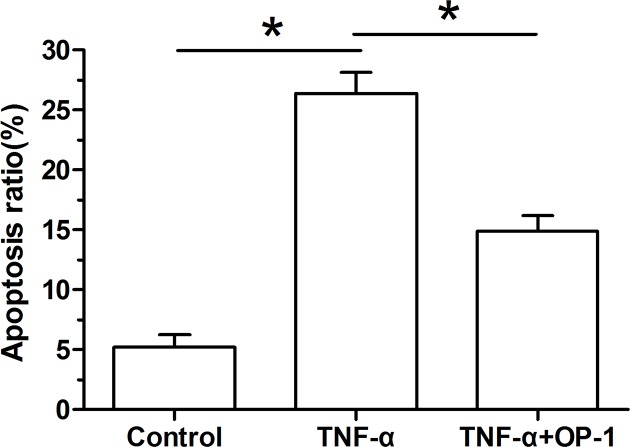
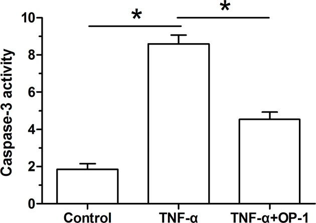
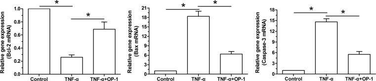
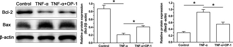
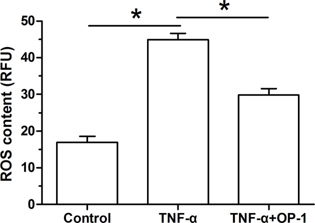
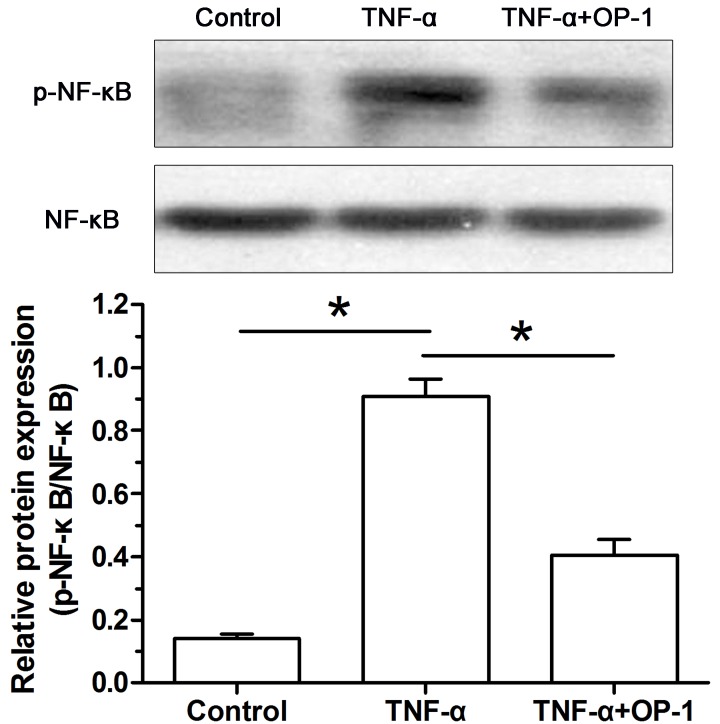
Methods: Rat NP cells were cultured in medium with or without inflammatory cytokine tumor necrosis factor (TNF)-α for 6 days. The exogenous TNF-α was added into the medium to investigate its protective effects. NP cell apoptosis was evaluated by cell apoptosis ratio, caspase-3 activity, gene/protein expression of apoptosis-related molecules (Bcl-2, Bax, and caspase-3). Additionally, the intracellular reactive oxygen species (ROS) content and activity of the NF-κB pathway were also analyzed.
Results: Compared with the control NP cells, TNF-α significantly increased cell apoptosis ratio, caspase-3 activity, gene/protein expression of Bcl-2, Bax and caspase-3, ROS content, and activity of the NF-κB pathway. However, OP-1 partly attenuated these effects in NP cells treated with TNF-α.
Conclusion: OP-1 is effective in attenuating TNF-α-caused NP cell apoptosis, and the ROS/NF-κB pathway may be the potential signaling transduction pathway. The present study indicates that OP-1 may be helpful to inhibit inflammation-mediated disc degeneration.
Keywords: TNF-α; apoptosis; disc degeneration; nucleus pulposus; osteogenic protein-1.
© 2018 The Author(s).
Conflict of interest statement
The authors declare that there are no competing interests associated with the manuscript.
Figures
Similar articles
-
NF-κB signalling pathways in nucleus pulposus cell function and intervertebral disc degeneration.Cell Prolif. 2021 Jul;54(7):e13057. doi: 10.1111/cpr.13057. Epub 2021 May 24. Cell Prolif. 2021. PMID: 34028920 Free PMC article. Review.
-
Nucleus pulposus cell apoptosis is attenuated by CDMP-2 through regulating oxidative damage under the hyperosmotic environment.Biosci Rep. 2018 Oct 9;38(5):BSR20181176. doi: 10.1042/BSR20181176. Print 2018 Oct 31. Biosci Rep. 2018. Retraction in: Biosci Rep. 2024 Aug 28;44(8):BSR-2018-1176_RET. doi: 10.1042/BSR-2018-1176_RET. PMID: 30177520 Free PMC article. Retracted.
-
Osteogenic protein-1 attenuates the inflammatory cytokine-induced NP cell senescence through regulating the ROS/NF-κB pathway.Biomed Pharmacother. 2018 Mar;99:431-437. doi: 10.1016/j.biopha.2018.01.053. Biomed Pharmacother. 2018. PMID: 29367112
-
Transforming growth factor-β1-regulated Fas/FasL pathway activation suppresses nucleus pulposus cell apoptosis in an inflammatory environment.Biosci Rep. 2020 Feb 28;40(2):BSR20191726. doi: 10.1042/BSR20191726. Biosci Rep. 2020. Retraction in: Biosci Rep. 2024 Aug 28;44(8):BSR-2019-1726_RET. doi: 10.1042/BSR-2019-1726_RET. PMID: 31808511 Free PMC article. Retracted.
-
Redox regulation of tumor necrosis factor signaling.Antioxid Redox Signal. 2009 Sep;11(9):2245-63. doi: 10.1089/ars.2009.2611. Antioxid Redox Signal. 2009. PMID: 19361274 Free PMC article. Review.
Cited by
-
Natural products can modulate inflammation in intervertebral disc degeneration.Front Pharmacol. 2023 Feb 16;14:1150835. doi: 10.3389/fphar.2023.1150835. eCollection 2023. Front Pharmacol. 2023. PMID: 36874009 Free PMC article. Review.
-
Regulatory Effect of Inflammatory Mediators in Intervertebral Disc Degeneration.Mediators Inflamm. 2023 Apr 17;2023:6210885. doi: 10.1155/2023/6210885. eCollection 2023. Mediators Inflamm. 2023. PMID: 37101594 Free PMC article. Review.
-
Application of single and cooperative different delivery systems for the treatment of intervertebral disc degeneration.Front Bioeng Biotechnol. 2022 Nov 14;10:1058251. doi: 10.3389/fbioe.2022.1058251. eCollection 2022. Front Bioeng Biotechnol. 2022. PMID: 36452213 Free PMC article. Review.
-
Mesenchymal stem cells-derived exosomes ameliorate intervertebral disc degeneration through inhibiting pyroptosis.J Cell Mol Med. 2020 Oct;24(20):11742-11754. doi: 10.1111/jcmm.15784. Epub 2020 Aug 29. J Cell Mol Med. 2020. PMID: 32860495 Free PMC article.
-
NF-κB signalling pathways in nucleus pulposus cell function and intervertebral disc degeneration.Cell Prolif. 2021 Jul;54(7):e13057. doi: 10.1111/cpr.13057. Epub 2021 May 24. Cell Prolif. 2021. PMID: 34028920 Free PMC article. Review.
References
-
- Antoniou J., Steffen T., Nelson F., Winterbottom N., Hollander A.P., Poole R.A.. et al. (1996) The human lumbar intervertebral disc: evidence for changes in the biosynthesis and denaturation of the extracellular matrix with growth, maturation, ageing, and degeneration. J. Clin. Invest. 98, 996–1003 10.1172/JCI118884 - DOI - PMC - PubMed
Publication types
MeSH terms
Substances
LinkOut - more resources
Full Text Sources
Research Materials
Miscellaneous