. 2018 Oct 19;9(11):1070.
doi: 10.1038/s41419-018-0950-x.

MicroRNA-495-3p inhibits multidrug resistance by modulating autophagy through GRP78/mTOR axis in gastric cancer

Affiliations

MicroRNA-495-3p inhibits multidrug resistance by modulating autophagy through GRP78/mTOR axis in gastric cancer

Sheng Chen et al. Cell Death Dis. .

Abstract

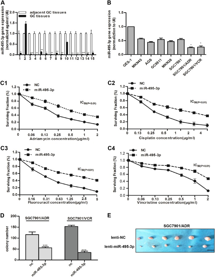
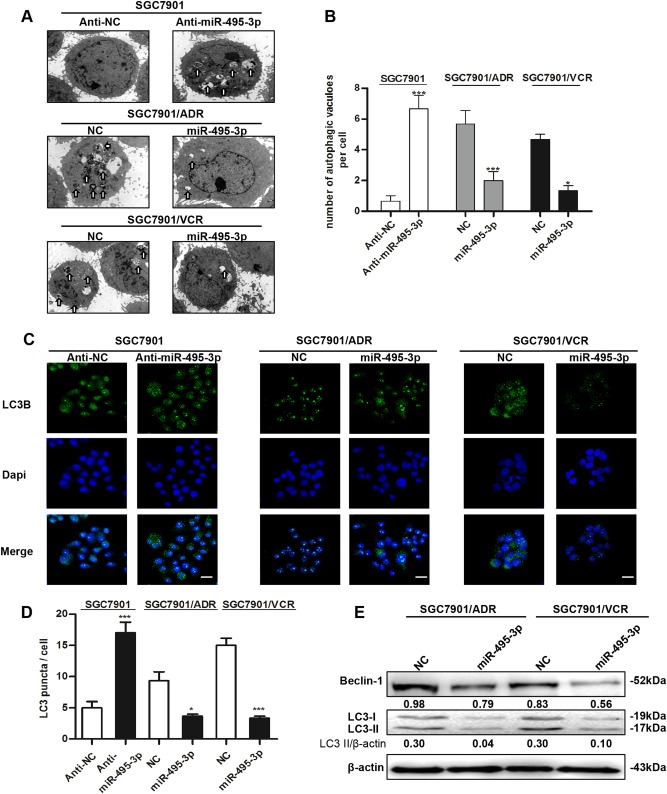
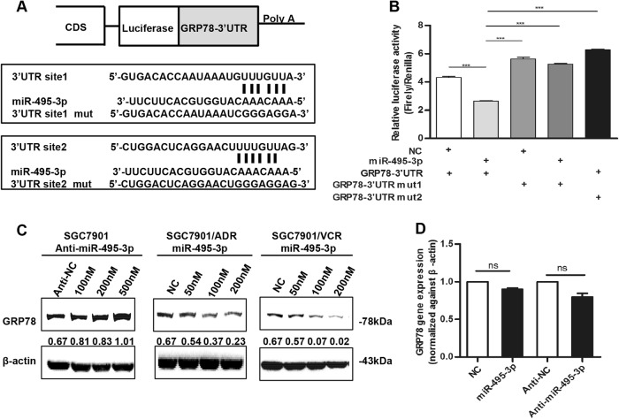
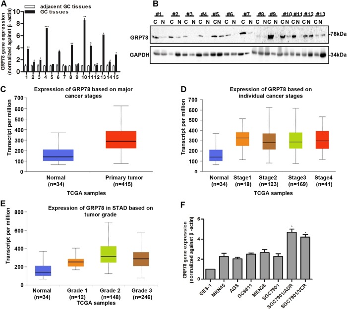
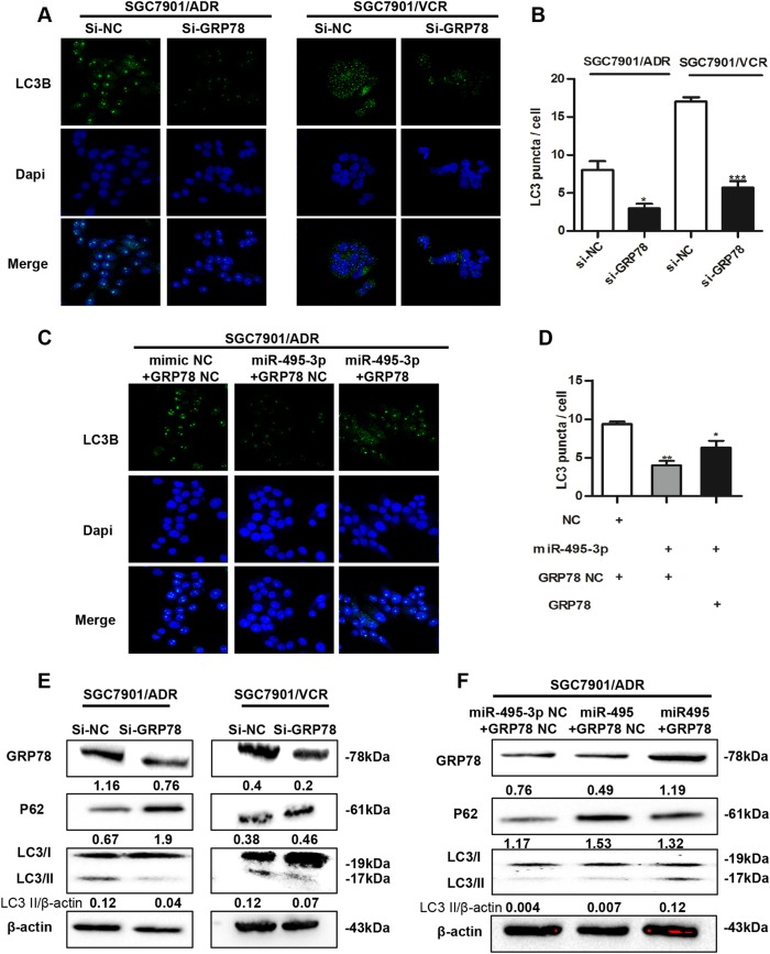
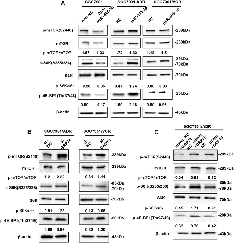
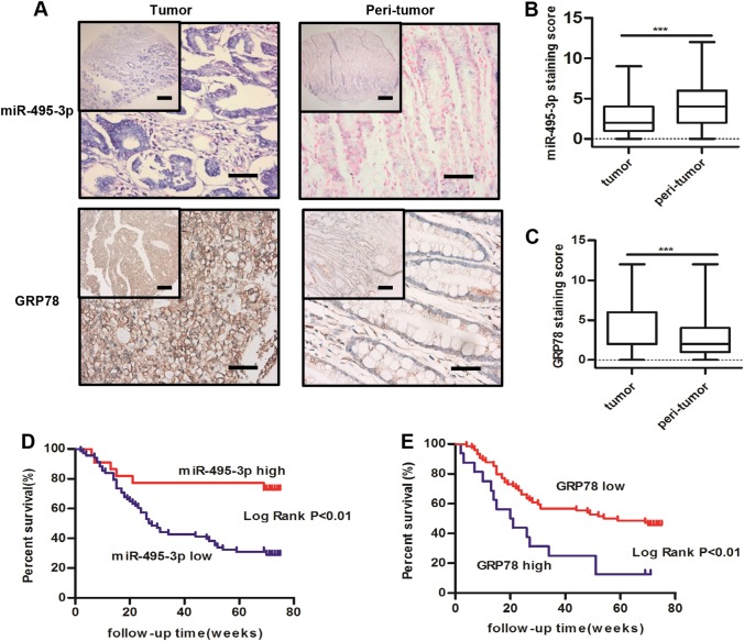
Multidrug resistance (MDR) accounts for poor prognosis in gastric cancer (GC). MicroRNAs (miRNAs) are critical regulators of MDR via modulation of the target genes. The present study revealed that miR-495-3p could act via a target gene, GRP78, to regulate the process of autophagy and inhibit MDR. Based on the in vitro and in vivo gain-of-function or loss-of-function experiments, overexpression of miR-495-3p was sufficient to reverse the MDR to four chemotherapeutics in vitro and inhibit the tumor growth in vivo. Moreover, GRP78 was positively associated with the occurrence of autophagy. Thus, reducing the expression of GRP78 by siRNA resulted in autophagy-suppressive activity similar to that of miR-495-3p on mammalian target of rapamycin (mTOR) and its substrates activation and autophagy inhibition, while restoring GRP78 attenuated the anti-autophagy effects caused by miR-495-3p. Clinically, either miR-495-3p downregulation or GRP78 upregulation was associated with malignant phenotypes in patients with GC. In conclusion, these findings demonstrate that miR-495-3p is an important regulator of autophagy balance and MDR by modulating the GRP78/mTOR axis. In addition, miR-495-3p and GRP78 could be used as prognostic factors for overall survival in GC, which implicates miR-495-3p as a therapeutic target in cancer.


Conflict of interest statement

The authors declare that they have no conflict of interest.

Figures

Fig. 1
Fig. 1. miR-495-3p is downregulated in GC and inhibits MDR and proliferation of GC MDR cells.
a Real-time PCR detected the relative expression level of miR-495-3p in peri-tumor tissue and gastric cancer tissue in 15 paired samples. b The expression level of miR-495-3p in normal gastric epithelial cell line, five gastric cancer cell lines and GC MDR cells. c Four chemotherapeutic drugs (c1 adriamycin; c2 cisplatin; c3 fluorouracil; c4 vincristine) with indicated concentration gradient were added to the medium of SGC7901/ADR transfected with miR-495-3p mimics or nc. Then, cell survival was measured using the MTT assay. d Colony formation assays were performed to evaluate the proliferative functions of GC MDR cells transfected with miR-495-3p mimic or nc. 103 cells per well were seeded initially in 6-well plates. e Representative images of tumors 4 weeks after subcutaneous injection of 107 SGC7901/ADR transfected with lenti-NC or lenti-miR-495-3p into the right flank of the nude mice. (Scale bar = 1 cm). All experiments were performed in triplicates. All values were expressed as mean ± SD, n = 3 for each group. *P < 0.05, **P < 0.01, ***P < 0.001
Fig. 2
Fig. 2. miR-495-3p inhibits autophagy in GC MDR cells.
a Transmission electron micrographs of morphological changes in SGC7901 transfected with miR-495-3p inhibitor or GC MDR cells transfected with miR-495-3p mimic. White arrows represent autophagosomes or autolysosomes. (Scale bar = 500 nm). b Quantitative analysis of the number of autophagic vesicles. c Representative images and d Quantification of LC3 puncta (green) in SGC7901 transfected with miR-495-3p inhibitor or GC MDR cells transfected with miR-495-3p mimics. Scale bars: 50 μM. e Western blot analysis of the protein levels of Beclin-1, and LC3I/II in GC MDR cells transfected with miR-495-3p mimic or nc. All values were expressed as mean ± SD, n = 4 for each group. *P < 0.05, **P < 0.01, ***P < 0.001
Fig. 3
Fig. 3. miR-495-3p directly targets GRP78 at the post-transcriptional level.
a Illustration of the GRP78 3′-UTR-containing reporter constructs. Mutations were generated at two predicted miR-495-3p binding sites located in the GRP78 3′-UTR. b Representative luciferase activity in SGC7901 cells co-transfected with wild-type or mutated reporter plasmids and nc, miR-495-3p mimic. c Western blot showed the changes in GRP78 protein levels after transient transfection of miR-495-3p inhibitor or mimic with indicated concentration gradient as compared to the negative controls (nc or anti-nc, respectively). d Real-time PCR determined the GRP78 level in SGC7901 after transfection with miR-495-3p inhibitor/mimic or anti-nc/nc. NS is no significance. All values were expressed as mean ± SD, n = 3 for each group. *P < 0.05, **P < 0.01, ***P < 0.001
Fig. 4
Fig. 4
GRP78 expression is increased and associated with poor outcomes in GC. a Real-time PCR shows the expression of GRP78 in 15 paired gastric cancer samples as compared to adjacent tumor tissues. b Western blotting indicates the protein level of GRP78 in 13 paired gastric cancer tissues and corresponding adjacent tumor tissues (C is carcinoma, N is normal tissues). Data from TCGA show the transcript per million of GRP78 in normal and primary GC tissues (c) based on individual cancer stages (d) and tumor grades (e). f The expression of GRP78 in normal gastric epithelial cell line and five gastric cancer cell lines and GC MDR cells by Real-time PCR. All values expressed as mean ± SD, n = 3 for each group. *P < 0.05, **P < 0.01, ***P < 0.001
Fig. 5
Fig. 5
Downregulation of GRP78 by miR-495-3p inhibits autophagy in GC MDR cells. a Representative images and b Quantification of LC3 puncta (green) in GC MDR cells transfected with si-GRP78 or si-nc. Scale bars: 50 μM. c Representative images and d Quantification of LC3 puncta (green) in SGC7901/ADR co-transfected with GRP78 3′-UTR-mutant overexpression vector/nc and miR-495-3p/nc. e The expression level of GRP78, LC3BI/II, and P62 in GC MDR cells transfected with si-GRP78 or si-NC were determined by western blotting. f The protein level of GRP78, LC3BI/II, and P62 in SGC7901/ADR co-transfected with GRP78 3′-UTR-mutant overexpression vector/nc and miR-495-3p/nc. All values expressed as mean ± SD, n = 3 for each group. *P < 0.05, **P < 0.01, ***P < 0.001
Fig. 6
Fig. 6. miR-495-3p inhibits autophagy by activating mTOR signaling.
a Western blot analysis of phosphorylated mTOR (p-mTOR) and its main substrates 4E-BP1 (p-4E-BP1) and S6 (p-S6) in SGC7901 transfected with anti-miR-495-3p and GC MDR cells transfected with miR-495-3p. b The phosphorylation status of mTOR, S6K (p-S6K), and 4E-BP1 (p-4E-BP1) transfected with si-GRP78 and si-NC were measured by western blotting (c). Western blotting determines the phosphorylation of mTOR, S6K (p-S6K), and 4E-BP1 (p-4E-BP1) in SGC7901/ADR co-transfected with GRP78 3′-UTR-mutant overexpression vector/nc and miR-495-3p/nc. All values expressed as mean ± SD, n = 3 for each group. *P < 0.05, **P < 0.01, ***P < 0.001
Fig. 7
Fig. 7
Expression levels of miR-495-3p and GRP78 in GC specimens. a The expression levels of miR-495-3p (upper) and GRP78 (lower) in peri-tumor and primary GC tissues, scale bars: 500 μm (top) and 200 μm (bottom). b miR-495-3p and c GRP78 expression staining score in peri-tumor and primary GC tissues. Kaplan–Meier survival curves of GC patients expressing miR-495-3p (d) and GRP78 (e) GC patients were ranked based on the staining score and divided into high-expression (>4) and low-expression (≤4) groups. *P < 0.05, **P < 0.01, ***P < 0.001

References

    1. Lordick F, Janjigian YY. Clinical impact of tumour biology in the management of gastroesophageal cancer. Nat. Rev. Clin. Oncol. 2016;13:348–360. doi: 10.1038/nrclinonc.2016.15. - DOI - PMC - PubMed
    1. Bouwman P, Jonkers J. The effects of deregulated DNA damage signalling on cancer chemotherapy response and resistance. Nat. Rev. Cancer. 2012;12:587–598. doi: 10.1038/nrc3342. - DOI - PubMed
    1. Szakács G, Paterson JK, Ludwig JA, Booth-Genthe C, Gottesman MM. Targeting multidrug resistance in cancer. Nat. Rev. Drug. Discov. 2006;5:219–234. doi: 10.1038/nrd1984. - DOI - PubMed
    1. Nieth C, Lage H. Induction of the ABC-transporters Mdr1/P-gp (Abcb1), mrpl (Abcc1), and bcrp (Abcg2) during establishment of multidrug resistance following exposure to mitoxantrone. J. Chemother. 2005;17:215–223. doi: 10.1179/joc.2005.17.2.215. - DOI - PubMed
    1. Liu Y, Peng H, Zhang JT. Expression profiling of ABC transporters in a drug-resistant breast cancer cell line using AmpArray. Mol. Pharmacol. 2005;68:430–438. - PubMed

Publication types

MeSH terms