. 2018 Oct 19;9(11):1073.
doi: 10.1038/s41419-018-1111-y.

Diesel exhaust particles induce autophagy and citrullination in Normal Human Bronchial Epithelial cells

Affiliations

Diesel exhaust particles induce autophagy and citrullination in Normal Human Bronchial Epithelial cells

Tania Colasanti et al. Cell Death Dis. .

Abstract

A variety of environmental agents has been found to influence the development of autoimmune diseases; in particular, the studies investigating the potential association of systemic autoimmune rheumatic diseases with environmental micro and nano-particulate matter are very few and contradictory. In this study, the role of diesel exhaust particles (DEPs), one of the most important components of environment particulate matter, emitted from Euro 4 and Euro 5 engines in altering the Normal Human Bronchial Epithelial (NHBE) cell biological activity was evaluated. NHBE cells were exposed in vitro to Euro 4 and Euro 5 particle carbon core, sampled upstream of the typical emission after-treatment systems (diesel oxidation catalyst and diesel particulate filter), whose surfaces have been washed from well-assessed harmful species, as polycyclic aromatic hydrocarbons (PAHs) to: (1) investigate their specific capacity to affect cell viability (flow cytometry); (2) stimulate the production of the pro-inflammatory cytokine IL-18 (Enzyme-Linked ImmunoSorbent Assay -ELISA-); (3) verify their specific ability to induce autophagy and elicit protein citrullination and peptidyl arginine deiminase (PAD) activity (confocal laser scanning microscopy, immunoprecipitation, Sodium Dodecyl Sulphate-PolyAcrylamide Gel Electrophoresis -SDS-PAGE- and Western blot, ELISA). In this study we demonstrated, for the first time, that both Euro 4 and Euro 5 carbon particles, deprived of PAHs possibly adsorbed on the soot surface, were able to: (1) significantly affect cell viability, inducing autophagy, apoptosis and necrosis; (2) stimulate the release of the pro-inflammatory cytokine IL-18; (3) elicit protein citrullination and PAD activity in NHBE cells. In particular, Euro 5 DEPs seem to have a more marked effect with respect to Euro 4 DEPs.


Conflict of interest statement

The authors declare that they have no conflict of interest.

Figures

Fig. 1
Fig. 1. Effects of DEPs on autophagy: induction of autophagic vacuoles in Normal Human Bronchial Epithelial (NHBE) cells by Euro 5 and Euro 4 carbon particles, before any after-treatment.
a Confocal microscopy of autophagy induction, evaluated by LC3 immunostaining after 48 h of treatment with the diesel particle carbon core, before any after-treatment. Single and merged images of differential interference contrast (DIC), LC3 immunofluorescent staining (green hue) and nuclei staining with Hoechst (blue hue) are shown. Bars 50 µm. b Quantitative assessment of autophagic LC3-positive (LC3+) cells, performed as described in Materials and Methods section; results are reported as percentage of LC3+ cells. For 3.3 μg/cm2: P = 0.0026 Euro 5 vs untreated; P = 0.0015 Euro 4 vs untreated; P = 0.004 Euro 5 vs Euro 4. For 6.6 μg/cm2: P = 0.0013 Euro 5 vs untreated; P = 0.0026 Euro 4 vs untreated; P = 0.002 Euro 5 vs Euro 4. For a dose-depending comparison: P = 0.002 Euro 5 3.3 vs 6.6 μg/cm2; P = 0.0026 Euro 4 3.3 vs 6.6 μg/cm2. P = 0.006 for Euro 4 3.3 μg/cm2 vs Euro 5 6.6 μg/cm2. Results are represented as the mean ± SD of three experiments (N = 3)
Fig. 2
Fig. 2. Effects of Euro 5 and Euro 4 DEPs, before any after-treatment, at 3.3 μg/cm2 on autophagy: evaluation of LC3-II and p62 levels as autophagic markers in Normal Human Bronchial Epithelial (NHBE) cells.
a Western blot analysis of LC3-II and p62 levels in lysates from NHBE cells (i) untreated and cultured for 48 h with (ii) lysosomal protease inhibitors E64d and Pepstatin A (PepA), (iii) Euro 5, (iv) Euro 5 + E64d + PepA, (v) Euro 4, (vi) Euro 4 + E64d + PepA. Blot shown is representative of nine experiments (N = 9). b Densitometry analysis of LC3-II levels relative to β-actin (mean ± SD). P = 0.0008 for Euro 5 vs untreated; P = 0.0101 for Euro 5 vs Euro 5 + E64d + PepA; P = 0.0022 for Euro 4 vs untreated. c Densitometry analysis of p62 levels relative to β-actin (mean ± SD). No significant (NS, P > 0.05) differences in p62 levels were found
Fig. 3
Fig. 3. Effects of Euro 5 and Euro 4 DEPs, before any after-treatment, at 6.6 μg/cm2 on autophagy: evaluation of LC3-II and p62 levels as autophagic markers in Normal Human Bronchial Epithelial (NHBE) cells.
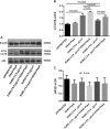
a Western blot analysis of LC3-II and p62 levels in lysates from NHBE cells (i) untreated and cultured for 48 h with (ii) E64d + PepA, (iii) Euro 5, (iv) Euro 5 + E64d + PepA, (v) Euro 4, (vi) Euro 4 + E64d + PepA. Blot shown is representative of nine experiments (N = 9). b Densitometry analysis of LC3-II levels relative to β-actin (mean ± SD). P = 0.0012 for Euro 5 vs untreated; P = 0.02 for Euro 5 vs Euro 5 + E64d + PepA; P = 0.0098 for Euro 4 vs untreated; P = 0.0407 for Euro 4 vs Euro 4 + E64d + PepA; P = 0.0107 for Euro 5 vs Euro 4. c Densitometry analysis of p62 levels relative to β-actin (mean ± SD). No significant (NS, P > 0.05) differences in p62 levels were found
Fig. 4
Fig. 4. Effect of Euro 5 and Euro 4 DEPs, before any after-treatment, on cell viability: evaluation of apoptosis and necrosis in Normal Human Bronchial Epithelial (NHBE) cells.
a Morphological characterization and analysis of apoptosis and necrosis by flow cytometry in NHBE cells untreated and treated for 24 h with Euro 5 and Euro 4 DEPs, before any after-treatment. Results shown are representative of three experiments in triplicate (N = 9). Numbers in upper and bottom right quadrants of each plot refer to Annexin V positive (AV+) cells, while numbers in the upper quadrants (left and right) represent Propidium Iodide positive (PI+) cells. b DEPs induce apoptosis in NHBE cells, reported as the percentage of AV+ cells. For 3.3 μg/cm2: P = 0.0003 for Euro 5 vs untreated; P = 0.0043 for Euro 4 vs untreated; P = 0.0032 for Euro 5 vs Euro 4. For 6.6 μg/cm2: P < 0.0001 for Euro 5 vs untreated; P = 0.0007 for Euro 4 vs untreated; P = 0.0006 for Euro 5 vs Euro 4. For a dose-depending comparison: P = 0.0018 for Euro 5 3.3 vs 6.6 μg/cm2; P = 0.0146 for Euro 4 3.3 vs 6.6 μg/cm2. c DEPs increase the percentage of necrotic (PI+) NHBE cells. For 3.3 μg/cm2: P = 0.0003 for Euro 5 vs untreated; P = 0.0028 for Euro 4 vs untreated; P = 0.0019 for Euro 5 vs Euro 4. For 6.6 μg/cm2: P < 0.0001 for Euro 5 vs untreated; P = 0.0048 for Euro 4 vs untreated; P < 0.0001 for Euro 5 vs Euro 4. For a dose-depending comparison: P = 0.0005 for Euro 5 3.3 vs 6.6 μg/cm2; P > 0.05 for Euro 4 3.3 vs 6.6 μg/cm2
Fig. 5
Fig. 5. Effect of Euro 5 and Euro 4 DEPs, before any after-treatment, in enhancing citrullinated protein levels in Normal Human Bronchial Epithelial (NHBE) cells.
a Western blot analysis of citrullinated protein levels in lysates from NHBE cells (i) untreated and cultured for 48 h with (ii) Euro 5 3.3 μg/cm2, (iii) Euro 4 3.3 μg/cm2, (iv) Euro 5 6.6 μg/cm2, (v) Euro 4 6.6 μg/cm2. Blots shown are representative of three experiments (N = 3). b Cells were lysed and vimentin was immunoprecipitated from lysates. An appropriate IgG isotypic control (irrelevant IgG, Irr. IgG) was employed to verify that immunoprecipitation was correctly performed. Blots shown are representative of nine experiments (N = 9). c Densitometry analysis of citrullinated vimentin levels relative to vimentin (mean ± SD). For 3.3 μg/cm2: P = 0.0073 Euro 5 vs untreated; P = 0.0025 Euro 4 vs untreated; P = 0.0467 Euro 5 vs Euro 4. For 6.6 μg/cm2: P < 0.0001 Euro 5 and Euro 4 vs untreated; P = 0.0119 Euro 5 vs Euro 4. For a dose-depending comparison: P = 0.0003 Euro 5 3.3 vs 6.6 μg/cm2; P = 0.0009 Euro 4 3.3 vs 6.6 μg/cm2
Fig. 6
Fig. 6. Effect of Euro 5 and Euro 4 DEPs, before any after-treatment, on PAD enzymatic activity induction.
PAD activity was assessed in lysates a from NHBE cells (i) untreated and cultured for 48 h with (ii) Euro 5 3.3 μg/cm2, (iii) Euro 4 3.3 μg/cm2, (iv) Euro 5 6.6 μg/cm2, (v) Euro 4 6.6 μg/cm2. Quadruplicate analyses (replicates) were conducted for all the lysates in three different assays (N = 3) and results obtained are shown as mean ± SD. For 3.3 μg/cm2: P = 0.0344 Euro 5 vs untreated; P = 0.0203 Euro 4 vs untreated. For 6.6 μg/cm2: P = 0.0051 Euro 5 vs untreated; P = 0.0025 Euro 4 vs untreated. For a dose-depending comparison: P = 0.0422 Euro 5 3.3 vs 6.6 μg/cm2; P = 0.0161 Euro 4 3.3 vs 6.6 μg/cm2. b Since Ca2 + is required for PAD activity, the background of the assay was set up by analyzing each lysate in the presence of the chelating agent EDTA. No increase in PAD activity was observed, following treatment with Euro 4 and Euro 5 DEPs (NS, P > 0.05 for all the experimental conditions). Quadruplicate analyses (replicates) were conducted for all the lysates in three different assays (N = 3) and results obtained are shown as mean ± SD
Fig. 7
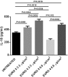
Fig. 7. Effect of Euro 5 and Euro 4 DEPs, before any after-treatment, on IL-18 production evaluated in Normal Human Bronchial Epithelial (NHBE) cell supernatants.
Cytokine release was analyzed using a commercially available ELISA kit. NHBE cells were stimulated for 24 h with Euro 5 and Euro 4 DEPs, before any after-treatment. After cell exposure to DEPs, supernatants were collected and analyzed. All the supernatants were run in quadruplicate in three different assays (N = 3) and the results are shown as mean ± SD. For 3.3 μg/cm2: P = 0.0089 Euro 5 vs untreated; P = 0.0004 Euro 4 vs untreated; P = 0.0059 Euro 5 vs Euro 4. For 6.6 μg/cm2: P = 0.0018 for Euro 5 vs untreated; P = 0.0016 for Euro 4 vs untreated; P = 0.0002 for Euro 5 vs Euro 4. For a dose-depending comparison: P = 0.0004 Euro 5 3.3 vs 6.6 μg/cm2; P = 0.032 Euro 4 3.3 vs 6.6 μg/cm2

Similar articles

Cited by

References

    1. Beelen R, et al. Long-term exposure to traffic-related air pollution and lung cancer risk. Epidemiology. 2008;19:702–710. doi: 10.1097/EDE.0b013e318181b3ca. - DOI - PubMed
    1. Donaldson K, et al. Combustion-derived nanoparticles: a review of their toxicology following inhalation exposure. Part. Fibre Toxicol. 2005;2:10. doi: 10.1186/1743-8977-2-10. - DOI - PMC - PubMed
    1. Nemmar A, Al-Maskari S, Ali BH, Al-Amri IS. Cardiovascular and lung inflammatory effects induced by systemically administered diesel exhaust particles in rats. Am. J. Physiol. Lung Cell. Mol. Physiol. 2007;292:L664–L670. doi: 10.1152/ajplung.00240.2006. - DOI - PubMed
    1. Nel A. Atmosphere. Air pollution-related illness: effects of particles. Science. 2005;308:804–806. doi: 10.1126/science.1108752. - DOI - PubMed
    1. Durga M, Nathiya S, Rajasekar A, Devasena T. Effects of ultrafine petrol exhaust particles on cytotoxicity, oxidative stress generation, DNA damage and inflammation in human A549 lung cells and murine RAW 264.7 macrophages. Environ. Toxicol. Pharmacol. 2014;38:518–530. doi: 10.1016/j.etap.2014.08.003. - DOI - PubMed

Publication types