. 2018 Oct 19;9(1):4346.
doi: 10.1038/s41467-018-06677-1.

DHX9 helicase promotes R-loop formation in cells with impaired RNA splicing

Affiliations

DHX9 helicase promotes R-loop formation in cells with impaired RNA splicing

Prasun Chakraborty et al. Nat Commun. .

Abstract

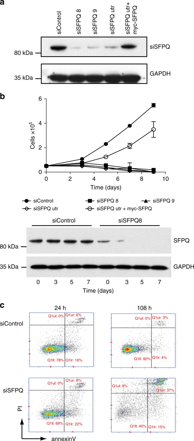
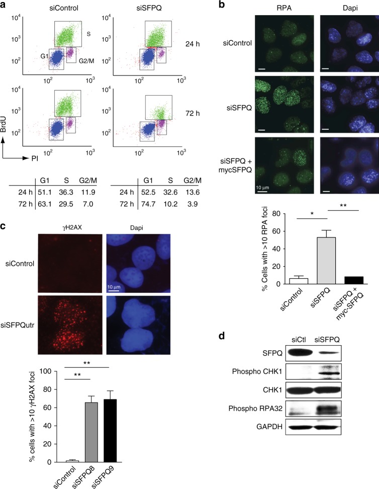
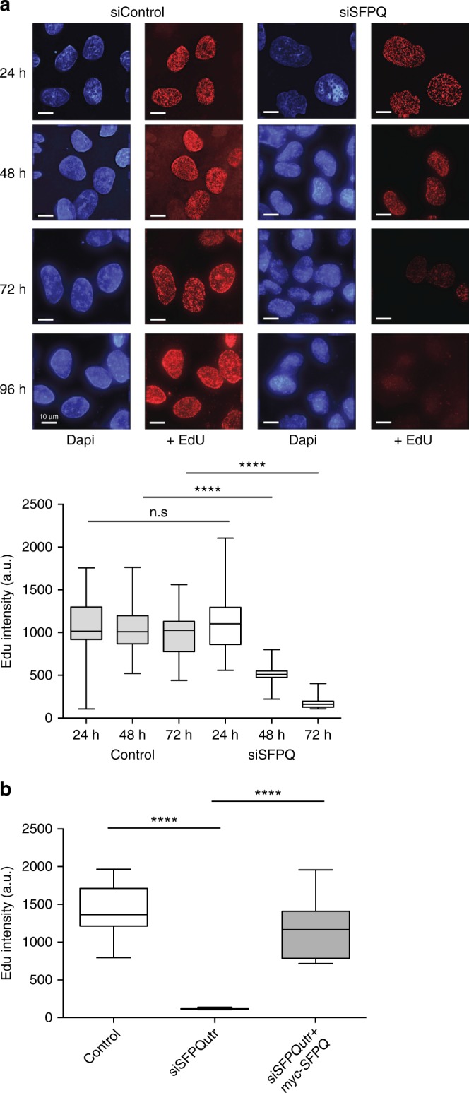
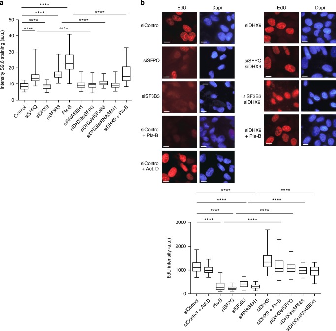
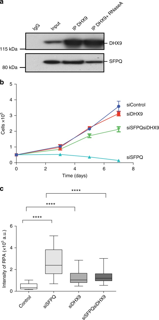
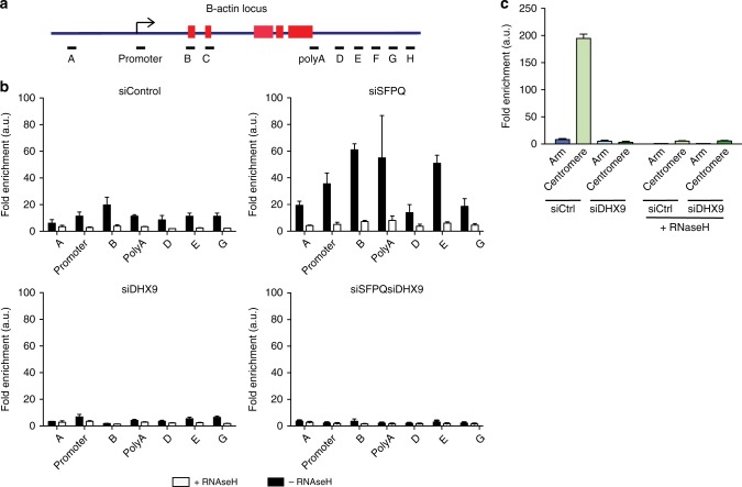
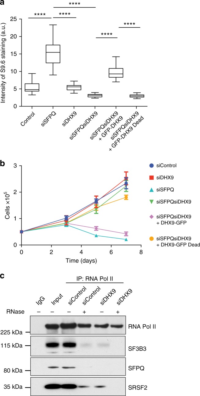
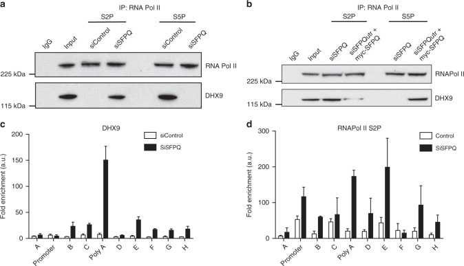
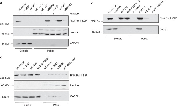
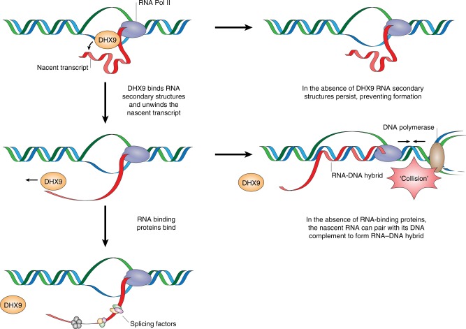
R-loops are stable nucleic acid structures that have important physiological functions, but which also pose a significant threat to genomic stability. Increased R-loops cause replication stress and chromosome fragility and have been associated with diseases such as neurodegeneration and cancer. Although excessive R-loops are a feature of cells that are defective in RNA processing, what causes them to form is unclear. Here, we demonstrate that DHX9 (RNA helicase A) promotes the formation of pathological and non-pathological R-loops. In the absence of splicing factors, formation of R-loops correlates with the prolonged association of DHX9 with RNA Polymerase II (RNA Pol II). This leads to the production of DNA-RNA hybrid, which traps RNA Pol II on chromatin with the potential to block DNA replication. Our data provide a molecular mechanism for the formation of R-loops that is relevant to neurodegenerative diseases and cancers in which deregulated RNA processing is a feature.


Conflict of interest statement

The authors declare no competing interests.

Figures

Fig. 1
Fig. 1
Depletion of SFPQ impairs cell growth. a Western blot showing knockdown of SFPQ using different siRNAs. Expression of myc-SFPQ is resistant to knockdown by siSFPQutr. b Cell proliferation in U2OS cells transfected with different siRNAs targeting SFPQ and a non-specific sequence (siControl). Cell number was measured 48 h after transfection with siRNA (time = 0) and then at time = 3, 5, 7, and 9 days. Overexpression of siRNA-resistant myc-SFPQ rescued cell growth in SFPQ knockdown cells. Data are an average n = 3 independent experiments ± s.d. Western blot (below) shows the level SFPQ protein over time. c Knockdown of SFPQ (siSFPQ9) leads to increased apoptosis, quantified by staining for Annexin-V
Fig. 2
Fig. 2
Defects in SFPQ cause replication stress. a FACS analysis showing that knockdown of SFPQ (siSFPQ9) leads to a reduction in the s-phase cells. Quantification of FACS data was averaged from three independent experiments. b Microscope images showing that knockdown of SFPQ with siSFPQ8 in U2OS cells leads to increased RPA foci. Expression of exogenous myc-SFPQ (indicated) resulted in partial suppression of RPA foci in SFPQ-depleted cells. Quantification is shown below and indicate mean percentage of cells with >10 RPA foci ± s.d. More than 100 cells were counted from each of three independent experiments. Statistical significance was determined using Student’s t-test (*<0.05 and **<0.01). c Images showing increased γH2AX foci in U2OS cells knocked down with siSFPQutr compared with control cells. Graph shows mean percentage of cells with >5 γH2AX foci ± s.d. Data are plotted from three independent experiments. Statistical significance was determined using Student’s t-test (**p < 0.01). d Western blot showing that knockdown of SFPQ with siSFPQ8 leads to the phosphorylation of CHK1 on serine 345 and phosphorylation of RPA32 on Ser4/Ser8
Fig. 3
Fig. 3
Defects in SFPQ cause impaired DNA synthesis. a Microscope images showing that, in U2OS cells, incorporation of EdU into DNA over time is diminished by knockdown of SFPQ (siSFPQ8). Immunofluorescence intensity of nuclear EdU was quantified from >100 cells and is presented graphically as a box and whiskers plot. Statistical significance was determined using Mann–Whitney test (****p < 0.0001). Scale bars represent 10 μm. b Expression of siRNA-resistant myc-SFPQ in U2OS cells restores DNA synthesis. EdU fluorescence intensity from >100 cells is depicted in arbitrary units (a.u.). Statistical significance was determined using Mann–Whitney test (****p < 0.0001). Box and whisker plots display median, upper and lower quartile range with whiskers depicting lowest and highest values
Fig. 4
Fig. 4
Defects in RNA splicing cause increased R-loops. a Inhibition of RNA splicing leads to elevated levels of RNA–DNA hybrids that are suppressed by knockdown of DHX9. Cells were transfected with siRNAs targeted to the indicated genes, individually and in combination. Where indicated, cells were treated with Pla-B (5μM) for 2 h to inhibit splicing. b Incorporation of EdU into DNA 72 h after knockdown of the indicated genes. Where indicated, cells were treated with Pla-B (5 μm) or Actinomycin D (0.5 μg/ml). All scale bars represent 10 μm. Graphical data from n > 90 cells are presented. Statistical significance for all graphs was determined using Mann–Whitney test (****p < 0.0001)
Fig. 5
Fig. 5
DHX9 interacts with SFPQ. a Western blot showing co-immunoprecipitation of SFPQ with DHX9 from Hela cell nuclear extract. Control sample treated with RNaseA is indicated. Input sample and IgG control are also shown. b Knockdown of DHX9 suppresses the growth defect in SFPQ-depleted cells (siSFPQ8). Cell number was measured 48 h after transfection with siRNA (time 0) and again after 3, 5, and 7 days. c Knockdown of DHX9 suppresses replication stress in SFPQ-depleted cells. Fluorescence intensity of RPA foci was measured in n > 90 cells, 72 h after transfection with the indicated siRNA. Statistical significance was determined using Mann–Whitney test (****p < 0.0001). Box and whisker plots display median, upper and lower quartile range with whiskers depicting lowest and highest values
Fig. 6
Fig. 6
DHX9 promotes the formation of pathological and non-pathological R-loops. a Diagram of the Β-actin locus depicting relative position of primer pairs used for qPCR in DNA–RNA immunoprecipitation (DRIP) and chromatin immunoprecipitation (ChIp) experiments. Exons are depicted as red boxes. b DRIP analysis at the Β-actin locus using S9.6 antibody. Quantitative PCR was performed using primers to the indicated regions of the Β-actin locus. Data are represented as fold enrichment compared to control immunoprecipitations. Data are an average of three independent experiments. c DRIP analysis of RNA–DNA hybrid at centromeric and flanking arm regions of chromosome 1 in cells knocked down for DHX9. Samples were treated with RNaseH1 to remove DNA–RNA hybrid are indicated
Fig. 7
Fig. 7
DHX9 helicase activity is required for R-loop formation and growth inhibition. a Expression of wild-type DHX9, but not a helicase-defective mutant (pGFP-DHX9dead), promotes R-loop formation in SFPQ-defective cells. Fluorescence intensity of S9.6 staining was measured for n > 50 cells. Statistical significance was determined using Mann–Whitney test (****p < 0.0001). b Expression of wild-type DHX9 but not a helicase-defective mutant confers impaired cell proliferation in SFPQ-depleted cells (siSFPQ8). Cell number was measured using a Casey Cell Counter 48 h after transfection with siRNA (time 0) and again after 3, 5, and 7 days. c RNA Pol II S2P was immunoprecipitated from Hela cell nuclear extracts and co-purification of different splicing factors was probed by western blot (as indicated). Duplicate samples were treated with RNaseA as indicated to demonstrate the requirement of RNA in these interactions
Fig. 8
Fig. 8
DHX9 associates with different phosphorylated forms of RNA Polymerase II. a Western blot of RNA Pol II and DHX9 immunoprecipitated from HeLa cells with antibodies specific for phosphor-serine 2 (S2P) and phosphor-serine 5 (S5P) forms of RNA Pol II. Where indicated cells were transfected with siRNA against a scrambled DNA sequence (siControl) or against SFPQ (siSFPQ8, siSFPQ9). b Expression of siRNA-resistant myc-SFPQ in SFPQ knockdown cells promotes the association of DHX9 with RNA Pol II S5P and diminishes its association with S2P. c Chromatin immunoprecipitation of DHX9 at the Β-actin locus. Data are depicted as fold enrichment over the control IP. qPCR of IP samples was performed for the primer pairs described in Fig. 6a. Means and s.e.m. are plotted and data are an average from three independent replicates. d Chromatin immunoprecipitation of RNA Pol II S2P at the Β-actin locus as described in c
Fig. 9
Fig. 9
DNA–RNA hybrid traps RNA Polymerase II on chromatin. a Depletion of SFPQ and SF3B3 causes retention of RNA Pol II on chromatin that is released by treatment with RNAseH1. Western blot of soluble (cytoplasmic) and insoluble (pellet) fractions of nuclear extracts prepared from HeLa cells that were knocked down with siRNAs against the indicated genes. Blots were also probed for GAPDH and LaminA to validate cytoplasmic and chromatin pellet fractions, respectively. b As in a showing that knockdown of DHX9 in cells depleted of SFPQ reverses the retention of RNA Pol II S2P on chromatin. c RNA Pol II (S2P) is not retained in the insoluble chromatin pellet from cells knocked down for XRN2
Fig. 10
Fig. 10
DHX9 promotes the generation of R-loops. Model showing how DHX9 promotes the formation of R-loops by unwinding the nascent RNA to generate the free RNA end, which is required for the invasion of duplex DNA and generation of DNA–RNA hybrid. RNA-binding proteins prevent R-loop formation by binding to the nascent RNA inhibiting its ability to pair with its complementary DNA template. In the absence of RNA-binding proteins, the free RNA end that is generated by DHX9 is available for R-loop formation. This may lead to RNA Pol II becoming trapped on chromatin where it can pose a barrier to DNA replication and increases the likelihood of transcription–replication conflicts. In the absence of DHX9, the formation of secondary structures in the nascent RNA prevent it from invading the DNA duplex to form R-loops

Similar articles

Cited by

References

    1. Roberts RW, Crothers DM. Stability and properties of double and triple helices: dramatic effects of RNA or DNA backbone composition. Science. 1992;258:1463–1466. doi: 10.1126/science.1279808. - DOI - PubMed
    1. Kabeche L, Nguyen HD, Buisson R, Zou L. A mitosis-specific and R loop-driven ATR pathway promotes faithful chromosome segregation. Science. 2018;359:108–114. doi: 10.1126/science.aan6490. - DOI - PMC - PubMed
    1. Aguilera A, Garcia-Muse T. R loops: from transcription byproducts to threats to genome stability. Mol. Cell. 2012;46:115–124. doi: 10.1016/j.molcel.2012.04.009. - DOI - PubMed
    1. Wongsurawat T, Jenjaroenpun P, Kwoh CK, Kuznetsov V. Quantitative model of R-loop forming structures reveals a novel level of RNA-DNA interactome complexity. Nucleic Acids Res. 2012;40:e16. doi: 10.1093/nar/gkr1075. - DOI - PMC - PubMed
    1. Ginno PA, Lott PL, Christensen HC, Korf I, Chedin F. R-loop formation is a distinctive characteristic of unmethylated human CpG island promoters. Mol. Cell. 2012;45:814–825. doi: 10.1016/j.molcel.2012.01.017. - DOI - PMC - PubMed

Publication types

MeSH terms

LinkOut - more resources