. 2018 Oct 19;9(1):4363.
doi: 10.1038/s41467-018-06554-x.

Mycobacterial DnaB helicase intein as oxidative stress sensor

Affiliations

Mycobacterial DnaB helicase intein as oxidative stress sensor

Danielle S Kelley et al. Nat Commun. .

Abstract

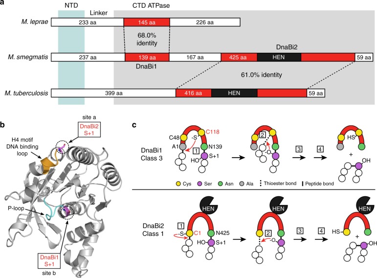
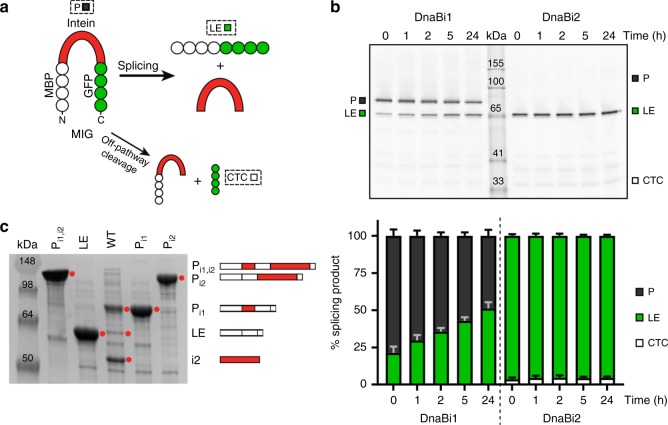
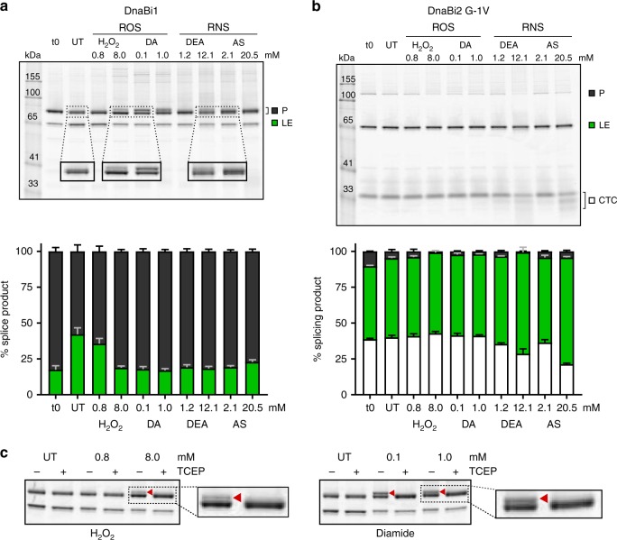
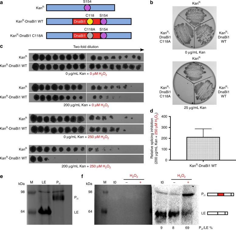
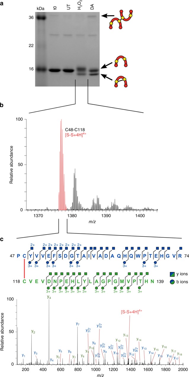
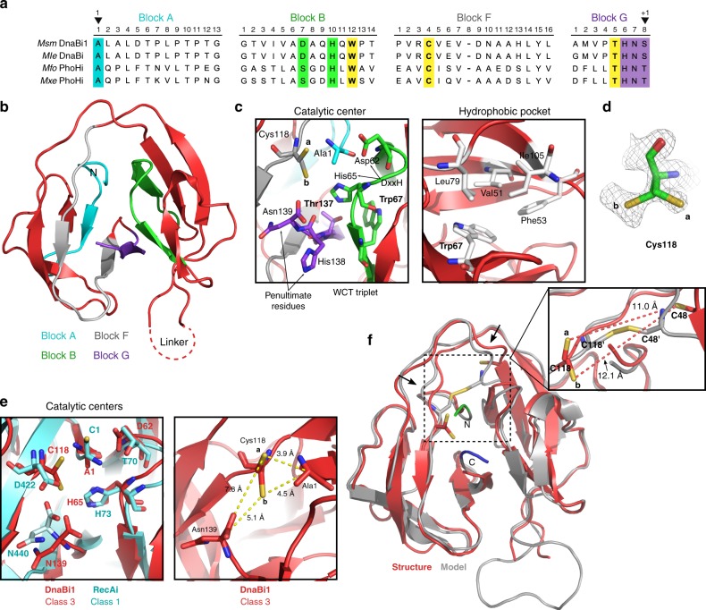
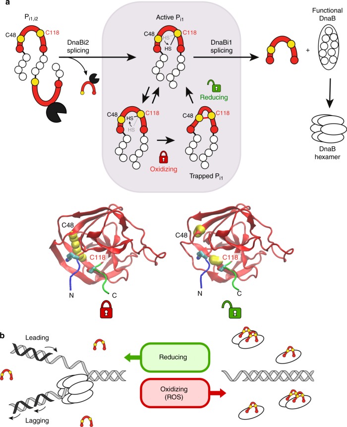
Inteins are widespread self-splicing protein elements emerging as potential post-translational environmental sensors. Here, we describe two inteins within one protein, the Mycobacterium smegmatis replicative helicase DnaB. These inteins, DnaBi1 and DnaBi2, have homology to inteins in pathogens, splice with vastly varied rates, and are differentially responsive to environmental stressors. Whereas DnaBi1 splicing is reversibly inhibited by oxidative and nitrosative insults, DnaBi2 is not. Using a reporter that measures splicing in a native intein-containing organism and western blotting, we show that H2O2 inhibits DnaBi1 splicing in M. smegmatis. Intriguingly, upon oxidation, the catalytic cysteine of DnaBi1 forms an intramolecular disulfide bond. We report a crystal structure of the class 3 DnaBi1 intein at 1.95 Å, supporting our findings and providing insight into this splicing mechanism. We propose that this cysteine toggle allows DnaBi1 to sense stress, pausing replication to maintain genome integrity, and then allowing splicing immediately when permissive conditions return.


Conflict of interest statement

The authors declare no competing interests.

Figures

Fig. 1
Fig. 1
Overview of mycobacterial DnaB inteins. a Relationship of DnaB inteins in three mycobacterial species. The two M. smegmatis (Msm) DnaB inteins (DnaBi1 and DnaBi2) were aligned to the single DnaB inteins in M. leprae (Mle) and M. tuberculosis (Mtu) using a protein pairwise alignment (EMBOSS Needle; http://www.ebi.ac.uk/Tools/psa/emboss_needle). The amino acid percent identity between inteins (red) are shown. The number of residues for the intein and extein fragments (white) are indicated and the homing endonuclease (HEN) is shown in black. The N-terminal domain of DnaB is indicated in light blue shading and the C-terminal ATPase is indicated in gray shading. NTD N-terminal domain, CTD C-terminal domain, aa amino acid. b Localization of DnaB intein insertions. A structure model of the DnaB ATPase domain (residues 200–461) is shown. The insertion sites of DnaBi1 and DnaBi2 are indicated by the S + 1 in purple. The P-loop, where DnaBi1 is inserted, is cyan and the H4 motif/DNA-binding loop, where DnaBi2 is found, is orange. c Intein splicing mechanisms. The class 3 mechanism (top) is used by Msm DnaBi1 and Mle DnaBi, whereas the canonical class 1 pathway (bottom) is used by Msm DnaBi2 and Mtu DnaBi. See the main text and Supplementary Figure 1 for detailed splicing description and steps (boxed numbers). Residue numbering refers to Msm inteins
Fig. 2
Fig. 2
Different splicing profiles of M. smegmatis DnaB inteins. a Schematic of MIG. The reporter construct maltose-binding protein (MBP)-intein-GFP (MIG) allows monitoring of splicing by in-gel fluorescence of GFP-containing products. The precursor molecule (P) can splice, yielding ligated exteins (LE) and free intein (I), or can undergo off-pathway cleavage reactions, such as C-terminal cleavage (CTC). b The two Msm inteins have distinct splicing. The gel of a splicing time course shows that MIG DnaBi1 splices slowly while MIG DnaBi2 splices almost instantaneously. Quantitation of MIG time course is shown below (stack plots), where the ratios of splice products were plotted. Data are representative of three biological replicates and values are expressed as mean ± s.d. c Splicing of Msm DnaB inteins with native exteins corresponds to splicing in MIG. Full-length DnaB protein constructs were made to understand how the inteins splice with native exteins. The wild-type (WT) lane represents lysate of overexpressed DnaB protein with splicing-competent inteins. The adjacent lanes are lysates containing splicing-inactive controls representing possible splicing outcomes and are schematically indicated. These splicing products include full precursor with both inteins present (Pi1,i2), alternative precursor 1 (Pi1), with only DnaBi1 present, alternative precursor 2 (Pi2), with only DnaBi2 present, and ligated exteins (LE), with no inteins present. In WT there is accumulation of Pi1, which indicates rapid splicing of DnaBi2. Consistent with this observation, abundant DnaBi2 is visible in the WT lane. Bands of interest are indicated by red circles. Data are representative of three biological replicates
Fig. 3
Fig. 3
M. smegmatis DnaB inteins are differentially sensitive to stressors. a MIG DnaBi1 accumulates precursor following exposure to stressors. After treatment with ROS and RNS agents there is an increase in the amount of precursor (P) compared to untreated (UT) (top). Additionally, the P band becomes diffuse (RNS) and secondary bands above P are apparent (ROS). The splicing product ratios were quantitated (stack plots below). All the treated samples, except 0.8 mM H2O2, had increased P compared to UT. DA diamide; DEA DEA NONOate; AS Angeli’s Salt. Data are representative of three biological replicates and values are expressed as mean ± s.d. b Splicing of MIG DnaBi2 is less responsive to stressors. A mutant version (G-1V) of MIG DnaBi2, which has slower splicing compared to WT (Fig. 2), was evaluated for changes in response to ROS and RNS treatment (top). Unlike MIG DnaBi1, there is no condition that results in precursor accumulation or visible differences in the appearance of the P bands. The splicing product ratios in response to stressors were quantitated (stack plots below). Data are representative of three biological replicates and values are expressed as mean ± s.d. c MIG DnaBi1 upper bands are reducible. Reducing agent TCEP was added to UT and ROS-treated samples. The upper bands seen in ROS-treated samples (H2O2 and DA; red arrowhead) resolved into single precursor bands following treatment, suggesting that a reversible modification is occurring in response to treatment. Data are representative of three biological replicates
Fig. 4
Fig. 4
DnaBi1 splicing is inhibited by H2O2 in M. smegmatis. a Overview of “Splice or Die” constructs. Kanamycin-resistance protein was left uninterrupted (KanR) or interrupted with either an active DnaBi1 (KanR-DnaBi1 WT) or inactive DnaBi1 with a C118A mutation (KanR-DnaBi1 C118A). The KanR Ser154 serves as the +1 nucleophile for DnaBi1 splicing. b DnaBi1 splicing is required to confer resistance to kanamycin in KanR-DnaBi1 fusion. M. smegmatis with KanR, KanR-DnaBi1 WT, or KanR-DnaBi1 C118A (splicing inactive) were spread onto media with either 0 (top) or 25 (bottom) µg/mL kanamycin. Biological replicates (n = 4) were performed under similar conditions. c Survival of M. smegmatis expressing the KanR-DnaBi1 fusion is selectively decreased compared to uninterrupted KanR in the presence of kanamycin and H2O2. Two-fold dilutions of early log cells, shown covering a range from ~3 × 10−1 to ~9.2 × 10−6, were spotted onto media with varying concentrations of kanamycin and H2O2, indicated below. Each experiment consisted of one culture for each strain. Biological replicates (n = 3) were performed under similar conditions. d Quantitation of the relative splicing inhibition of KanR-DnaBi1 WT in the presence of kanamycin and H2O2. The relative splicing inhibition of KanR-DnaBi1 WT was found to be a 213.3-fold effect. Data are representative of biological replicates (n = 3) and are expressed as mean ± s.d. e Migration pattern of DnaB ligated exteins and Pi1 precursor. Overexpressed DnaB ligated exteins, Pi1 precursor, and a prestained ladder as a size marker (M) were separated by SDS-PAGE and stained by Coomassie. The apparent higher molecular weight of products compared to Fig. 2c is a result of different buffers between the experiments. f Western blot analysis shows DnaB Pi1 precursor accumulates following H2O2 treatment in M. smegmatis. Stationary phase cells (t0) were diluted tenfold in fresh media without (H2O2, −) or with 5 mM H2O2 (H2O2, +) and grown for 1 h. Left, Western blot membrane with prestained ladder post-transfer. Right, chemiluminescence detection of DnaB ligated extein and Pi1 precursor products, using an anti-DnaB extein 1 antibody. Biological replicates (n = 3) were performed under similar conditions
Fig. 5
Fig. 5
DnaBi1 forms a disulfide bond via its catalytic cysteine in response to ROS. a DnaBi1 is modified by ROS. Purified DnaBi1 was treated with ROS reagents under anaerobic conditions. Samples were then run anaerobically on a non-reducing SDS-PAGE gel. The identity of the various products is shown schematically as reduced, intra- or intermolecular disulfide-bonded intein. Gel is representative of three technical replicates of experiments prepared for mass spectrometry analysis. UT untreated; DA diamide. b Mass spectrometry identifies intramolecular disulfide bond peak. Following H2O2 treatment, mass spectrometry showed peaks corresponding to an intramolecular disulfide link between fragments containing Cys48 and Cys118, represented here by the most prominent S–S peak at m/z = 1376.891784+ in red. c Fragmentation confirmed the peak identity as an intramolecular bond between the two cysteines. The indicated disulfide peak from panel b was isolated and tandem mass spectrometry was performed. Fragmentation confirmed the peptide identities, with coverage indicated for both the Cys48-containing peptide (blue) and the Cys118 peptide (green) by the y ions (squares) and b ions (circles). The y series for both peptides are shown on the spectra. The fragmentation ions are colored to match the peptide from which they originated
Fig. 6
Fig. 6
Structure reveals conformational variability of Cys118. a Mycobacterial class 3 intein alignment. Four class 3 mycobacterial inteins from two intein-containing proteins, DnaB and PhoH (phosphate starvation-inducible protein) show the four splicing blocks and class 3 features. The WCT triplet residues are yellow. Important residues are highlighted, and the 1 and +1 residues noted (arrowheads). Msm M. smegmatis; Mle M. leprae; Mfo M. fortuitum; Mxe M. xenopi. b DnaBi1 structure provides insight into class 3 inteins. The class 3 intein crystal structure was solved to 1.95 Å. The four splicing blocks are in cyan (block A), green (block B), gray (block F), and purple (block G). Amino (N) terminus is annotated. c Class 3 intein structural features. The catalytic center is shown (left). The WCT triplet residues (Trp67, Cys118, Thr137) are indicated. Cys118 shows two orientations, a and b, where a faces away from the catalytic center and b is oriented towards it. Important residues include the B block DxxH motif (Asp62 and His65) and the G-block penultimate His138 and terminal Asn139. The hydrophobic pocket containing Trp67 is presented (right) and hydrophobic packing residues are indicated (white). d Electron density map of Cys118 showing the distinct a and b orientations. e Overlay of class 3 and class 1 active sites. Residues involved in the splicing mechanism for both intein classes are shown (left). Class 3 DnaBi1 residues are red and class 1 RecAi residues are cyan. Cys118 is in the same location as Asp422, a residue proposed to coordinate the N- and C-junction reactions, . The right panel shows distances between centrally located Cys118 to the N- and C-extein junctions. f DnaBi1 structure and an optimized disulfide-bonded model overlay show minor conformational differences. The structure (red) was overlaid with a model (gray) optimized for a disulfide linkage between Cys48 and Cys118. The a and b catalytic Cys118 conformations are too distant from Cys48 for an intramolecular disulfide bond (a 11.0 Å; b 12.1 Å; inset). The disulfide-bonded model undergoes minor structural changes (arrows) and includes two N- (green) and C-extein (blue) residues
Fig. 7
Fig. 7
Model for impact of ROS/RNS stress on DnaB splicing and DNA replication. a Model for DnaBi1 splicing regulation by ROS. The full-length precursor (Pi1,i2) is expressed and DnaBi2 rapidly splices out, leaving an active alternative precursor (Pi1) with DnaBi1 still present (shaded box). Cys118 has conformational freedom, alternating between the a and b orientations (arrow). In an oxidizing environment, such as that resulting from ROS, an intramolecular disulfide bond forms between Cys48 and catalytic Cys118 of DnaBi1 (red line), locking Pi1 in a splicing-inactive state. Once the cell restores a reducing environment, the disulfide bridge is resolved and Cys118 toggles to initiate splicing, whereupon DnaB is immediately functional and hexamerizes to assume its role in replication. The structural changes are shown below in two DnaBi1 models with several extein residues (N-extein, blue; C-extein, green), indicating the movement of Cys118 from a disulfide-bonded state with Cys48 to a reduced state, where Cys118 is positioned to initiate splicing. b Model for replication arrest through splicing modulation. In the presence of ROS, intein splicing is inhibited through cysteine oxidation (red horseshoe as in panel a). This prevents some, but not necessarily all, DnaB functions and prevents replication fork formation (right). When the environment becomes favorable, the cysteines are reduced, enabling DnaBi1 splicing to proceed. This produces fully active DnaB protein, which is able to participate in replication fork formation and progression

Similar articles

Cited by

References

    1. Belfort M. Mobile self-splicing introns and inteins as environmental sensors. Curr. Opin. Microbiol. 2017;38:51–58. doi: 10.1016/j.mib.2017.04.003. - DOI - PMC - PubMed
    1. Lennon CW, Stanger M, Belfort M. Protein splicing of a recombinase intein induced by ssDNA and DNA damage. Genes Dev. 2016;30:2663–2668. doi: 10.1101/gad.289280.116. - DOI - PMC - PubMed
    1. Topilina NI, et al. SufB intein of Mycobacterium tuberculosis as a sensor for oxidative and nitrosative stresses. Proc. Natl. Acad. Sci. USA. 2015;112:10348–10353. doi: 10.1073/pnas.1512777112. - DOI - PMC - PubMed
    1. Topilina NI, Novikova O, Stanger M, Banavali NK, Belfort M. Post-translational environmental switch of RadA activity by extein-intein interactions in protein splicing. Nucleic Acids Res. 2015;43:6631–6648. doi: 10.1093/nar/gkv612. - DOI - PMC - PubMed
    1. Reitter JN, Cousin CE, Nicastri MC, Jaramillo MV, Mills KV. Salt-dependent conditional protein splicing of an intein from Halobacterium salinarum. Biochemistry. 2016;55:1279–1282. doi: 10.1021/acs.biochem.6b00128. - DOI - PubMed

Publication types