

Deep sequencing of primary human lung epithelial cells challenged with H5N1 influenza virus reveals a proviral role for CEACAM1
- PMID: 30341336
- PMCID: PMC6195505
- DOI: 10.1038/s41598-018-33605-6
Deep sequencing of primary human lung epithelial cells challenged with H5N1 influenza virus reveals a proviral role for CEACAM1
Abstract
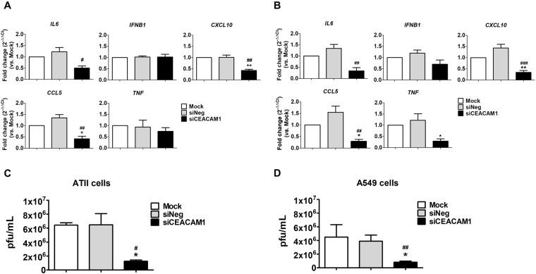
Current prophylactic and therapeutic strategies targeting human influenza viruses include vaccines and antivirals. Given variable rates of vaccine efficacy and antiviral resistance, alternative strategies are urgently required to improve disease outcomes. Here we describe the use of HiSeq deep sequencing to analyze host gene expression in primary human alveolar epithelial type II cells infected with highly pathogenic avian influenza H5N1 virus. At 24 hours post-infection, 623 host genes were significantly upregulated, including the cell adhesion molecule CEACAM1. H5N1 virus infection stimulated significantly higher CEACAM1 protein expression when compared to influenza A PR8 (H1N1) virus, suggesting a key role for CEACAM1 in influenza virus pathogenicity. Furthermore, silencing of endogenous CEACAM1 resulted in reduced levels of proinflammatory cytokine/chemokine production, as well as reduced levels of virus replication following H5N1 infection. Our study provides evidence for the involvement of CEACAM1 in a clinically relevant model of H5N1 infection and may assist in the development of host-oriented antiviral strategies.
Conflict of interest statement
The authors declare no competing interests.
Figures
Similar articles
-
Human microRNAs profiling in response to influenza A viruses (subtypes pH1N1, H3N2, and H5N1).Exp Biol Med (Maywood). 2016 Feb;241(4):409-20. doi: 10.1177/1535370215611764. Epub 2015 Oct 29. Exp Biol Med (Maywood). 2016. PMID: 26518627 Free PMC article.
-
Infection with influenza A viruses causes changes in promoter DNA methylation of inflammatory genes.Influenza Other Respir Viruses. 2013 Nov;7(6):979-86. doi: 10.1111/irv.12127. Epub 2013 Jun 11. Influenza Other Respir Viruses. 2013. PMID: 23758996 Free PMC article.
-
Antiviral activity of the proteasome inhibitor VL-01 against influenza A viruses.Antiviral Res. 2011 Sep;91(3):304-13. doi: 10.1016/j.antiviral.2011.07.006. Epub 2011 Jul 13. Antiviral Res. 2011. PMID: 21777621
-
CLEC5A-Mediated Enhancement of the Inflammatory Response in Myeloid Cells Contributes to Influenza Virus Pathogenicity In Vivo.J Virol. 2016 Dec 16;91(1):e01813-16. doi: 10.1128/JVI.01813-16. Print 2017 Jan 1. J Virol. 2016. PMID: 27795434 Free PMC article.
-
Comparison of the pathology caused by H1N1, H5N1, and H3N2 influenza viruses.Arch Med Res. 2009 Nov;40(8):655-61. doi: 10.1016/j.arcmed.2009.10.001. Epub 2010 Jan 6. Arch Med Res. 2009. PMID: 20304252 Review.
Cited by
-
Bioinformatics analyses of significant genes, related pathways, and candidate diagnostic biomarkers and molecular targets in SARS-CoV-2/COVID-19.Gene Rep. 2020 Dec;21:100956. doi: 10.1016/j.genrep.2020.100956. Epub 2020 Nov 4. Gene Rep. 2020. PMID: 33553808 Free PMC article.
-
Echovirus-30 Infection Alters Host Proteins in Lipid Rafts at the Cerebrospinal Fluid Barrier In Vitro.Microorganisms. 2020 Dec 10;8(12):1958. doi: 10.3390/microorganisms8121958. Microorganisms. 2020. PMID: 33321840 Free PMC article.
-
Enhanced expression of immune checkpoint receptors during SARS-CoV-2 viral infection.Mol Ther Methods Clin Dev. 2021 Mar 12;20:109-121. doi: 10.1016/j.omtm.2020.11.002. Epub 2020 Nov 12. Mol Ther Methods Clin Dev. 2021. PMID: 33200082 Free PMC article.
-
Pathogenic interactions between Helicobacter pylori adhesion protein HopQ and human cell surface adhesion molecules CEACAMs in gastric epithelial cells.Iran J Basic Med Sci. 2019 Jul;22(7):710-715. doi: 10.22038/ijbms.2019.34237.81. Iran J Basic Med Sci. 2019. PMID: 32373290 Free PMC article. Review.
-
Alternative Experimental Models for Studying Influenza Proteins, Host-Virus Interactions and Anti-Influenza Drugs.Pharmaceuticals (Basel). 2019 Sep 30;12(4):147. doi: 10.3390/ph12040147. Pharmaceuticals (Basel). 2019. PMID: 31575020 Free PMC article. Review.
References
Publication types
MeSH terms
Substances
LinkOut - more resources
Full Text Sources
Medical
Molecular Biology Databases
Miscellaneous
