. 2018 Oct 19;8(1):15501.
doi: 10.1038/s41598-018-33319-9.

Host lipid sensing promotes invasion of cells with pathogenic Salmonella

Affiliations

Host lipid sensing promotes invasion of cells with pathogenic Salmonella

Sonia Shivcharan et al. Sci Rep. .

Abstract

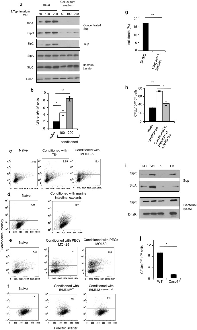
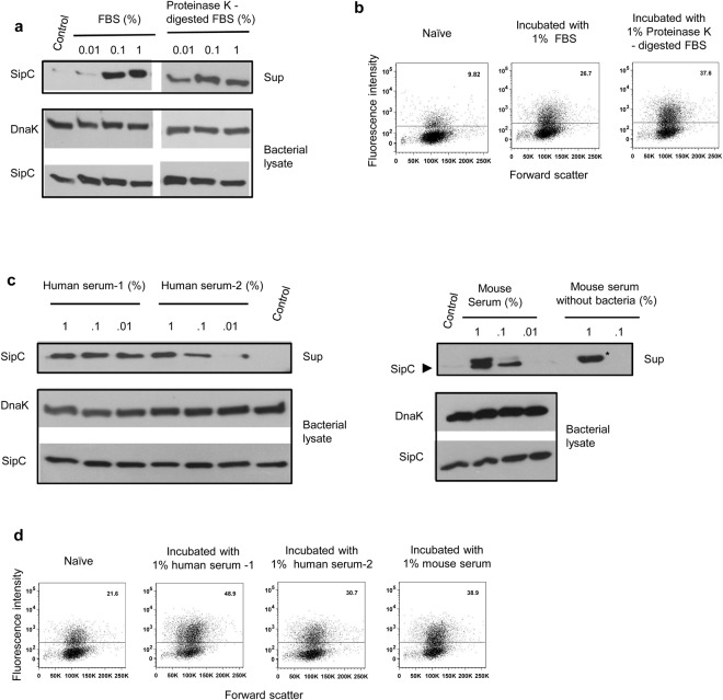
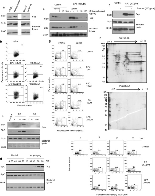
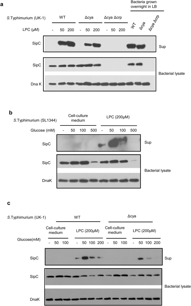
Pathogenic Salmonella species initiate infection by invading non-phagocytic intestinal epithelial cells (IEC). This invasion is brought about by a number of Salmonella invasion promoting molecules (Sips) encoded by the Salmonella Pathogenicity Island - 1 (SPI-1). Intracellular delivery of some of these molecules also brings about caspase-1 - mediated pyroptotic cell death that contributes to pathogen clearance. These molecules are secreted and delivered inside cells upon contact of Salmonella with one or more host signals whose identity has not been established. We show that lysophosphatidylcholine (LPC) released following activation of caspase-1 in Salmonella - infected cells and abundant in plasma amplifies production of Sips from this pathogen and promotes its cellular invasion. LPC brings about adenylate cyclase and cAMP receptor protein (CRP) - dependent de novo synthesis of SipC that is accompanied by its translocation to bacterial cell surface and release into the outside milieu. Treatment of Salmonella with LPC produces sustained induction of SPI - 1 transcriptional regulator, hilA. Our findings reveal a novel host lipid sensing - driven regulatory mechanism for Salmonella invasion.


Conflict of interest statement

The authors declare no competing interests.

Figures

Figure 1
Figure 1
Caspase-1 – mediated cell death-dependent stimulus from host cells enhances invasion ability of pathogenic Salmonella. (a) SipA and SipC expression in cell-free supernatants (sup) and corresponding bacterial lysates from S. Typhimurium incubated for 1 h either with serum free cell culture medium (control) or with Hela cells at a pathogen to cell ratio of 50:1, 100:1 and 200:1 (conditioned). Concentrated supernatant represents TCA precipitated cell-free supernatants from bacteria cultured with serum-free medium or with Hela cells, and sup represents cell-free supernatants without concentration. DnaK was used as a loading control for cell lysates throughout the study. (b) Bacterial load in Hela cells infected for 1 h with S. Typhimurium (naïve or conditioned as indicated) at 50 multiplicity of infection (MOI). (c–f) Flow cytometric analysis of Hela cells infected at MOI 100 with GFP-expressing S. Typhimurium, naïve or conditioned for 1 h with human intestinal epithelial cell line T84 (c), murine intestinal epithelial cell line MODE-K at MOI 100 (c), ileal explants prepared from C57BL/6 mice (d), PECs (e) or with iBMDM lines (f). Numbers indicate percent cells containing GFP-positive Salmonella. (g) Percentage cell death in Hela infected with S. Typhimurium at MOI 100 in the presence of caspase-1 inhibitor (z-YVAD; 100 µM) or vehicle control (DMSO). (h) Intracellular bacterial load in Hela cells infected for 1 h at MOI 50 with S. Typhimurium, naïve or conditioned with Hela in the absence or presence of caspase-1 inhibitor. (i) SipA and SipC expression in cell-free supernatants from bacteria cultured in serum-free cell culture medium (c) or conditioned with iBMDMs, wild-type (WT) and caspase-1-deficient (KO). Sips in supernatants and lysates from bacteria grown overnight in LB are shown as positive control. Bacterial lysates were probed for DnaK and SipC. (j) Bacterial load in Hela cells infected at MOI 20 with bacteria recovered from WT or caspase-1 deficient iBMDMs after 6 h of infection. Error bars in (b,g,h,j) represent mean ± standard deviation (s.d.). Statistical significance was determined by two tailed student’s t-test. *p < 0.05, **p < 0.01. Immunoblot data within a marked box represents cropped images from one membrane probed with one or more antibodies. The full length immunoblots are presented in the supplementary figures with cropped regions marked by boxes.
Figure 2
Figure 2
Serum borne lipids induce secretion of SipC and increase invasion capability of Salmonella. (a) Secretion of SipC in the supernatants of S. Typhimurium incubated with different concentrations of undigested or Proteinase-K digested FBS. Bacterial lysates were probed for SipC and DnaK antibodies. (b) Flow cytometric analysis of Hela cells infected with GFP-expressing S. Typhimurium stimulated with 1% Proteinase K-digested or undigested FBS. (c) Release of SipC by S. Typhimurium incubated with increasing concentrations of human or mice sera for 1 h at 37 °C. Bacterial lysates were probed for SipC and DnaK. A non-specific band of higher molecular weight (denoted by asterisk) in addition to SipC (denoted by filled black arrowhead) was also observed in the supernatant with higher concentration of mouse serum. (d) Flow cytometric analysis of Hela cells infected with GFP-expressing S. Typhimurium stimulated with human (two healthy donors) or mice sera for 1 h at 37 °C. Numbers indicate percent cells containing GFP positive bacteria. Data are representative of 2–3 independent experiments. Immunoblot data within a marked box represents cropped images from one membrane probed with one or more antibodies. The full length immunoblots are presented in the supplementary figures with cropped regions marked by boxes.
Figure 3
Figure 3
Activation with lysophosphatidylcholine (LPC) induces expression and release of Salmonella invasion-promoting molecules, and increases invasion ability of Salmonella. (a) SipC expression in cell-free supernatants from S. Typhimurium co-cultured with Hela cells in the absence or presence of cPLA2 inhibitor, Pyrrophenone (0.5 µM) or iPLA2 inhibitor, FKGK11 (30 µM) or their respective vehicle controls. Bacterial lysates were also probed for SipC and Dnak. (b) Flow cytometric analysis of Hela cells infected at MOI 100 with GFP-labeled S. Typhimurium stimulated with indicated concentrations of LPC or PC for 1 h. Numbers indicate percent cells containing GFP-positive Salmonella. (c) Expression of SipA, SipC and DnaK in cell-free supernatants and corresponding bacterial lysates derived from S. Typhimurium incubated with increasing concentrations of LPC or PC. (d) Expression of SipC in the supernatants and lysates of S. Typhimurium cultured in absence or presence of LPC (200 μM) for increasing time durations. Bacterial lysates were also probed for DnaK. (e,f) The expression of SipC in supernatants and lysates of S. Typhimurium stimulated with LPC (200 μM) in presence of indicated concentrations of Chloramphenicol or vehicle control (e), or in the presence of suramin (200 μg/ml) or vehicle control (f). (g) Flow cytometric analysis of expression of SipC on the surface of S. Typhimurium following activation with different concentrations of LPC for 30 and 60 minutes. Numbers indicate percent SipC-expressing bacteria. (h) 108 S. Typhimurium were stimulated with LPC or PC (200 µM) and after 1 h, bacteria-free supernatants were subjected to 2D gel electrophoresis. The spots in the gel were visualized by silver staining. (i) Flow cytometric analysis of GFP expression in S. Typhimurium expressing GFP driven by Hil-A promoter upon treatment with LPC or PC (200 µM) for different time durations. Data are representative of 3 independent experiments. Immunoblot data within a marked box represents cropped images from one membrane probed with one or more antibodies. The full length immunoblots are presented in the Supplementary Figures with cropped regions marked by boxes.
Figure 4
Figure 4
LPC activates release of SipC from Salmonella through cAMP-CRP dependent signaling pathway. (a) SipC levels were analyzed in the culture supernatants of Wild-type S. Typhimurium (UK-1 strain), its adenylate cyclase knock-out derivative (Δcya-27) and adenylate cyclase – CRP double knock-out strain (Δcya-Δcrp), treated with different concentrations of LPC. Expression levels from bacteria grown overnight in LB are shown as positive controls. Bacterial lysates were also probed for SipC and DnaK. (b) SipC levels were determined in (b) S. Typhimurium SL 1344 and (c) S. Typhimurium UK-1 and its Δcya-27 derivative stimulated with LPC (200 µM) in presence of increasing concentrations of glucose. Bacterial lysates were probed for SipC and DnaK. Data are representative of two independent experiments. Immunoblot data within a marked box represents cropped images from one membrane probed with one or more antibodies. The full length immunoblots are presented in the supplementary figures with cropped regions marked by boxes.

Similar articles

Cited by

References

    1. Galán JE. Interaction of Salmonella with host cells through the centisome 63 type III secretion system. Curr. Opin. Microbiol. 1999;2:46–50. doi: 10.1016/S1369-5274(99)80008-3. - DOI - PubMed
    1. Galán JE, Curtiss R. Cloning and molecular characterization of genes whose products allow Salmonella typhimurium to penetrate tissue culture cells. Proc. Natl. Acad. Sci. USA. 1989;86:6383–7. doi: 10.1073/pnas.86.16.6383. - DOI - PMC - PubMed
    1. Kubori T, et al. Supramolecular structure of the Salmonella typhimurium type III protein secretion system. Science. 1998;280:602–605. doi: 10.1126/science.280.5363.602. - DOI - PubMed
    1. Collazo CM, Galán JE. The invasion-associated type III system of Salmonella typhimurium directs the translocation of Sip proteins into the host cell. Mol. Microbiol. 1997;24:747–756. doi: 10.1046/j.1365-2958.1997.3781740.x. - DOI - PubMed
    1. Lara-Tejero M, Galan JE. Salmonella enterica serovar typhimurium pathogenicity island 1-encoded type III secretion system translocases mediate intimate attachment to nonphagocytic cells. Infect. Immun. 2009;77:2635–2642. doi: 10.1128/IAI.00077-09. - DOI - PMC - PubMed

Publication types