. 2018 Oct 19;8(1):15508.
doi: 10.1038/s41598-018-33975-x.

White-nose syndrome is associated with increased replication of a naturally persisting coronaviruses in bats

Affiliations

White-nose syndrome is associated with increased replication of a naturally persisting coronaviruses in bats

Christina M Davy et al. Sci Rep. .

Abstract

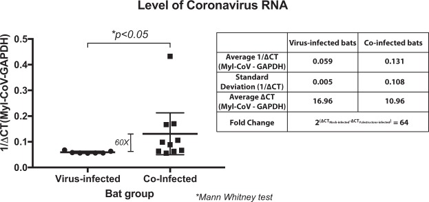
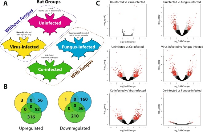
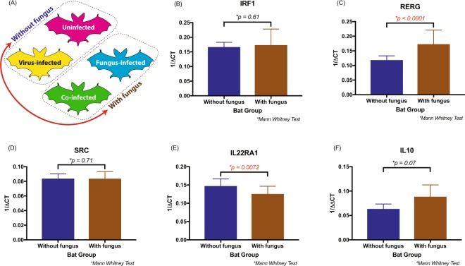
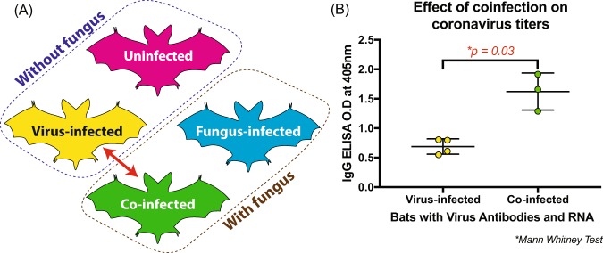
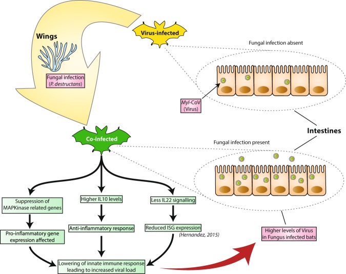
Spillover of viruses from bats to other animals may be associated with increased contact between them, as well as increased shedding of viruses by bats. Here, we tested the prediction that little brown bats (Myotis lucifugus) co-infected with the M. lucifugus coronavirus (Myl-CoV) and with Pseudogymnoascus destructans (Pd), the fungus that causes bat white-nose syndrome (WNS), exhibit different disease severity, viral shedding and molecular responses than bats infected with only Myl-CoV or only P. destructans. We took advantage of the natural persistence of Myl-CoV in bats that were experimentally inoculated with P. destructans in a previous study. Here, we show that the intestines of virus-infected bats that were also infected with fungus contained on average 60-fold more viral RNA than bats with virus alone. Increased viral RNA in the intestines correlated with the severity of fungus-related pathology. Additionally, the intestines of bats infected with fungus exhibited different expression of mitogen-activated protein kinase pathway and cytokine related transcripts, irrespective of viral presence. Levels of coronavirus antibodies were also higher in fungal-infected bats. Our results suggest that the systemic effects of WNS may down-regulate anti-viral responses in bats persistently infected with M. lucifugus coronavirus and increase the potential of virus shedding.


Conflict of interest statement

The authors declare no competing interests.

Figures

Figure 1
Figure 1
Effect of white-nose syndrome on level of Myotis lucifugus coronavirus (Myl-CoV) RNA in hibernating little brown bats (M. lucifugus). Relative transcript levels for the coronavirus RNA polymerase gene for each bat are depicted as reciprocal of Cycle threshold (Ct) normalized separately (ΔCt) for levels of glyceraldehyde 3-phosphate dehydrogenase (GAPDH) transcripts in each sample. The horizontal bar represents the mean while the vertical bar indicates standard deviation from the mean. Significance (p value) is as calculated with an independent Mann-Whitney test. Virus-infected bats had lower 1/ΔCt values for coronavirus RNA than co-infected bats. The average fold-differences between virus-infected and co-infected bats were calculated from the difference between the average ΔCt values.
Figure 2
Figure 2
Co-infection of little brown bats (Myotis lucifugus) with M. lucifugus coronavirus (Myl-CoV) and Pseudogymnoascus destructans results in non-additive patterns of gene expression compared to sole infection with the virus or fungus. (A) Experimental design, showing the four treatments of little brown bat (Myotis lucifugus) established by experimental inoculation with Pseudogymnoascus destructans and by qPCR detection of persistent Myl-CoV infections: uninfected, virus-infected, fungus-infected and co-infected. (B) Differential gene expression identified by DESeq2 among virus-infected, fungus-infected and Co-infected bats as compared to the change each exhibited relative to uninfected bats. (C) Differential gene expression among the four treatments, detected by DESEQ2 and visualized in volcano plots. The log of the adjusted p-value is plotted as a function of the log ratio of differential expression. Colored data points represent different groups of genes based on fold change and false discovery rate (FDR) cutoff; red (>2 fold change, FDR <0.05), dark grey (>2 fold change, FDR > 0.05), light grey (<2 fold change, FDR < 0.05), black (<2 fold change, FDR > 0.05).
Figure 3
Figure 3
Effect of white-nose syndrome (WNS) on the levels of immune genes IRF1, RERG, SRC, IL22RA1 and IL10 expressed in the ileum of little brown bats (Myotis lucifugus). (A) Summary of the four treatments, with a red arrow indicating the two groups (“with fungus” and “without fungus”) that were compared. (BF) The relative transcript levels of each gene for bats with and without WNS, depicted as reciprocal of Cycle threshold (Ct) normalized separately (ΔCt) for levels of transcripts for GAPDH in each sample. Statistical significance was calculated based on the independent Mann Whitney test. The difference in the two groups was significant for RERG and IL22RA1 genes.
Figure 4
Figure 4
Little brown bats (Myotis lucifugus) coinfected with M. lucifugus coronavirus (Myl-CoV) and Pseudogymnoascus destructans produce more antibodies against Myl-CoV than bats infected only with Myl-CoV. (A) Diagram summarizes the four treatments; the red arrow shows the two groups between which antibody levels were compared. (B) Antibody levels against the Myl-CoV N protein detected by antibody capture ELISA expressed as optical density (O.D.) values at 405 nm. Co-infected bats had significantly higher antibody levels than bats infected only with Myl-CoV (independent Mann Whitney test; p value = 0.03).
Figure 5
Figure 5
Hypothesized model of pathways involved in increased coronavirus shedding and white-nose syndrome (WNS) severity in little brown bats (Myotis lucifugus) co-infected with M. lucifugus coronavirus (Myl-CoV) and Pseudogymnoascus destructans. Diagram summarizes the changes observed by comparing co-infected bats with virus-infected bats. Bats with persistent Myl-CoV infection exhibit relatively low viral shedding. When bats are also infected with P. destructans (shown in yellow arrow) and develop WNS, the level of coronavirus increases. There is a change in the level of some immune genes, such as IL22, RERG and possibly IL10, which may have an effect on immune response and cell proliferation. The increase in coronavirus levels in co-infected bats is possibly due to the bats’ systemic response to WNS reducing innate anti-viral responses.

Similar articles

Cited by

References

    1. Ge XY, et al. Isolation and characterization of a bat SARS-like coronavirus that uses the ACE2 receptor. Nature. 2013;503:535–538. doi: 10.1038/nature12711. - DOI - PMC - PubMed
    1. Corman VM, et al. Rooting the phylogenetic tree of middle East respiratory syndrome coronavirus by characterization of a conspecific virus from an African bat. J Virol. 2014;88:11297–11303. doi: 10.1128/JVI.01498-14. - DOI - PMC - PubMed
    1. Ithete NL, et al. Close relative of human Middle East respiratory syndrome coronavirus in bat, South Africa. Emerg Infect Dis. 2013;19:1697–1699. doi: 10.3201/eid1910.130946. - DOI - PMC - PubMed
    1. Yang L, et al. MERS-related betacoronavirus in Vespertilio superans bats, China. Emerg Infect Dis. 2014;20:1260–1262. doi: 10.3201/eid2007.140318. - DOI - PMC - PubMed
    1. Anthony, S. J. et al. Further Evidence for Bats as the Evolutionary Source of Middle East Respiratory Syndrome Coronavirus. MBio8, 10.1128/mBio.00373-17 (2017). - PMC - PubMed

Publication types