. 2018 Oct 19;9(1):4354.
doi: 10.1038/s41467-018-06774-1.

TRIP13 and APC15 drive mitotic exit by turnover of interphase- and unattached kinetochore-produced MCC

Affiliations

TRIP13 and APC15 drive mitotic exit by turnover of interphase- and unattached kinetochore-produced MCC

Dong Hyun Kim et al. Nat Commun. .

Abstract

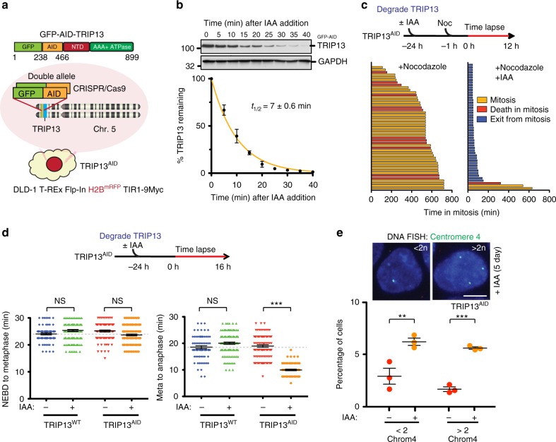
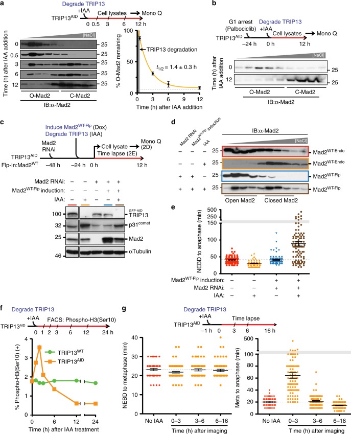
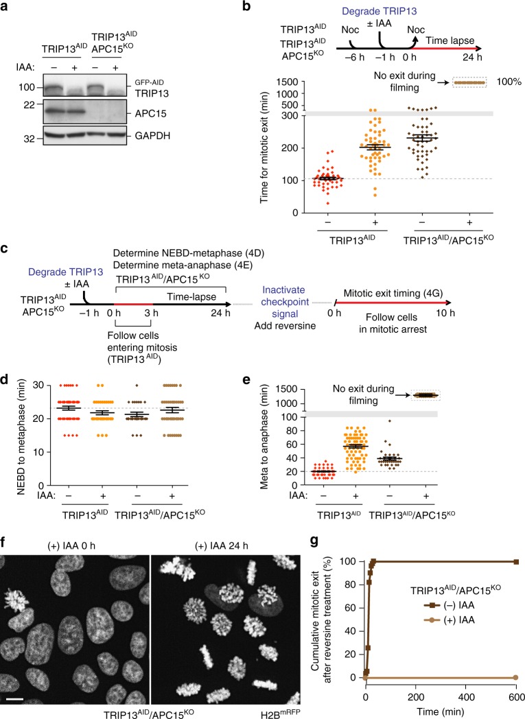
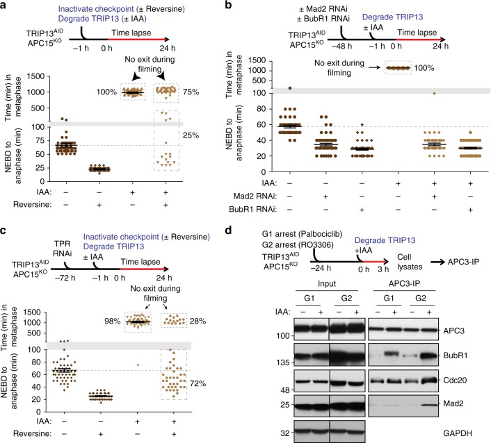
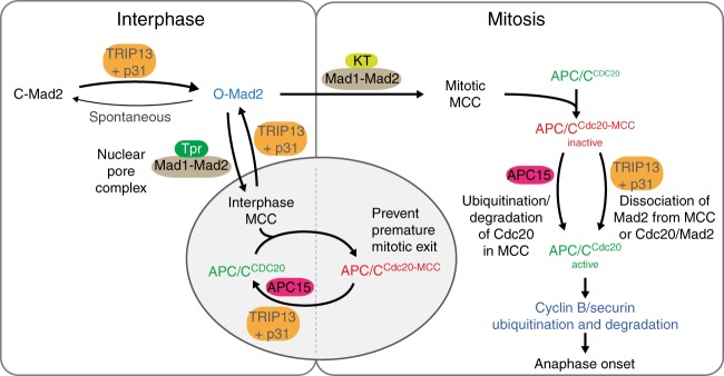
The mitotic checkpoint ensures accurate chromosome segregation through assembly of the mitotic checkpoint complex (MCC), a soluble inhibitor of the anaphase-promoting complex/cyclosome (APC/C) produced by unattached kinetochores. MCC is also assembled during interphase by Mad1/Mad2 bound at nuclear pores, thereby preventing premature mitotic exit prior to kinetochore maturation and checkpoint activation. Using degron tagging to rapidly deplete the AAA+ ATPase TRIP13, we show that its catalytic activity is required to maintain a pool of open-state Mad2 for MCC assembly, thereby supporting mitotic checkpoint activation, but is also required for timely mitotic exit through catalytic disassembly of MCC. Strikingly, combining TRIP13 depletion with elimination of APC15-dependent Cdc20 ubiquitination/degradation results in a complete inability to exit mitosis, even when MCC assembly at unattached kinetochores is prevented. Thus, mitotic exit requires MCC produced either in interphase or mitosis to be disassembled by TRIP13-catalyzed removal of Mad2 or APC15-driven ubiquitination/degradation of its Cdc20 subunit.


Conflict of interest statement

The authors declare no competing interests.

Figures

Fig. 1
Fig. 1
Abrupt TRIP13 depletion compromises mitotic checkpoint activation. a Schematic illustration of CRISPR-Cas9 mediated genome editing of DLD-1 T-REx Flp-In H2BmRFP + TIR1-9Myc+ human colon cancer cell line to tag both endogenous TRIP13 alleles with green fluorescent protein (GFP) and an auxin-inducible degron (AID). b Upper: Indole-3-acetic acid (IAA) was added to engineered TRIP13AID cells and level of GFP-AID-TRIP13 was measured by quantitative immunoblot analysis. Lower: Degradation kinetics of GFP-AID-TRIP13 from triplicate measurements, fit using a single-exponential decay function (yellow line). c TRIP13 function in mitotic checkpoint. Upper: Schematic of experiment to test the role of TRIP13 in mitotic checkpoint activation. Lower: Timing of mitotic exit and cell fate as measured by time-lapse live-cell imaging. (n = 57 for +Nocodazole, n = 54 for +Nocodazole, +IAA). d TRIP13 function in unperturbed mitosis. Upper: Schematic of experiment to test the contribution of TRIP13 in mitotic checkpoint activation. Lower: For each cell, timing of nuclear envelope breakdown (NEBD) to metaphase (left panel) and metaphase-to-anaphase (right panel) were measured. Cells monitored: n = 78, 137, 100, 120, 79, 129, 102, 81. e Upper: Representative images of TRIP13AID cells treated with IAA for 5 days and probed by FISH for the centromere of chromosome 4. Lower: Percentage of cells with the indicated number of chromosome 4 centromere foci after 5-day IAA treatment. Total cells counted: 1156 for control, 1119 for IAA treatment over three separate experiments. Scale bar, 5 μm. P-values for d and e were calculated using an unpaired two-tailed t-test (**P < 0.01, ***P < 0.001, NS not significant). Lines in b, d, and e represent the mean ± s.e.m.
Fig. 2
Fig. 2
TRIP13 regulates mitotic checkpoint activation and silencing through Mad2. a Upper: Schematic of experiment to test kinetics of Mad2 conformational conversion after TRIP13 depletion. Lower left: Mono-Q ion-exchange chromatography analysis of soluble Mad2 in TRIP13AID cells after IAA treatment for the indicated times. Lower right: Kinetics of O-Mad2 to C-Mad2 conversion was calculated from triplicate measurements, fit using a single-exponential decay function (yellow line). t1/2 = 1.4 ± 0.3 h, plateau = 8% O-Mad2 (gray dashed line). b Mad2 conformation in G1 after TRIP13 depletion. TRIP13AID cells were arrested in G1 with Palbociclib and Mad2 conformation was measured by Mono-Q ion-exchange chromatography at 0 or 12 h after addition of IAA to degrade TRIP13. c Upper: Schematic of the approach to replace endogenous Mad2 in TRIP13-depleted cells (used in d and e). Lower: Immunoblot analysis of the replacement of endogenous Mad2 with Mad2WT-Flp induced by doxycycline. d Mono-Q ion-exchange chromatography analysis of Mad2 conformation after replacement of endogenous Mad2 with Mad2WT-Flp, in the presence (blue box) or absence (brown box) of TRIP13. High-level expression of Mad2WT-Flp resulted in a significant O-Mad2 population even after TRIP13 depletion. e Mitotic timing (NEBD-to-anaphase) after replacement of endogenous Mad2 with Mad2WT-Flp, measured by time-lapse live-cell imaging. (n = 100 for each condition). f Upper: Schematic of experiment to measure population of mitotic TRIP13WT and TRIP13AID cells upon IAA treatment. Lower: Percentage of phospho-histone H3 (Ser10) positive cells was measured using flow cytometry at different time points after IAA treatment. Detailed FACS profiles in Supplementary Fig. 2D. g Upper: Schematic of experiment to measure mitotic timing in TRIP13AID cells upon IAA treatment. Lower: NEBD-to-metaphase (left) and metaphase-to-anaphase (right) timing of TRIP13AID cells at different time points after IAA addition. (From left to right panel, n = 50, 69, 56, 58, 176, 93, 70, and 110). Lines in e and g represent the mean ± s.e.m.
Fig. 3
Fig. 3
TRIP13 and APC15 synergistically act on kinetochore-generated MCC. a Western blot showing effective depletion of GFP-AID-TRIP13 in TRIP13AID and TRIP13AID/APC15KO cells. b Upper: Schematic of experiment to measure timing of mitotic exit after TRIP13 depletion by IAA addition. Lower: Timing of mitotic exit after 6-h nocodazole treatment, followed by release into DMEM. (n = 50 for each experimental condition). c Schematic of approach to monitor unperturbed mitosis after depletion of TRIP13 (by IAA addition) in TRIP13AID/APC15KO cells (used in df). d Unperturbed mitotic timing NEBD-to-metaphase; (n = 50 for each experimental condition). e Metaphase-to-anaphase of TRIP13AID and TRIP13AID/APC15KO cells entering mitosis 3 h after depletion of TRIP13. (n = 70 for each experimental condition). f Representative images of chromosomes in TRIP13AID/APC15KO cells after before IAA treatment (left) and 24 h after IAA treatment (right). Scale bar, 5 μm. g Mitotic exit timing after inhibition of kinetochore signaling. Reversine was added 24 h after the start of imaging to inactivate kinetochore signaling, and mitotic exit of cells previously arrested in mitosis was measured for 10 h. (n = 70 for each experimental condition). Lines in b, d, and e represent the mean ± s.e.m.
Fig. 4
Fig. 4
TRIP13 and APC15 are required for turnover of MCC produced in interphase. a Upper: Schematic of experiment to measure the dependence of the observed mitotic exit delay on kinetochore-catalyzed MCC production. Lower: mitotic timing in reversine-treated TRIP13AID /APC15KO cells in the presence or absence of IAA. (n = 50 for each experimental condition). b Upper: Schematic of experiment to measure the dependence of the observed mitotic exit delay on Mad2 and BubR1. Lower: mitotic timing in Mad2 or BubR1 RNAi treated TRIP13AID/APC15KO cells in the presence or absence of IAA. (n = 50 for each experimental condition). c Upper: Schematic of experiment to measure the dependence of the observed mitotic exit delay on Tpr. Lower: mitotic timing in Tpr RNAi treated TRIP13AID /APC15KO cells in the presence or absence of reversine and IAA. (n = 50 for each experimental condition). d Upper: Schematic of experiment to measure APC/CMCC formation in TRIP13AID/APC15KO. APC3 was immunoprecipitated from cells in G1 with Palbociclib and in late G2 with RO-3306. Lower: APC/C interacting BubR1 and Cdc20 levels were measured by quantitative immunoblotting. Lines in ac represent the mean ± s.e.m.
Fig. 5
Fig. 5
Model for TRIP13 and APC15 contributions to mitotic checkpoint signaling. In interphase cells, TRIP13 and p31comet counteract the spontaneous conversion of O-Mad2 to C-Mad2 in order to maintain a pool of O-Mad2 for checkpoint activation and regulate the homeostasis of both Mad2 and p31comet. Additionally, TRIP13 and APC15 together prevent formation of excess MCC during interphase for proper mitotic exit in the next mitosis. In prometaphase, conversion of O-Mad2 to Cdc20-bound C-Mad2 at unattached kinetochores catalyzes assembly of MCC, which binds and inhibits APC/CCdc20. Checkpoint silencing at metaphase can occur through two pathways: First, TRIP13 and p31comet can catalyze the disassembly of free MCC and the removal/disassembly of MCC bound to APC/CCdc20. Second, APC15-mediated conformational changes within the APC/C can allow ubiquitination of Cdc20 in MCC, followed by reactivation of APC/CCdc20

References

    1. Irniger S, Piatti S, Michaelis C, Nasmyth K. Genes involved in sister chromatid separation are needed for B-type cyclin proteolysis in budding yeast. Cell. 1995;81:269–278. doi: 10.1016/0092-8674(95)90337-2. - DOI - PubMed
    1. Sudakin V, et al. The cyclosome, a large complex containing cyclin-selective ubiquitin ligase activity, targets cyclins for destruction at the end of mitosis. Mol. Biol. Cell. 1995;6:185–197. doi: 10.1091/mbc.6.2.185. - DOI - PMC - PubMed
    1. King RW, et al. A 20S complex containing CDC27 and CDC16 catalyzes the mitosis-specific conjugation of ubiquitin to cyclin B. Cell. 1995;81:279–288. doi: 10.1016/0092-8674(95)90338-0. - DOI - PubMed
    1. Tugendreich S, Tomkiel J, Earnshaw W, Hieter P. CDC27Hs colocalizes with CDC16Hs to the centrosome and mitotic spindle and is essential for the metaphase to anaphase transition. Cell. 1995;81:261–268. doi: 10.1016/0092-8674(95)90336-4. - DOI - PubMed
    1. Pines J. Cubism and the cell cycle: the many faces of the APC/C. Nat. Rev. Mol. Cell Biol. 2011;12:427–438. doi: 10.1038/nrm3132. - DOI - PubMed

Publication types

MeSH terms