Review
. 2019 Jan;26(1):99-114.
doi: 10.1038/s41418-018-0212-6. Epub 2018 Oct 19.

Pyroptosis versus necroptosis: similarities, differences, and crosstalk

Affiliations
Review

Pyroptosis versus necroptosis: similarities, differences, and crosstalk

Daniel Frank et al. Cell Death Differ. 2019 Jan.

Abstract

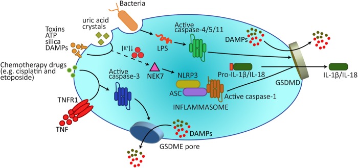
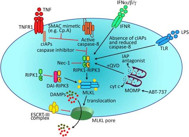
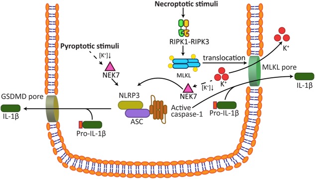
Pyroptosis and necroptosis represent two pathways of genetically encoded necrotic cell death. Although these cell death programmes can protect the host against microbial pathogens, their dysregulation has been implicated in a variety of autoimmune and auto-inflammatory conditions. The disease-promoting potential of necroptosis and pyroptosis is likely a consequence of their ability to induce a lytic cell death. This cell suicide mechanism, distinct from apoptosis, allows the release of immunogenic cellular content, including damage-associated molecular patterns (DAMPs), and inflammatory cytokines such as interleukin-1β (IL-1β), to trigger inflammation. In this Review, we discuss recent discoveries that have advanced our understanding on the primary functions of pyroptosis and necroptosis, including evidence for the specific cytokines and DAMPs responsible for driving inflammation. We compare the similar and unique aspects of pyroptotic- and necroptotic-induced membrane damage, and explore how these may functionally impact distinct intracellular organelles and signalling pathways. We also examine studies highlighting the crosstalk that can occur between necroptosis and pyroptosis signalling, and evidence supporting the physiological significance of this convergence. Ultimately, a better understanding of the similarities, unique aspects and crosstalk of pyroptosis and necroptosis will inform as to how these cell death pathways might be manipulated for therapeutic benefit.


Conflict of interest statement

The authors declare that they have no conflict of interest.

Figures

Fig. 1
Fig. 1
Gasdermins form membrane pores to cause pyroptosis. A wide array of extracellular stimuli can drive pyroptosis. In the canonical model of pyroptosis, inflammasome sensor proteins, such as NLRP3, recognize cellular stressors, including those from bacteria, viruses, toxins, ATP, uric acid crystals, silica, and DAMPs. These stressors activate NLRP3 indirectly through potassium efflux, which leads to NEK7 binding NLRP3 to trigger its oligomerization. NLRP3 subsequently activates caspase-1 via the adaptor protein ASC. Caspase-1 processes and activates IL-1β and IL-18, and also cleaves GSDMD to release the membrane pore-forming GSDMD-N domain. GSDMD-N pores promote the release of activated IL-1β and IL-18 and, most likely, DAMPs that can be accommodated by the 10–20 nm pore diameter. Additional DAMPs will be released following the collapse of the plasma membrane. Cytosolic LPS binds Caspase-4/5/11 to trigger their cleavage of GSDMD, but not IL-1β and IL-18. In addition, recent research has revealed how the apoptotic effector caspase, caspase-3, can cleave GSDME to also cause pyroptotic death. DAMPs damage-associated molecular patterns, LPS lipopolysaccharide
Fig. 2
Fig. 2
Membrane-associated MLKL induces necroptosis. Necroptosis can be triggered by death receptors (e.g. TNFR1), the IFNR, and TLR3/4, which promote the assembly of a RIPK1-RIPK3-MLKL signalling complex. RIPK3-mediated phosphorylation of MLKL results in MLKL translocation to the plasma membrane to induce membrane damage. Damaged membrane shedding, and cell death, is limited by the ESCRT-III complex. Recent findings have documented additional necroptotic triggers, such as BH3-mimetic (ABT-737)-induced MOMP or ZBP1/DAI-induced RIPK3 dimerization. cIAPs cellular inhibitor of apoptosis proteins, DAI DNA-dependent activator of IFN-regulatory factors, DAMPs damage-associated molecular patterns, ESCRT-III endosomal sorting complex required for transport (ESCRT) complex III, INFR interferon receptor, LPS lipopolysaccharide, MOMP mitochondrial outer membrane permeabilization, Nec-1 Necrostatin-1, Smac mimetic; an IAP antagonist, QVD; a pan-caspase inhibitor
Fig. 3
Fig. 3
Necroptotic signalling activates the NLRP3 inflammasome to drive inflammation. Both the pyroptosis and the necroptosis pathways can activate the NLRP3-IL-1β signalling axis. Data support the hypothesis that both caspase-4/5/11 induction of GSDMD pores, or MLKL-induced membrane damage, cause potassium efflux. As a result, NEK7 binds and triggers NLRP3 inflammasome formation. Activated IL-1β is subsequently released from pre-lytic GSDMD and MLKL membrane perforations that may also accommodate appropriately sized DAMPs. Subsequent cellular rupture allows the release of larger DAMPs. DAMPs; damage-associated molecular patterns

References

    1. Lockshin RA, Williams CM. Programmed cell death--I. Cytology of degeneration in the intersegmental muscles of the Pernyi silkmoth. J Insect Physiol. 1965;11:123–33. - PubMed
    1. Kerr JF, Wyllie AH, Currie AR. Apoptosis: a basic biological phenomenon with wide-ranging implications in tissue kinetics. Br J Cancer. 1972;26:239–57. - PMC - PubMed
    1. Lindqvist LM, Frank D, McArthur K, Dite TA, Lazarou M, Oakhill JS, et al. Autophagy induced during apoptosis degrades mitochondria and inhibits type I interferon secretion. Cell Death Differ 2017;25:782–794. - PMC - PubMed
    1. White MJ, McArthur K, Metcalf D, Lane RM, Cambier JC, Herold MJ, et al. Apoptotic caspases suppress mtDNA-induced STING-mediated type I IFN production. Cell. 2014;159:1549–62. - PMC - PubMed
    1. Rongvaux A, Jackson R, Harman CC, Li T, West AP, de Zoete MR, et al. Apoptotic caspases prevent the induction of type I interferons by mitochondrial DNA. Cell. 2014;159:1563–77. - PMC - PubMed

Publication types