Review
. 2019 Apr;48(4):517-525.
doi: 10.1007/s00256-018-3086-2. Epub 2018 Oct 19.

Thalidomide and neurotrophism

Affiliations
Review

Thalidomide and neurotrophism

Judith R Soper et al. Skeletal Radiol. 2019 Apr.

Abstract

Background: Following the thalidomide disaster (1958-62), Henkel and Willert analysed the pattern of dysmelia in the long bones (J Bone Joint Surg Br. 51:399-414, 1969) and the extremities, Willert and Henkel (Z Orthop Ihre Grenzgeb. 107:663-75, 1970). Willert's material from deformed extremities is re-examined here asking "How does thalidomide reduce the skeleton?"

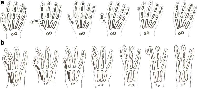
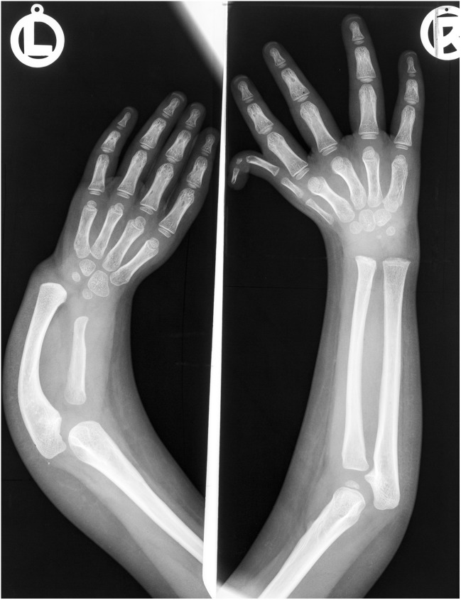
Materials and methods: We reviewed the original data collection of Willert and Henkel (Z Orthop Ihre Grenzgeb. 107:663-75, 1970), comprising musculoskeletal histology slides from 30 children affected by thalidomide with radiographs of hands (19 cases) and feet (4 cases).

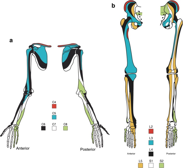
Results: All original observations by Willert and Henkel (Z Orthop Ihre Grenzgeb. 107:663-75, 1970), were verified. Radial rays of the hand disappeared early, but the foot was spared until late. Radiology confirms that bone reduction in the hand (aplasia or hypoplasia in the thumb and index finger) coincides with sensory segmental nerve C6. In the foot, reduction of the toes is rare, but mesenchymal excess (polydactyly) occurs in the hallux (L5 sclerotome), usually associated with absent tibia (L4 sclerotome). Histology confirms skeletal mesenchymal components to be unremarkable, contrasting with grossly abnormal bony architecture, a striking discordance between microscopic and macroscopic findings. No necrosis or vascular pathology was seen.

Conclusion: The basic lesion was an abnormal quantity rather than quality of mesenchyme. Cell populations result from cellular proliferation, controlled in early limb bud formation by neurotrophism. Thalidomide is a known sensory neurotoxin in adults. In the embryo, sensorineural injury alters neurotrophism, causing increased or diminished cell proliferation in undifferentiated mesenchyme. Differentiation into normal cartilage occurs later, but within an altered mesenchymal mass. Reduction or excess deformity results, with normal histology, a significant finding. The primary pathological condition is not in the skeleton, but in the nerves.

Keywords: Embryonic sensorineural osteoarthropathy; Embryonic sensory neuropathy; Neurotrophism; Quantitative neuropathology; Thalidomide.


Conflict of interest statement

The authors have no conflicts of interests.

Figures

Fig. 1
Fig. 1
a Thumb reduction in thalidomide embryopathy. As the severity of damage increases, the thumb and first metacarpal are reduced from normal to total absence, through hypoplastic and triphalangeal morphology to pedunculated remnants, and then no thumb at all [2]. A non-functional pedunculated digital remnant was usually surgically removed in infancy. b Index finger reduction from normal to nil. The second metacarpal disappears before the phalanges. Two or three phalanges may be reduced in size or fused to the adjacent third ray by the syndactyly of soft tissues or by a narrow pedicle (flail digit) [2]
Fig. 2
Fig. 2
Forearms of a child affected by thalidomide. Absent left thumb, short hypoplastic radius, compensatory curved ulna. Hypoplastic right triphalangeal thumb with short middle phalanx. The first metacarpal is duplicated. Damage to sclerotome C6 bilaterally, but more severe on the left
Fig. 3
Fig. 3
a Sclerotomes of the upper limb: cervical segmental sensory nerve supply to the skeleton, derived from studies of referred pain [20]. A sclerotome is defined as a band of skeletal structures supplied by one spinal segmental sensory nerve. It is the somatic equivalent of a dermatome. It crosses through soft tissues, bones and joints, irrespective of the usual anatomical structures such as joint spaces. b Sclerotomes of the lower limb: lumbar and sacral sensory nerve supply to the lower limbs [20], incorporating the repositioning of the hallux from a superior to medial position during development of leg length in utero, demonstrated by medial twisting of sclerotomes. L4 was most vulnerable to thalidomide with sclerotome deletion; L5 could show addition/polydactyly
Fig. 4
Fig. 4
a Normal hyaline cartilage and bony components at low power. b Epiphyseal plates were unremarkable with reserve, proliferative and hypertrophic zones with endochondral ossification. c Wholemount section and d specimen radiograph showing a small middle phalanx and fused incomplete proximal interphalangeal joint replaced by a notch (black arrow). A cleft is visible on the radiograph (white arrow). e In some cases, complete lack of ossification of the proximal phalanx was present, f illustrating apparent discontinuity of the structures on imaging
Fig. 5
Fig. 5
a Photograph shows infant with bilateral polydactyly of the toes. b Left foot with duplicated first metatarsal and triphalangeal hallux. An additional partial digit is seen in the next webspace. Polydactyly in the L5 sclerotome. Single long bone in the lower leg. c Right foot with duplication of the distal first metatarsal and triphalangeal hallux. Single long bone in the lower leg. Partial polydactyly in the L5 distribution. d Both lower legs of another patient. The right side shows a small bone representing residual tibia of the distal L3 sclerotome (black arrows). L4 sclerotome is absent. Wide fibula is due to fusion of the distal tibia and fibula (sometimes called “tibula”). Four metatarsals with only the head of the first metatarsal present. The left side shows partial resection of a duplicated hallux with residual partial duplication of the first metatarsal
Fig. 6
Fig. 6
TN Portion of transverse section of the sural nerve from a woman with thalidomide neuropathy of 5 years’ duration, microscopy and histogram of fibre diameters. The large diameter fibres have been destroyed [9]. C Normal control subject of the same age [9]. Normal bimodal histogram of fibre diameters
Fig. 7
Fig. 7
Timelines [4] compiled from Langman [22], Nowack [36], and Knapp et al. [37]. The neural crest appears at day 18 in the human embryo. The upper limb bud appears at day 28 in the human embryo. Thalidomide acts between days 21 and 42. The first arm defects follow ingestion of thalidomide at day 24, when the neural crest is present, but 4 days before the limb bud exists. This was confirmed in laboratory primates by Neubert and Neubert [39]

References

    1. Henkel H-L, Willert H-G. Dysmelia. A classification and a pattern of malformation in a group of congenital defects of the limbs. J Bone Joint Surg Br. 1969;51:399–414. doi: 10.1302/0301-620X.51B3.399. - DOI - PubMed
    1. Willert HG, Henkel HL. Pathologisch-anatomische Prinzipien bei Extremitäten Feldbildungen, dargestellt am Beispiel der Finger. Z Orthop Ihre Grenzgeb. 1970;107:663–675. - PubMed
    1. McCredie J, Willert HG. Longitudinal limb deficiencies and the sclerotomes: an analysis of 378 dysmelic malformations induced by thalidomide. J Bone Joint Surg Br. 1999;81:9–23. doi: 10.1302/0301-620X.81B1.8448. - DOI - PubMed
    1. McCredie J. Beyond thalidomide: birth defects explained. London: Royal Society of Medicine Press; 2007.
    1. Fullerton P, Kremer M. Neuropathy after intake of thalidomide (Distaval) BMJ. 1961;ii:855–858. doi: 10.1136/bmj.2.5256.855. - DOI - PMC - PubMed

MeSH terms