. 2019 Jan;1863(1):232-240.
doi: 10.1016/j.bbagen.2018.10.006. Epub 2018 Oct 17.

Dimerization of an aptamer generated from Ligand-guided selection (LIGS) yields a high affinity scaffold against B-cells

Affiliations

Dimerization of an aptamer generated from Ligand-guided selection (LIGS) yields a high affinity scaffold against B-cells

Sana Batool et al. Biochim Biophys Acta Gen Subj. 2019 Jan.

Abstract

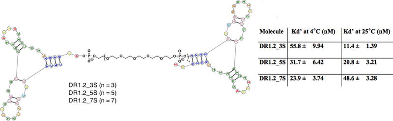
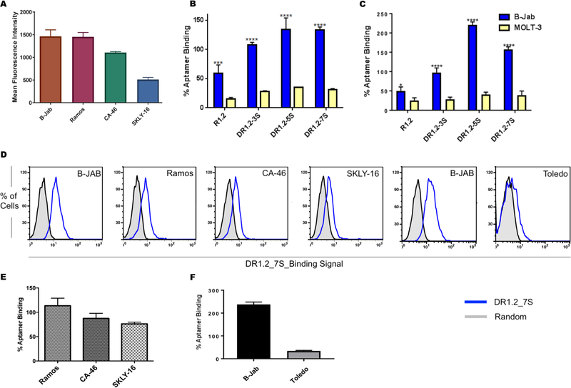
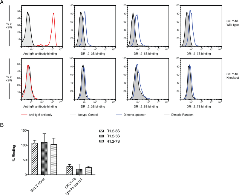
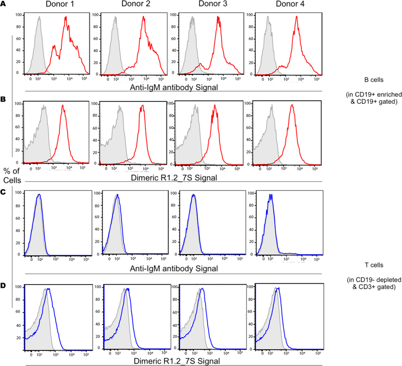
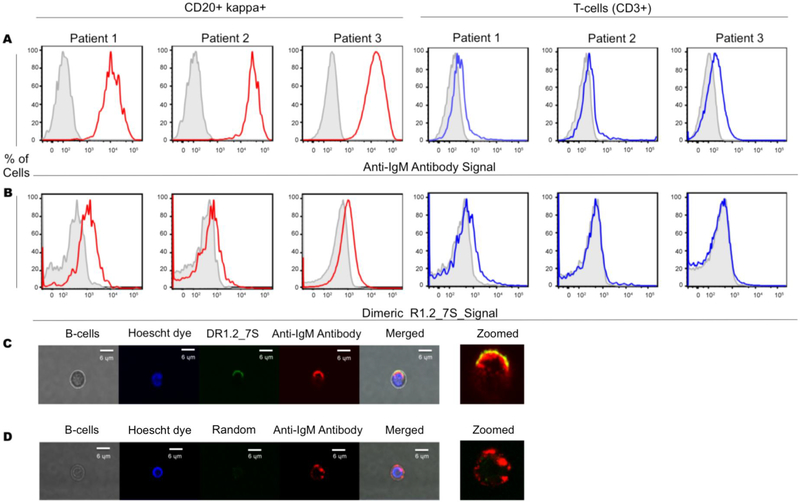
Nucleic Acid Aptamers (NAAs) are a class of synthetic DNA or RNA molecules that bind specifically to their target. We recently introduced an aptamer termed R1.2 against membrane Immunoglobulin M (mIgM) expressing B-cell neoplasms using Ligand Guided Selection (LIGS). While LIGS-generated aptamers are highly specific, their lower affinity prevents aptamers from being used for translational applications. Highly specific aptamers with higher affinity can increase targetability, boosting the application of aptamers as diagnostic and therapeutic molecules. Herein, we report that dimerization of R1.2, an aptamer generated from LIGS, leads to high affinity variants without compromising the specificity. Three dimeric aptamer analogues with variable linker lengths were designed to evaluate the effect of linker length in affinity. The optimized dimeric R1.2 against cultured B-cell neoplasms, four donor B-cell samples and mIgM-positive Waldenström's Macroglobulinemia (WM) showed specificity. Furthermore, confocal imaging of dimeric aptamer and anti-IgM antibody in purified B-cells suggests co-localization. Binding assays against IgM knockout Burkitt's Lymphoma cells utilizing CRISPR/Cas9 further validated specificity of dimeric R1.2. Collectively, our findings show that LIGS-generated aptamers can be re-engineered into dimeric aptamers with high specificity and affinity, demonstrating wide-range of applicability of LIGS in developing clinically practical diagnostic and therapeutic aptamers.

Keywords: Aptamer; B-cell lymphoma; Dimerization; Ligand-guided selection; mIgM.


Conflict of interest statement

Disclosures/Conflict of Interest

Authors declare no competing interests

Figures

Figure 1.
Figure 1.
Design of dimeric assemblies of aptamer R1.2 and their affinity. Dimeric aptamer R1.2 linked by spacer molecule comprised of Polyethylene glycol (PEG). Three analogues were designed with 3, 5 or 7 spacer molecules. All constructs were synthesized using standard solid-state phosphoramid ite chemistry, and affinity was evaluated against mIgM positive BJAB cells (a Burkitt’s Lymphoma cell line). Monomeric R1.2 showed an affinity of 35.5 ± 8.94 nM at 4°C.
Figure 2.
Figure 2.
Anti-IgM antibody binding (A) and conclusion from three independent specificity analyses of dimeric aptamers towards BJAB and MOLT3 cells at 4°C (B) and at 25°C (C). Binding of DR1.2_7S against Ramos, CA-46, SKLY-16, BJAB and Toledo cells (D). Conclusion from three independent analyses of specificity of DR1.2_7S with Ramos, CA46, SKLY16 (E). Overall conclusion from three independent specificity analyses against mIgM negative Toledo cells (F). ( 2way ANOVA test **** : P ≤ 0.0001).
Figure 3.
Figure 3.
Analysis of specificity of dimeric R1.2 constructs against mIgM negative SKLY16 knockout cells. Evaluation of negative mIgM in CRISPR/CAS9 SKLY-16 knockout cells against SKLY16 wild type and analysis of binding of dimeric aptamer constructs to SKLY-16 knockout and wild-type (A). Overall conclusion from three independent binding studies against knockout SKLY-16 cells (B).
Figure 4.
Figure 4.
Analysis of DR1.2_7S binding to mIgM-positive B-cells against mIgM-negative Tcells from peripheral blood mononuclear cells (PBMCs). Anti-IgM antibody was used as a positive control. Gray, tinted histograms depict an isotype control for anti-IgM antibody stained samples or a random sequence control for anti-IgM aptamer control stained samples. Colored lines represent staining with either binding of anti-IgM antibody or binding of DR1.2_7S.
Figure 5.
Figure 5.
Specificity of DR1.2_7S towards B-cells gated from WM samples. Anti-IgM and DR1.2_7S binding to B-cells and T-cells (A and B). Confocal microscopic analysis of DR1.2_7S and anti-IgM binding to purified B-cells from PBMCs (C) and dimeric random sequence and anti-IgM binding to purified B-cells from PBMCs (D).

Similar articles

Cited by

References

    1. Borowitz MJ, Bousvaros A, Brynes RK, Cousar JB, Crissman JD, Whitcomb CC, Kerns BJ, Byrne GE Jr., Monoclonal antibody phenotyping of B-cell non-Hodgkin’s lymphomas. The Southeastern Cancer Study Group experience, Am J Pathol, 121 (1985) 514–521. - PMC - PubMed
    1. Swerdlow SH, Campo E, Pileri SA, Harris NL, Stein H, Siebert R, Advani R, Ghielmini M, Salles GA, Zelenetz AD, Jaffe ES, The 2016 revision of the World Health Organization classification of lymphoid neoplasms, Blood, 127 (2016) 2375–2390. - PMC - PubMed
    1. Harris NL, Jaffe ES, Stein H, Banks PM, Chan JK, Cleary ML, Delsol G, De Wolf-Peeters C, Falini B, Gatter KC, et al., A revised European-American classification of lymphoid neoplasms: a proposal from the International Lymphoma Study Group, Blood, 84 (1994) 1361–1392. - PubMed
    1. Campo E, Swerdlow SH, Harris NL, Pileri S, Stein H, Jaffe ES, The 2008 WHO classification of lymphoid neoplasms and beyond: evolving concepts and practical applications, Blood, 117 (2011) 5019–5032. - PMC - PubMed
    1. Perry AM, Diebold J, Nathwani BN, MacLennan KA, Muller-Hermelink HK, Bast M, Boilesen E, Armitage JO, Weisenburger DD, Non-Hodgkin lymphoma in the developing world: review of 4539 cases from the International Non-Hodgkin Lymphoma Classification Project, Haematologica, 101 (2016) 1244–1250. - PMC - PubMed

Publication types

MeSH terms