Review
. 2019 Feb;76(4):667-680.
doi: 10.1007/s00018-018-2951-4. Epub 2018 Oct 20.

Revisiting Dscam diversity: lessons from clustered protocadherins

Affiliations
Review

Revisiting Dscam diversity: lessons from clustered protocadherins

Yongfeng Jin et al. Cell Mol Life Sci. 2019 Feb.

Abstract

The complexity of neuronal wiring relies on the extraordinary recognition diversity of cell surface molecules. Drosophila Dscam1 and vertebrate clustered protocadherins (Pcdhs) are two classic examples of the striking diversity from a complex genomic locus, wherein the former encodes more than 10,000 distinct isoforms via alternative splicing, while the latter employs alternative promoters to attain isoform diversity. These structurally unrelated families show remarkably striking molecular parallels and even similar functions. Recent studies revealed a novel Dscam gene family with tandemly arrayed 5' cassettes in Chelicerata (e.g., the scorpion Mesobuthus martensii and the tick Ixodes scapularis), similar to vertebrate clustered Pcdhs. Likewise, octopus shows a more remarkable expansion of the Pcdh isoform repertoire than human. These discoveries of Dscam and Pcdh diversification reshape the evolutionary landscape of recognition molecule diversity and provide a greater understanding of convergent molecular strategies for isoform diversity. This article reviews new insights into the evolution, regulatory mechanisms, and functions of Dscam and Pcdh isoform diversity. In particular, the convergence of clustered Dscams and Pcdhs is highlighted.

Keywords: Down syndrome cell adhesion molecule; Duplication; Homophilic binding; Neural circuit; Pcdh; Regulatory mechanism.


Conflict of interest statement

The authors declare that they have no conflict of interest.

Figures
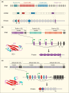
Fig. 1
Fig. 1
Gene organisation and isoform diversity of fly Dscam1, human Pcdhs, and tick sDscam. aDrosophila melanogaster Dscam1 gene structure and mRNA and protein architecture. Mutually exclusive alternative splicing occurs for exon clusters 4, 6, 9, and 17, creating the diversity of the immunoglobulin (Ig) 2, Ig3, Ig7, and transmembrane (TM) domains [3]. The resulting Dscam1 protein has ten Ig domains (circles), six fibronectin type III domains (hexagons), and one TM domain. b Schematic diagram and isoform diversity of the human Pcdh locus. Human Pcdhs are tandemly arrayed in three groups, Pcdhα, Pcdhβ, and Pcdhγ, which have 15, 15, and 22 repeats, respectively, in their 5′ variable regions [2]. Each repeat contains a large exon encoding six extracellular domains, a TM domain, and a partial cytoplasmic region. Each variable repeat is preceded by an alternative promoter. The tertiary structure of the first EC1 domain of Pcdhα has a β-sandwich structure similar to that of the Ig1 domain of sDscam. c Schematic diagram of Ixodes scapularis sDscam gene organisation [9]. Each variable cassette comprises four exons (coloured boxes) encoding the N-terminal Ig1–2 domains (coloured). Each variable cassette is preceded by a promoter. Left, tertiary structure model of the first Ig1 domain
Fig. 2
Fig. 2
Classification of bilaterian Dscams. Circle, immunoglobulin domains; hexagon, fibronectin III domains. Small boxes at the N-terminus, signal peptides; grey and green boxes, TM and cytoplasmic domains, respectively. Five Dscam types (LDscam, mDscam, sDscamα, sDscamβ, and sDscamγ) are classified based on their size and structure. LDscam contains a canonical ectodomain comprising ten Ig domains and six fibronectin type III (FNIII) domains; the tenth Ig domain is located between the fourth and fifth FNIII domains. mDscam lacks the Ig10 and FNIII 3–4 domains of classical Dscam. sDscamα contains variable N-terminal Ig1 domains (blue), which correspond to the variable Ig7 domain of Drosophila Dscam1. sDscamβ contains variable N-terminal Ig1 + 2 domains (coloured), corresponding to the variable Ig7 + 8 domains of Drosophila Dscam1. sDscamγ has domains similar to sDscamα and sDscamβ but without 5′ tandemly arrayed cassettes
Fig. 3
Fig. 3
Metazoan evolution and phylogenetic distribution of Dscams and Pcdhs. Blue and red lines, taxa with Dscams and Pcdhs, respectively; dashed lines and boxes, loss of Dscams and Pcdhs. Mutually exclusive splicing of four exon clusters generates Dscam diversity in all insect and crustacean species investigated [19, 81], while Strigamia uses gene duplications combined with mutually exclusive splicing of the exon 9 cluster. In contrast, Dscam isoforms are predominantly generated by alternative promoters in Chelicerata [9]. Putative numbers of Dscam isoforms are estimated based on the number of Ig7 domains or their orthologues caused by gene and exon duplication. Vertebrates employ alternative promoters to generate Pcdh diversity, while octopus uses tandem gene duplication [13]. The numbers of Pcdh isoforms are estimated by assessing gene and exon duplication
Fig. 4
Fig. 4
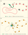
Models of the regulation of Drosophila Dscam1 and vertebrate Pcdh isoforms. a Alternative splicing regulation of Dscam1 pre-mRNA [39, 40, 45, 46]. In this model, pairing of a selector sequence with the docking site brings together the locus control region enhancers and their binding proteins to form a splicing-activation complex. The proximal alternative exon was specifically activated by promoting the recognition of the weak splice site coupled with inactivation of the splicing repressor (repressive mark). b Model for alternative transcription of clustered Pcdhs. This model is based on studies of clustered Pcdh genes [53]. In this model, the targeted promoters were brought into a transcriptional activation complex through CTCF/cohesin-mediated long-range DNA looping
Fig. 5
Fig. 5
Biological functions of fly Dscam1 isoforms. aDrosophila Dscam1 controls self-avoidance in dendrites of da sensory neurons [–71]. The branches of wild-type (WT) neurons exhibit little self-crossing. Deletion of Dscam1 leads to a loss of self-avoidance in all classes. Single-neuron mutant clones exhibit extensive self-crossing and even bundling of dendritic branches. Expression of a Dscam1 isoform or a reduction in their diversity in all da neurons prevents overlap of their dendrites. b Dscam1 isoform diversity mediates the patterning of axonal arborisations [32, 78]. In WT mechanosensory neurons, axonal projections are characterised by an elaborate branch pattern, whose growth cones exhibit an excess of long filopodia-like extensions in various directions. In flies with RNA interference-mediated knockdown of Dscam1, the growth cones exhibited abnormally dense and short filopodia-like extensions. By contrast, mechanosensory neurons with a decreased number of Dscam1 isoforms show retraction of nascent axon branches and reduction of growth cone sprouting, with fewer and shorter extensions

Similar articles

Cited by

References

    1. Sperry RW. Chemoaffinity in the orderly growth of nerve fiber patterns and connections. Proc Natl Acad Sci USA. 1963;50(4):703–710. doi: 10.1073/pnas.50.4.703. - DOI - PMC - PubMed
    1. Wu Q, Maniatis T. A striking organization of a large family of human neural cadherin-like cell adhesion genes. Cell. 1999;97(6):779–790. doi: 10.1016/S0092-8674(00)80789-8. - DOI - PubMed
    1. Schmucker D, Clemens JC, Shu H, Worby CA, Xiao J, Muda M, Dixon JE, Zipursky SL. Drosophila Dscam is an axon guidance receptor exhibiting extraordinary molecular diversity. Cell. 2000;101(6):671–684. doi: 10.1016/S0092-8674(00)80878-8. - DOI - PubMed
    1. Zipursky SL, Sanes JR. Chemoaffinity revisited: dscams, protocadherins, and neural circuit assembly. Cell. 2010;143(3):343–353. doi: 10.1016/j.cell.2010.10.009. - DOI - PubMed
    1. Tasic B, Nabholz CE, Baldwin KK, Kim Y, Rueckert EH, Ribich SA, Cramer P, Wu Q, Axel R, Maniatis T. Promoter choice determines splice site selection in protocadherin alpha and gamma pre-mRNA splicing. Mol Cell. 2002;10(1):21–33. doi: 10.1016/S1097-2765(02)00578-6. - DOI - PubMed

MeSH terms

LinkOut - more resources