Review
. 2019 Apr:198:3-26.
doi: 10.1016/j.biomaterials.2018.09.036. Epub 2018 Oct 1.

Cardiovascular disease models: A game changing paradigm in drug discovery and screening

Affiliations
Review

Cardiovascular disease models: A game changing paradigm in drug discovery and screening

Houman Savoji et al. Biomaterials. 2019 Apr.

Abstract

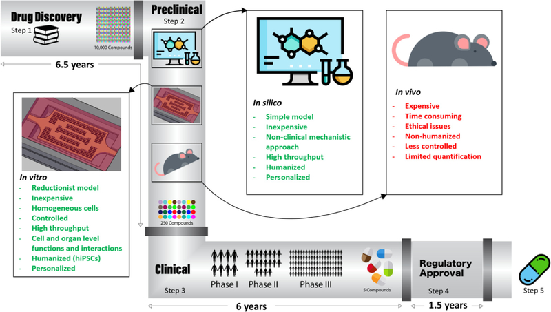
Cardiovascular disease is the leading cause of death worldwide. Although investment in drug discovery and development has been sky-rocketing, the number of approved drugs has been declining. Cardiovascular toxicity due to therapeutic drug use claims the highest incidence and severity of adverse drug reactions in late-stage clinical development. Therefore, to address this issue, new, additional, replacement and combinatorial approaches are needed to fill the gap in effective drug discovery and screening. The motivation for developing accurate, predictive models is twofold: first, to study and discover new treatments for cardiac pathologies which are leading in worldwide morbidity and mortality rates; and second, to screen for adverse drug reactions on the heart, a primary risk in drug development. In addition to in vivo animal models, in vitro and in silico models have been recently proposed to mimic the physiological conditions of heart and vasculature. Here, we describe current in vitro, in vivo, and in silico platforms for modelling healthy and pathological cardiac tissues and their advantages and disadvantages for drug screening and discovery applications. We review the pathophysiology and the underlying pathways of different cardiac diseases, as well as the new tools being developed to facilitate their study. We finally suggest a roadmap for employing these non-animal platforms in assessing drug cardiotoxicity and safety.

Keywords: Cardiomyocyte; Cardiovascular diseases; Drug discovery; Human induced pluripotent stem cells; In silico disease models; In vitro disease models; In vivo disease models; Organ-on-a-chip.


Figures

Fig. 1.
Fig. 1.
Schematic of lipoprotein metabolism and conversion in the body. Dietary lipid and cholesterol, digested from food with the help of cholesterol and bile acids in the intestines, pass through enterocytes and enter into circulation as chylomicrons. The molecules go through a series of lipolysis steps in the peripheral tissue and interact with nascent HDL on their way to the liver. They loose TG and get more cholesterol as they become denser and form remnants. Remnants enter the liver by interacting with LDL receptor-related protein 1 and LDL receptor, being modified to VLDL. VLDL communicates with HDL in circulation by Cholesterylester transfer protein (CETP) enzymatic activity and through lipolysis becomes IDL. Hepatic acid lipase (LIPC) converts IDL to LDL. IDL and LDL can go to the liver or peripheral tissues for further metabolism. In the liver and in circulation, LDL can be modified to form Lp(a). HDL acts as an acceptor of TG in circulation and from peripheral tissues, carrying TG to the liver for further metabolism and excretion. The concept of the figure is adapted from Ref. [39].
Fig. 2.
Fig. 2.
Schematic of the consequences of myocardial infarction in the heart at a cellular level. Infarction restricts blood supply to the surrounding cells, leading to apoptosis and necrosis. After clog removal or reperfusion, surrounding live cells, which are at hypoxic states, start to generate ROS. ROS can trigger another phase of apoptosis and necrosis, as well as inflammation. Apoptotic and necrotic bodies release DAMPS. DAMPS, similar to ROS, are recognized by cells inside the heart (ECs, CMs, fibroblasts and immune cells) through a series of receptors (PRRs) and TLRs, triggering the expression of inflammatory cytokines and adhesion molecules. Enhanced inflammatory state is sensed by the surrounding cells, including CMs, thus leading to further expression of inflammatory molecules.
Fig. 3.
Fig. 3.
Different approaches to fabricate engineered heart/cardiac tissue: A) Organ on chip [250], B) Cardiac patch; scale bar: 2.5 mm [126], C) Circular EHT to apply mechanical stimulation [251], D) Rod-shaped Biowire to apply electrical stimulation; scale bar: 0.5 mm [252]. (Reprinted with permission from Ref. [,–252]).
Fig. 4.
Fig. 4.
Vasculature-on-a-chip (A–C), SEM of lumen networks for A) a 1D tube (scale bar = 1.5 mm and 500 μm), B) a 2D AngioChip scaffold (scale bar = 1 mm and 300 μm) and C) a multi-layer 3D AngioChip scaffold with 20 μm micro-holes (scale bar = 1 mm and 400 μm) created with the 3D stamping technique. SEM of parenchymal spaces of D) an AngioChip scaffold with 10 μm micro-holes on the channel walls (Scale bar = 200 μm); E) the 3D lattice matrix in between the microchannels (scale bar = 100 μm); F) the cross-section of a 10 μm micro-hole on the channel wall (scale bar: 50 μm). Red arrows point to the micro-holes. (G–H) SEM of the parenchymal space of an AngioChip scaffolds with 20 μm micro-holes on the top and side walls of the micro-channels. Red arrows point to the micro-holes on the top and side walls. Scale bar: G) 400μm, and H) 100 μm. I), Schematic of the assembly of the bioreactor showing inlet, main and outlet wells. (Reprinted with permission from Ref. [266]). J) schematic diagram that summarizes the key aspects of the InVADE platform (Reprinted with permission from Ref. [274]).
Fig. 5.
Fig. 5.
Thrombosis on-a-chip. A) Endothelialized perfusable microchannels fabricated with collagen hydrogel to study whole blood interactions with ECs, B) Z-stack confocal image of endothelialized microfluidic vessels showing endothelial sprouting from the walls, Red, CD31; blue, nuclei. (Scale bar: 100 μm), C) Leukocytes and platelet adhesion on stimulated microfluidic vessels after perfusion with of whole blood for 1 h. Red, CD31; green, CD41a; white, CD45; and blue, nuclei. (Scale bar: 100 μm), D) SEM of leukocyte adhesion on and migration through stimulated microfluidic vessels after 1 h of whole blood perfusion (Scale bar: 10 μm) (Reprinted with permission from Ref. [305]), E) Schematic of the bioprinting process: i, ii) bioprinting of a Pluronic mold; iii) assembly of the dried mold on PDMS; iv) filling the mold with GelMA followed by UV crosslinking; v) washing off the sacrificial channels to create vi) the final construct with hollow channels, F) Optical image representing the formed thrombus in a microchannel, where aggregated RBCs were clearly observed (Scale bar: 50 μm), (G) Optical image showing H&E-stained transverse sections of (i) a thrombus in control channel and (ii) a thrombus in endothelialized channel with HUVECs, both after 7 days, and (iii) a thrombus formed in vivo at 7 days (Scale bar: 50 μm), (H) Time-lapse photographs showing the thrombolysis of (i) a 1 day clot and (ii) a 7 day clot (Scale bar: 2 mm) (Reprinted with permission from Ref. [296]).
Fig. 6.
Fig. 6.
Engineered disease models of cardiac fibrosis. A) An engineered cardiac tissue model of fibrotic myocardium based on modulation of CM and myofibroblast volume fractions. The fibrotic EHTs were constructed by encapsulating chicken embryonic cardiomyocytes and myofibroblasts in Type I rat tail collagen. The fibrotic tissue models were generated by replacing cardiomyocytes with myofibroblasts. B) The effects of cellular composition on impulse conduction in this fibrotic model. The CM and myofibroblast volume fractions determined the impulse propagation velocity [309]. C) A simplified 3D hydrogel platform to study cardiac fibrosis. Primary neonatal rat CMs and CFs were encapsulated within a GelMA-based pre-polymer solution to generate in vitro EHTs. Cell-laden hydrogel was placed into a customized UV-chamber and subsequently crosslinked by UV light. The fibrotic EHTs were treated by TGF-β1 [310]. (Reprinted with permission from Ref. [309,310]).
Fig. 7.
Fig. 7.
Drug development pipeline. Step 1: Discovery: Search for a drug candidate starts in the R&D setting; Step 2: Preclinical Research: Drugs undergo in vitro and in vivo testing to address basic concerns about safety; Step 3: Clinical Research: Drugs are evaluated on people in terms of their safety and efficacy; Step 4: FDA Review: Regulatory teams meticulously evaluate all master files related to a new drug or device and decide on their approval; Step 5: FDA Post-Market Safety Monitoring: FDA monitors all device and drug safety once products are marketed.

References

    1. Laverty H, Benson C, Cartwright E, Cross M, Garland C, Hammond T, Holloway C, McMahon N, Milligan J, Park B, How can we improve our understanding of cardiovascular safety liabilities to develop safer medicines? Br. J. Pharmacol. 163 (4) (2011) 675–693. - PMC - PubMed
    1. Ross JS, Krumholz HM, Bringing Vioxx Back to Market, British Medical Journal Publishing Group, 2018. - PubMed
    1. Waxman HA, The lessons of Vioxx—drug safety and sales, N. Engl. J. Med 352 (25) (2005) 2576–2578. - PubMed
    1. Onakpoya IJ, Heneghan CJ, Aronson JK, Worldwide withdrawal of medicinal products because of adverse drug reactions: a systematic review and analysis, Crit. Rev. Toxicol 46 (6) (2016) 477–489. - PubMed
    1. Shah RR, Can Pharmacogenetics Help Rescue Drugs Withdrawn from the Market? (2006). - PubMed

Publication types

MeSH terms