. 2018 Nov 13;11(5):1040-1050.
doi: 10.1016/j.stemcr.2018.09.008. Epub 2018 Oct 18.

The SCRIB Paralog LANO/LRRC1 Regulates Breast Cancer Stem Cell Fate through WNT/β-Catenin Signaling

Affiliations

The SCRIB Paralog LANO/LRRC1 Regulates Breast Cancer Stem Cell Fate through WNT/β-Catenin Signaling

Leonor Lopez Almeida et al. Stem Cell Reports. .

Abstract

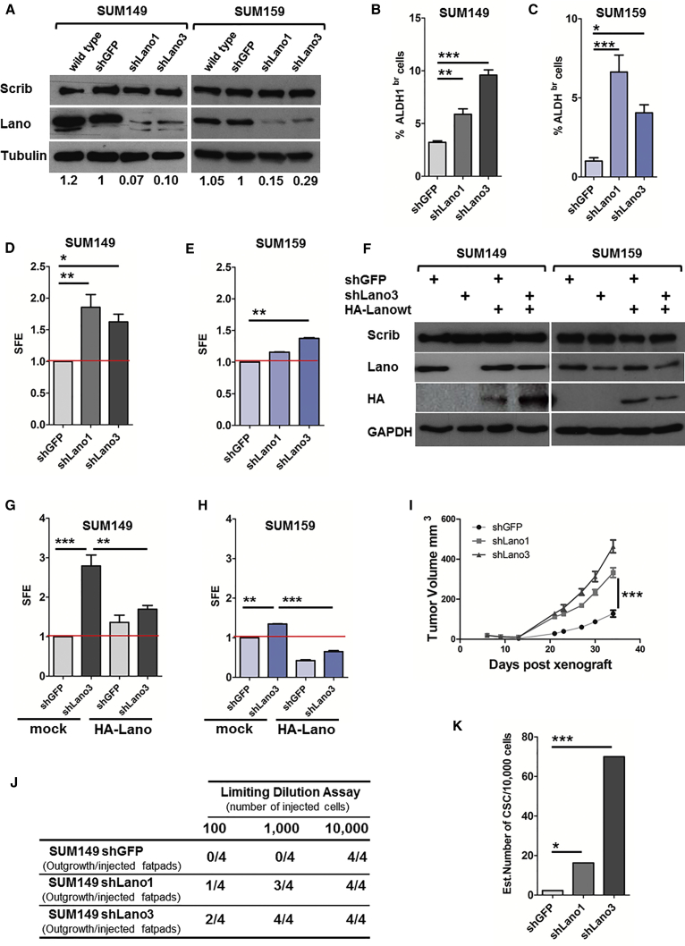
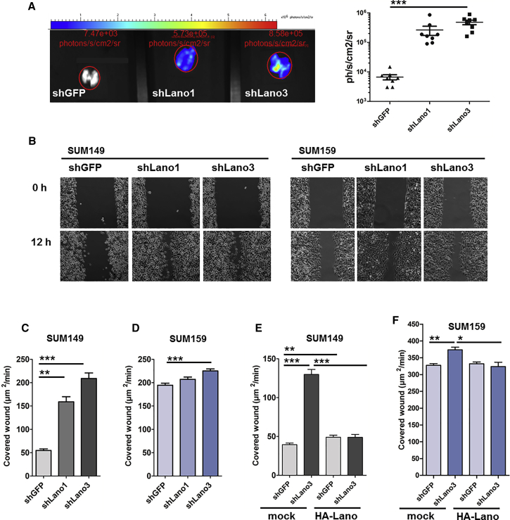
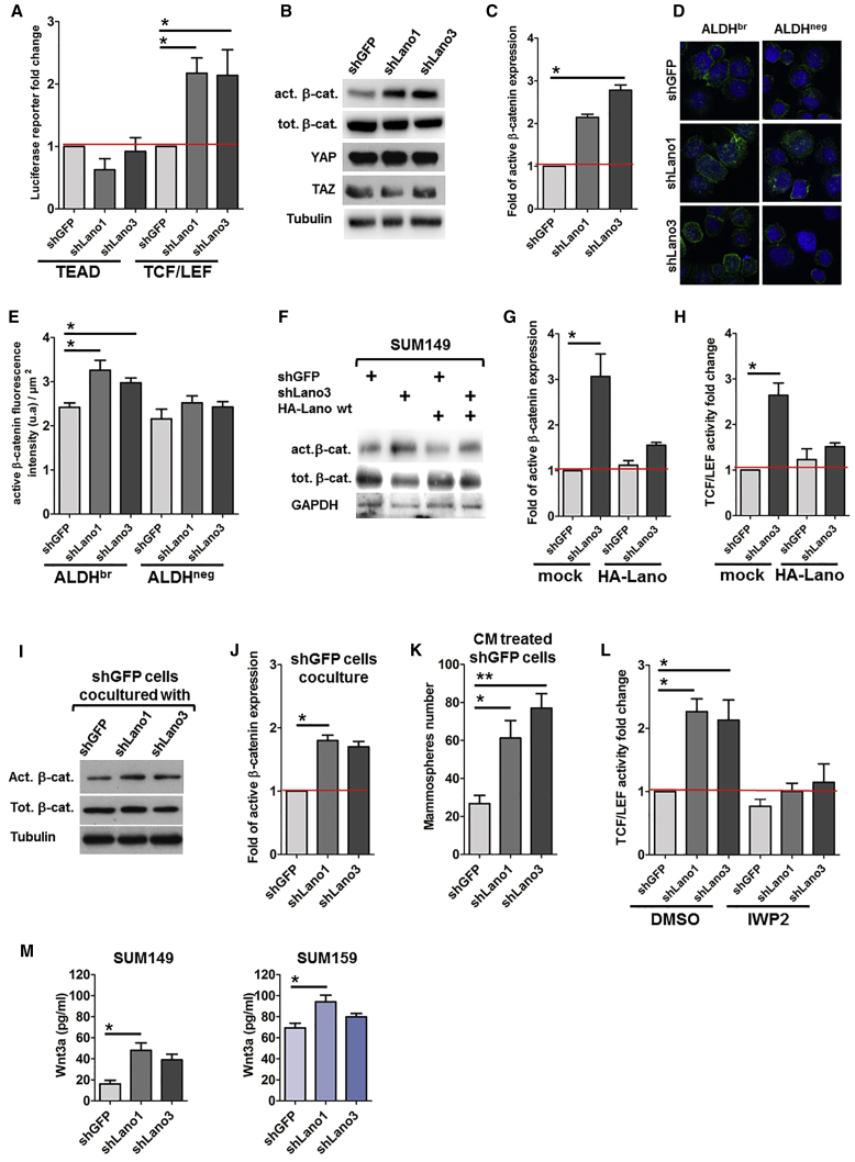
Tumor initiation, progression, and therapeutic resistance have been proposed to originate from a subset of tumor cells, cancer stem cells (CSCs). However, the current understanding of the mechanisms involved in their self-renewal and tumor initiation capacity remains limited. Here, we report that expression of LANO/LRRC1, the vertebrate paralog of SCRIB tumor suppressor, is associated with a stem cell signature in normal and tumoral mammary epithelia. Through in vitro and in vivo experiments including a Lano/Lrrc1 knockout mouse model, we demonstrate its involvement in the regulation of breast CSC (bCSC) fate. Mechanistically, we demonstrate that Lano/LRRC1-depleted cells secrete increased levels of WNT ligands, which act in a paracrine manner to positively deregulate the WNT/β-catenin pathway in bCSCs. In addition to describing the first function of LANO/LRRC1, our results suggest that its expression level could be used as a biomarker to stratify breast cancer patients who could benefit from WNT/β-catenin signaling inhibitors.

Keywords: LANO/LRRC1; SCRIB; Wnt/β-catenin; breast cancer; stem cell; tumor suppressor.


Figures

Figure 1
Figure 1
SCRIB and LANO Expression in Human and Murine Mammary Gland (A) Scrib and Lano immunofluorescence staining in normal human mammary gland sections with the indicated antibodies. Scale bar, 5 μm. (B) Localization profiles of Lano and Scrib (right panel) along the red line are depicted in the left panel. (C–F) (C and D) Expression levels of SCRIB and LANO/LRRC1 in human normal breast reported as a box plot. MaSC, mammary stem cell subsets; LP, luminal progenitors; mL, mature luminal cells. Statistical analysis was performed using one-way ANOVA with Tukey’s post test. p < 0.05, ∗∗p < 0.01, ∗∗∗p < 0.001. Pubescent 6-week-old mouse inguinal mammary glands of wild-type (wt) (n = 2) or Lano knockout mice (n = 4) were analyzed (E) by immunoblot for Scrib and Lano protein expression, with α-tubulin as loading control, and (F) by Mayer's hemalum whole-mount staining to visualize the epithelial tree. Lymph node (LN) is used as a marker (left panel). Quantifications of percentage of relative duct area (middle panel) and TEBs number (right panel) are shown. (G) Bar plots represent significance of univariate linear regression analysis of LANO/LRRC1 (blue) and SCRIB (red) with stem cell and stroma signatures. Each bar score was defined as the log-transformed p value (−log10) and weighted by direction of association for analysis. Thus, at 5% risk, a score above 1.3 or under −1.3 was considered significant. ALDges, ALDH gene expression signature; MS-like, mammosphere-like; PLAU, plasminogen activator urokinase.
Figure 2
Figure 2
LANO Downregulation Increases Stemness Properties of bCSC Models In Vitro and In Vivo (A) Immunoblot analysis of shGFP, shLano1, and shLano3 cell populations probed with the antibodies indicated. Quantification of Lano expression is indicated below. (B and C) Percentage of ALDHbr cells for (B) SUM149 and (C) SUM159 (n = 3 for both). (D and E) Sphere-forming efficiency (SFE) compared with the control of cell populations expressed as fold, for SUM149 (D; n = 3) and SUM159 (E; n = 4). (F–H) SFE of SUM149 (G) and SUM159 (H) cells defined in (F) expressed as fold (n = 3). (I) Kinetics of tumor growth of SUM149 cells of each population orthotopically xenografted in NSG mice. (J and K) Table (J) showing the number of outgrowths generated as a function of the amount of injected cells (K). bCSC frequencies were calculated using an extreme limiting dilution analysis algorithm (shLano1 versus shGFP, p = 0.0235; shLano3 versus shGFP, p = 0.0002). Results are expressed as mean ± SD, n for independent experiment, statistical significance using Kruskal-Wallis ANOVA with Dunnett's post test. p < 0.05, ∗∗p < 0.01, ∗∗∗p < 0.001.
Figure 3
Figure 3
LANO Knockdown Contributes to Metastatic Spreading (A) Representative microphotographs of metastatic lungs of xenografted mice described in Figure 2I, and chemoluminescence measurements by PhotonIMAGER in rainbow scale and related graph plotted. (B) Representative images of wound-healing experiments for the shGFP, shLano1, and shLano3 SUM149 and SUM159 cell populations. (C and D) Speed of wound closure represented in μm2/min for SUM149 (C) and SUM159 (D) (n = 4). (E and F) Same as in (C) and (D) for cells expressing, or not, HA-tagged wild-type human Lano (n = 3). Results are expressed as mean ± SD, n for independent experiment, statistical significance using Kruskal-Wallis ANOVA with Dunnett's post test. p < 0.05, ∗∗p < 0.01, ∗∗∗p < 0.001.
Figure 4
Figure 4
LANO Represses Wnt Ligand Secretion (A) Fold induction of TEAD and TCF/LEF transcriptional activity by luciferase reporter assays (n = 6). (B) Immunoblot analysis for the indicated antibodies. (C) Fold induction of active β-catenin (act-β-cat) quantified by immunoblot (n = 3) related to shGFP. (D) Representative microphotographs of SUM149 cell populations sorted for either ALDHbr or ALDHneg status cytospinned and immunolabeled for active β-catenin antibody (green) and nucleus (blue). (E) Quantification as fluorescent mean intensity of active β-catenin normalized by cell area (n = 2). (F–H) (F) and (G) as in (B) and (C) for Lano rescued cell populations, and (H) their use in TCF/LEF transcriptional activity assay (n = 5). (I and J) shGFP SUM149 cells were co-cultured with indicated cell population growing in Transwell insert prior immunoblot for active β-catenin analysis (I) and quantified as fold related to shGFP (J) (n = 3). (K) Overnight cultured shGFP SUM149 cells were treated with the indicated conditioned medium (CM) prior to mammosphere formation assay (n = 3). (L) TCF/LEF transcriptional assay, same as in (A) for cells treated for 15 hr with DMSO (vehicle) or 20 μM IWP2 prior analysis (n = 4). (M) Amount of WNT3a in culture medium measured by ELISA (n = 5). Results are expressed as mean ± SD, n for independent experiment, statistical significance using Kruskal-Wallis ANOVA with Dunnett's post test. p < 0.05, ∗∗p < 0.01.

References

    1. Charafe-Jauffret E., Ginestier C., Iovino F., Wicinski J., Cervera N., Finetti P., Hur M.H., Diebel M.E., Monville F., Dutcher J. Breast cancer cell lines contain functional cancer stem cells with metastatic capacity and a distinct molecular signature. Cancer Res. 2009;69:1302–1313. - PMC - PubMed
    1. Cordenonsi M., Zanconato F., Azzolin L., Forcato M., Rosato A., Frasson C., Inui M., Montagner M., Parenti A.R., Poletti A. The Hippo transducer TAZ confers cancer stem cell-related traits on breast cancer cells. Cell. 2011;147:759–772. - PubMed
    1. Creighton C.J., Li X., Landis M., Dixon J.M., Neumeister V.M., Sjolund A., Rimm D.L., Wong H., Rodriguez A., Herschkowitz J.I. Residual breast cancers after conventional therapy display mesenchymal as well as tumor-initiating features. Proc. Natl. Acad. Sci. U S A. 2009;106:13820–13825. - PMC - PubMed
    1. Dow L.E., Kauffman J.S., Caddy J., Zarbalis K., Peterson A.S., Jane S.M., Russell S.M., Humbert P.O. The tumour-suppressor Scribble dictates cell polarity during directed epithelial migration: regulation of Rho GTPase recruitment to the leading edge. Oncogene. 2007;26:2272–2282. - PubMed
    1. El Helou R., Pinna G., Cabaud O., Wicinski J., Bhajun R., Guyon L., Rioualen C., Finetti P., Gros A., Mari B. miR-600 acts as a bimodal switch that regulates breast cancer stem cell fate through WNT signaling. Cell Rep. 2017;18:2256–2268. - PubMed

Publication types

MeSH terms