. 2018 Dec 5;100(5):1180-1193.e6.
doi: 10.1016/j.neuron.2018.09.049. Epub 2018 Oct 18.

Chd2 Is Necessary for Neural Circuit Development and Long-Term Memory

Affiliations

Chd2 Is Necessary for Neural Circuit Development and Long-Term Memory

Young J Kim et al. Neuron. .

Abstract

Considerable evidence suggests loss-of-function mutations in the chromatin remodeler CHD2 contribute to a broad spectrum of human neurodevelopmental disorders. However, it is unknown how CHD2 mutations lead to impaired brain function. Here we report mice with heterozygous mutations in Chd2 exhibit deficits in neuron proliferation and a shift in neuronal excitability that included divergent changes in excitatory and inhibitory synaptic function. Further in vivo experiments show that Chd2+/- mice displayed aberrant cortical rhythmogenesis and severe deficits in long-term memory, consistent with phenotypes observed in humans. We identified broad, age-dependent transcriptional changes in Chd2+/- mice, including alterations in neurogenesis, synaptic transmission, and disease-related genes. Deficits in interneuron density and memory caused by Chd2+/- were reproduced by Chd2 mutation restricted to a subset of inhibitory neurons and corrected by interneuron transplantation. Our results provide initial insight into how Chd2 haploinsufficiency leads to aberrant cortical network function and impaired memory.

Keywords: MGE transplantation; autism; epigenetics; epilepsy; hippocampus; intellectual disability; interneuron; synaptic transmission.


Conflict of interest statement

Declaration of Interests

The authors declare no competing interests.

Figures

Figure 1.
Figure 1.. Generation of Chd2+/− mice.
A. At P30, CHD2 (green) co-labeled with NEUN, GAD67 and OLIG2, but not GFAP (all in magenta). B. Quantification of CHD2 expression in each cell type (n=3 mice per marker). C. Schematic of the conditional allele for Chd2 (tm1c). Cre deletes the floxed exon 3 of Chd2 to generate a frame shift mutation (tm1d). D, E. Western blot analysis for CHD2 and Actin showed reduction of CHD2 protein in Chd2+/− brain at P30 (P= 0.027, n=3 mice per genotype). F. Mean body weight of male and female Chd2+/− mice was reduced compared to WT littermates (n=17–23 mice per genotype). G. Immunostaining for BRN2 (magenta), CTIP2 (green) and DAPI in somatosensory cortex (left panels) and hippocampus (right panels) at P30. H. Thickness of individual cell layers in somatosensory cortex (SS Ctx) was not altered by Chd2+/− (n = 4 mice per genotype). I. Width of granule cell layer (GCL) or CA1 pyramidal cell layer (CA1) was not altered by Chd2+/− (n = 4 mice per genotype). Error bars, s.e.m.; ∗ p < 0.05. ∗∗ p < 0.01; scale bars, 75 μm in A and 150 μm in G. See also Figure S1.
Figure 2.
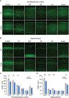
Figure 2.. Chd2+/− mice exhibit decreased density of GABAergic interneurons.
A, B. Immunostaining of coronal sections through somatosensory neocortex (A) and CA1 region of hippocampus (B) for GAD67, PV, SST, CR, VIP and reelin at P30. C, D. Quantification of each subtype marker shows Chd2+/− mice had decreased density of cells expressing GAD67, PV and SST in somatosensory cortex (C) and decreased density of cells expressing GAD67 PV, SST and reeling in hippocampus (D) compared to WT littermates (n=3–6 mice per genotype). Error bars, s.e.m.; ∗ p < 0.05. ∗∗ p < 0.01; scale bars, 150 μm in A and B. See also Figure S2 and S3.
Figure 3.
Figure 3.. Chd2 regulates proliferation of neural progenitors in developing forebrain.
A. Immunostaining for CHD2 (magenta) and Ki67, Pax6, Tbr2, Tbr1 and GAD67-GFP (all green) in neocortex at E14.5. B. Immunostaining for CHD2 (red), Nkx2.1 (blue) and GAD67-GFP (green) in ventral telencephalon shows CHD2 expression in GABAergic progenitor domains at E14.5. C. At E14.5, CHD2 (magenta) co-labeled with Ki67 and Nkx2.1, but not GAD67-GFP (all in green) in MGE. Lower magnification images of dorsal and ventral telencephalon are shown in Figure S4. D, E. At E14.5, the density of Ki67+ cells were reduced in MGE and cortex of Chd2+/− mice (n=3–5 mice per genotype). F, G. At E14.5, the density of Nkx2.1+ GABA progenitors were reduced in MGE of Chd2+/− mice (n=5–6 mice per genotype). H, I. At E14.5, the density of GAD67-GFP progenitors were reduced in cortex of Chd2+/− mice (n=4–6 mice per genotype). J-L. At P7, immunostaining analysis revealed a reduction in GAD67-GFP progenitors in somatosensory neocortex, but the density of cells expressing caspase-3 (CASP3) was not different between genotypes (n=5 mice per genotype). Error bars, s.e.m.; ∗ p < 0.05; ∗∗ p < 0.01; scale bars, 10 μm in A and C, 200 μm in B, and 20 μm in D, F, H, J. See also Figure S4.
Figure 4.
Figure 4.. Chd2 haploinsufficiency disrupts genes necessary for early cortical development and synaptic function.
A. Schematic showing the experimental approach used for RNA-sequencing. B. Volcano plot displaying genes that were significantly differentially expressed (Adj. P < 0.1) in Chd2+/− mice (n=3 mice per genotype). C. qRT-PCR validation of DE genes predicted by RNA-sequencing. D. Gene ontology for down-regulated genes at P45. Complete gene ontology is provided in Table S3. E. Plot of z-score by -log10 of the adjusted P-value for human diseases identified by disease ontology. The complete disease ontology is provided in Table S5. F. Heatmaps of genes associated with childhood epilepsy, intellectual disability and autism spectrum disorder that were differentially expressed in E13.5 MGE, E13.5 Neocortex and/or P45 Hippocampus.
Figure 5.
Figure 5.. Changes in neuronal excitability and synaptic function in Chd2+/− mice.
A. Voltage responses to hyperpolarizing (−80 pA) and depolarizing (+140 pA) current pulses in CA1 pyramidal neurons from a WT (black) and Chd2+/− mouse (blue). B. Plot of action potential firing frequency (Hz) as a function of current intensity shows increased firing in pyramidal neurons of Chd2+/− mice. C. Summary current–voltage plot shows Rinput was not different between groups. Intrinsic electrophysiological properties are summarized in Table S6. D. Miniature EPSCs (mEPSCs) recorded from pyramidal neurons in a WT (black) and Chd2+/− mouse (blue). E, F. At P30–35, mEPSC frequency was unchanged (E), but mEPSC amplitudes were increased (F) in pyramidal neurons of Chd2+/− mice. G. Averaged mEPSCs recorded from a pyramidal neuron in a WT (black) and Chd2+/− mouse (blue). H, I. At P30–35, mEPSC 10–90% rise time (RT) was unchanged (H), but mEPSC decay time constant was decreased (I) in pyramidal neurons of Chd2+/− mice. J. Miniature IPSCs (mIPSCs) recorded from pyramidal neurons in a WT (black) and Chd2+/− mouse (blue). K, L. At P30–35, mIPSC frequency was decreased (K), but mIPSC amplitudes were unchanged (L) in pyramidal neurons of Chd2+/− mice. M. Averaged mIPSCs recorded from a pyramidal neuron in a WT (black) and Chd2+/− mouse (blue). N, O. At P30–35, mIPSC 10–90% rise time (RT) and decay time constant (tau) were not different between genotypes. Error bars, s.e.m.; ∗ p < 0.05; ∗∗ p < 0.01. See also Figure S5.
Figure 6.
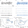
Figure 6.. Abnormal rhythmogenesis in Chd2+/− mice.
A, B. Example of each frequency band isolated from the local field potential (LFP) in a WT (A) and Chd2+/− mouse (B). C. Normalized EEG power spectra. Inset shows mean power for each frequency band (n=5 mice per genotype). D. Cross-cortical coherence across the EEG power spectra. Inset shows mean coherence for each frequency band (n=5 mice per genotype). Error bars, s.e.m.; ∗ p < 0.05; ∗∗ p < 0.01.
Figure 7.
Figure 7.. Chd2+/− mice exhibit deficits in long-term memory.
A. Heat map showing the location of WT and Chd2+/− littermates during the entire training and testing phases of the Object Location Memory (OLM) assay. B, C. Discrimination index during training and testing phases of OLM (n=7 WT mice and n=6 Chd2+/− mice). D. Heat map showing the location of WT and Chd2+/− littermates during the entire training and testing phases of the Object Recognition Memory (ORM) assay. E, F. Discrimination index during training and testing phases of ORM (n=9 WT mice and n=7 Chd2+/ mice). Error bars, s.e.m.; ∗∗ p < 0.01. See also Figure S6 and 7.
Figure 8.
Figure 8.. MGE transplantation rescues hippocampal memory problems in Chd2+/− mice.
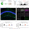
A. Schematic showing the experimental approach used for MGE transplantation. B. Volcano plot of differentially expressed genes between E13.5 MGE and neocortex (n = 3 mice per genotype). C. Hippocampus of a Chd2+/− mouse (45 DAT) labeled for NEUN (blue) and transplanted GFP-labeled inhibitory neurons (green). D At 45 DAT, GFP-labeled cells (green) co-expressed PV and SST, but did not express VIP (all in magenta). E, F. Discrimination index during training and testing phases of OLM (E) and ORM (F) assays shows MGE transplantation rescues spatial memory deficits in Chd2+/− mice (n=5–7 mice per treatment group). Arrowheads, co-labeled cells; error bars, s.e.m.; ∗∗ p < 0.01; scale bars, 100 µm (C) and 50 µm (D). See also Figure S8.

Comment in

  • CHD2: One Gene, Many Roles.
    Nieto-Estevez V, Hsieh J. Nieto-Estevez V, et al. Neuron. 2018 Dec 5;100(5):1014-1016. doi: 10.1016/j.neuron.2018.11.036. Neuron. 2018. PMID: 30521773

References

    1. Atallah BV, Scanziani M (2009) Instaneous modulation of gamma oscillation frequency by balancing excitation with inhibition. Neuron 62, 566–577. - PMC - PubMed
    1. Buckmaster PS (2012) Mossy fiber sprouting in the dentate gyrus. Jasper’s Basic Mechanisms of the Epilepsies 4th edn Eds: Noebels JL, Avoli M, Rogawski MA, Olsen RW, Delgado-Escueta AV (National Center for Biotechnology, Bethesda Maryland: ). - PubMed
    1. Cain-Hom C, Splinter E, Min MV, Simonis M, van de Heigning M, Martinez M, Asghari V, Cox JC, Warmin S (2017) Efficient mapping of transgene integration sites and local structural changes in Cre transgenic mice using targeted locus amplification. Nuc Acid Res 45, e62. - PMC - PubMed
    1. Carlén M, Meletis K, Siegle JH, Cardin JA, Futai K, Vierling-Claassen D, Rühlmann C, Jones SR, Deisseroth K, Sheng M, Moore CI, Tsai LH (2012) A critical role for NMDA receptors in parvalbumin interneurons for gamma rhythm induction and behavior. Mol. Psychiatry 17, 537–548. - PMC - PubMed
    1. Carvill GL, Heavin SB, Yendle SC, McMahon JM, O’Roak BJ, Cook J, Khan A, Dorschner MO, Weaver M, Calvert S, Malone S, Wallace G, Stanley T, Bye AM, Bleasel A, Howell KB, Kivity S, Mackay MT, Rodriguez-Casero V, Webster R, Korczyn A, Afawi Z, Zelnick N, Lerman-Sagie T, Lev D, Møller RS, Gill D, Andrade DM, Freeman JL, S adleir LG, Shendure J, Berkovic SF, Scheffer IE, Mefford H (2013) Targeted resequencing in epileptic encephalopathies identifies de novo mutations in CHD2 and SYNGAP1. Nat Genet 45, 825–830. - PMC - PubMed

Publication types

MeSH terms