

Endoscopic Sclerotherapy with a Large Volume of High Concentration of Cyanoacrylate for Jejunal Variceal Bleeding bys Single-Balloon Enteroscopy
- PMID: 30344299
- PMCID: PMC6262278
- DOI: 10.3390/medicina54050068
Endoscopic Sclerotherapy with a Large Volume of High Concentration of Cyanoacrylate for Jejunal Variceal Bleeding bys Single-Balloon Enteroscopy
Abstract
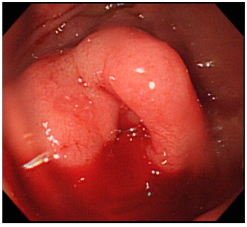
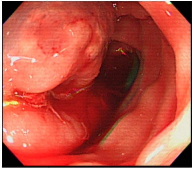
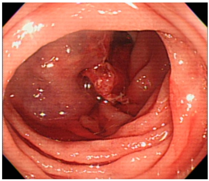
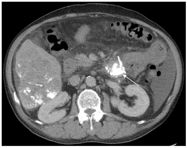
Jejunal varices are a rare manifestation of portal hypertension, and they are associated with a high mortality and poor prognosis when bleeding occurs. A bleeding jejunal varix is much more challenging to diagnose and manage because of its anatomic location. Herein, we describe the case of a 62-year-old man with active jejunal variceal bleeding who presented with massive hematochezia and hypovolemic shock. He was treated successfully with a high volume and concentration of a glue mixture as endoscopic sclerotherapy using single-balloon enteroscopy in the intensive care unit. Enteroscopic sclerotherapy is an effective option for jejunal variceal bleeding.
Keywords: jejunal varices; sclerotherapy; single-balloon enteroscopy.
Conflict of interest statement
The authors declare no conflicts of interest.
Figures
References
-
- Lebrec D., Benhamou J.P. Ectopic varices in portal hypertension. Clin. Gastroenterol. 1985;14:105–121. - PubMed
-
- Watanabe N., Toyonaga A., Kojima S., Takashimizu S., Oho K., Kokubu S., Nakamura K., Hasumi A., Murashima N., Tajiri T. Current status of ectopic varices in Japan: Results of a survey by the Japan Society for Portal Hypertension. Hepatol. Res. 2010;40:763–776. doi: 10.1111/j.1872-034X.2010.00690.x. - DOI - PubMed
Publication types
MeSH terms
Substances
LinkOut - more resources
Full Text Sources
Medical
