

Interplay Between Phosphorylation and O-GlcNAcylation of Sarcomeric Proteins in Ischemic Heart Failure
- PMID: 30344511
- PMCID: PMC6182077
- DOI: 10.3389/fendo.2018.00598
Interplay Between Phosphorylation and O-GlcNAcylation of Sarcomeric Proteins in Ischemic Heart Failure
Abstract
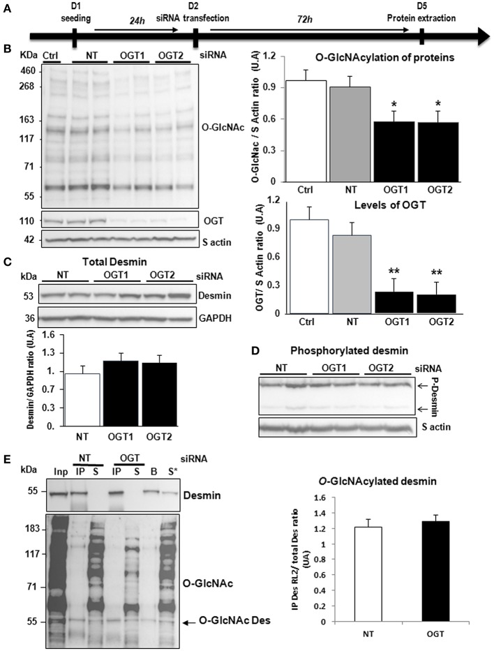
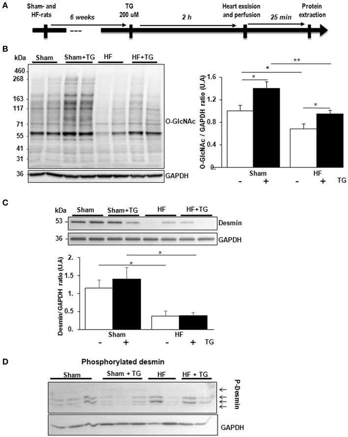
Post-translational modifications (PTMs) of sarcomeric proteins could participate to left ventricular (LV) remodeling and contractile dysfunction leading in advanced heart failure (HF) with altered ejection fraction. Using an experimental rat model of HF (ligation of left coronary artery) and phosphoproteomic analysis, we identified an increase of desmin phosphorylation and a decrease of desmin O-N-acetylglucosaminylation (O-GlcNAcylation). We aim to characterize interplay between phosphorylation and O-GlcNAcylation for desmin in primary cultures of cardiomyocyte by specific O-GlcNAcase (OGA) inhibition with thiamet G and silencing O-GlcNAc transferase (OGT) and, in perfused heart perfused with thiamet G in sham- and HF-rats. In each model, we found an efficiency of O-GlcNAcylation modulation characterized by the levels of O-GlcNAcylated proteins and OGT expression (for silencing experiments in cells). In perfused heart, we found an improvement of cardiac function under OGA inhibition. But none of the treatments either in in vitro or ex vivo cardiac models, induced a modulation of desmin, phosphorylated and O-GlcNAcylated desmin expression, despite the presence of O-GlcNAc moities in cardiac desmin. Our data suggests no interplay between phosphorylation and O-GlcNAcylation of desmin in HF post-myocardial infarction. The future requires finding the targets in heart involved in cardiac improvement under thiamet G treatment.
Keywords: O-GlcNAcylation; desmin; heart failure; interplay; phosphorylation; rat models; systolic.
Figures


Similar articles
-
Interplay between troponin T phosphorylation and O-N-acetylglucosaminylation in ischaemic heart failure.Cardiovasc Res. 2015 Jul 1;107(1):56-65. doi: 10.1093/cvr/cvv136. Epub 2015 Apr 26. Cardiovasc Res. 2015. PMID: 25916824
-
E2f1 deletion attenuates infarct-induced ventricular remodeling without affecting O-GlcNAcylation.Basic Res Cardiol. 2019 May 31;114(4):28. doi: 10.1007/s00395-019-0737-y. Basic Res Cardiol. 2019. PMID: 31152247 Free PMC article.
-
O-GlcNAcylation is a key modulator of skeletal muscle sarcomeric morphometry associated to modulation of protein-protein interactions.Biochim Biophys Acta. 2016 Sep;1860(9):2017-30. doi: 10.1016/j.bbagen.2016.06.011. Epub 2016 Jun 11. Biochim Biophys Acta. 2016. PMID: 27301331
-
O-GlcNAcylation, contractile protein modifications and calcium affinity in skeletal muscle.Front Physiol. 2014 Oct 30;5:421. doi: 10.3389/fphys.2014.00421. eCollection 2014. Front Physiol. 2014. PMID: 25400587 Free PMC article. Review.
-
Roles of ten-eleven translocation family proteins and their O-linked β-N-acetylglucosaminylated forms in cancer development.Oncol Lett. 2021 Jan;21(1):1. doi: 10.3892/ol.2020.12262. Epub 2020 Nov 3. Oncol Lett. 2021. PMID: 33240407 Free PMC article. Review.
Cited by
-
Desmin aggrephagy in rat and human ischemic heart failure through PKCζ and GSK3β as upstream signaling pathways.Cell Death Discov. 2021 Jun 26;7(1):153. doi: 10.1038/s41420-021-00549-2. Cell Death Discov. 2021. PMID: 34226534 Free PMC article.
-
Increased O-GlcNAcylation by Upregulation of Mitochondrial O-GlcNAc Transferase (mOGT) Inhibits the Activity of Respiratory Chain Complexes and Controls Cellular Bioenergetics.Cancers (Basel). 2024 Mar 5;16(5):1048. doi: 10.3390/cancers16051048. Cancers (Basel). 2024. PMID: 38473405 Free PMC article.
-
Global O-GlcNAcylation changes impact desmin phosphorylation and its partition toward cytoskeleton in C2C12 skeletal muscle cells differentiated into myotubes.Sci Rep. 2022 Jun 14;12(1):9831. doi: 10.1038/s41598-022-14033-z. Sci Rep. 2022. PMID: 35701470 Free PMC article.
-
The dual role of the hexosamine biosynthetic pathway in cardiac physiology and pathophysiology.Front Endocrinol (Lausanne). 2022 Oct 24;13:984342. doi: 10.3389/fendo.2022.984342. eCollection 2022. Front Endocrinol (Lausanne). 2022. PMID: 36353238 Free PMC article. Review.
-
O-GlcNAcylation: the "stress and nutrition receptor" in cell stress response.Cell Stress Chaperones. 2021 Mar;26(2):297-309. doi: 10.1007/s12192-020-01177-y. Epub 2020 Nov 7. Cell Stress Chaperones. 2021. PMID: 33159661 Free PMC article. Review.
References
-
- Savoye C, Equine O, Tricot O, Nugue O, Segrestin B, Sautière K, et al. . Left ventricular remodeling after anterior wall acute myocardial infarction in modern clinical practice (from the REmodelage VEntriculaire [REVE] Study Group). Am J Cardiol. (2006) 98:1144–9. 10.1016/j.amjcard.2006.06.011 - DOI - PubMed
-
- Fertin M, Hennache B, Hamon M, Ennezat PV, Biausque F, Elkohen M, et al. . Usefulness of serial assessment of B-type natriuretic peptide, troponin I, and C-reactive protein to predict left ventricular remodeling after acute myocardial infarction (from the REVE-2 Study). Am J Cardiol. (2010) 106:1410–6. 10.1016/j.amjcard.2010.06.071 - DOI - PubMed
LinkOut - more resources
Full Text Sources
Molecular Biology Databases
Research Materials
Miscellaneous