

A Subset of CXCR5+CD8+ T Cells in the Germinal Centers From Human Tonsils and Lymph Nodes Help B Cells Produce Immunoglobulins
- PMID: 30344522
- PMCID: PMC6183281
- DOI: 10.3389/fimmu.2018.02287
A Subset of CXCR5+CD8+ T Cells in the Germinal Centers From Human Tonsils and Lymph Nodes Help B Cells Produce Immunoglobulins
Abstract
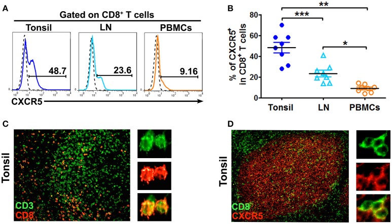
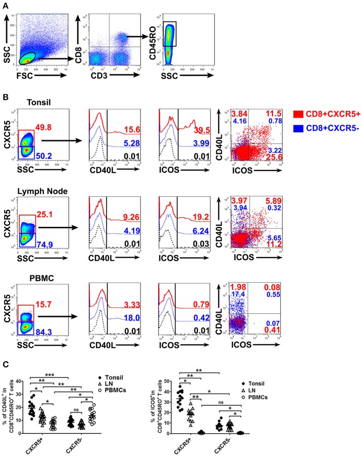
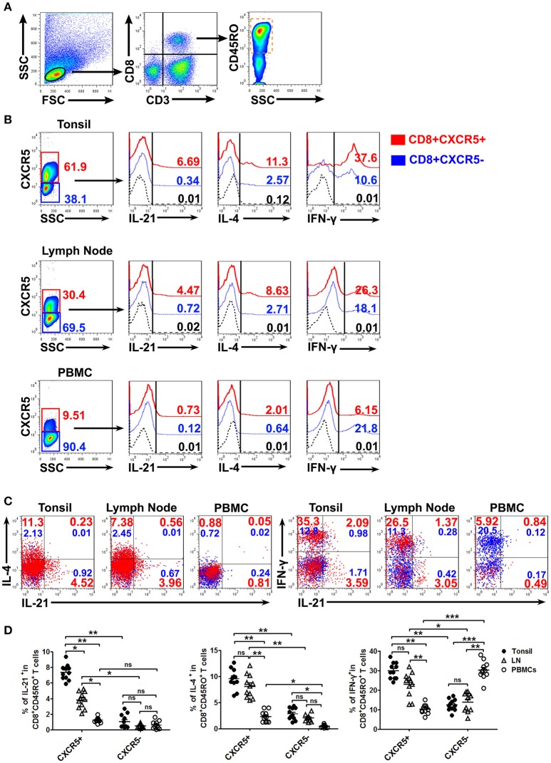
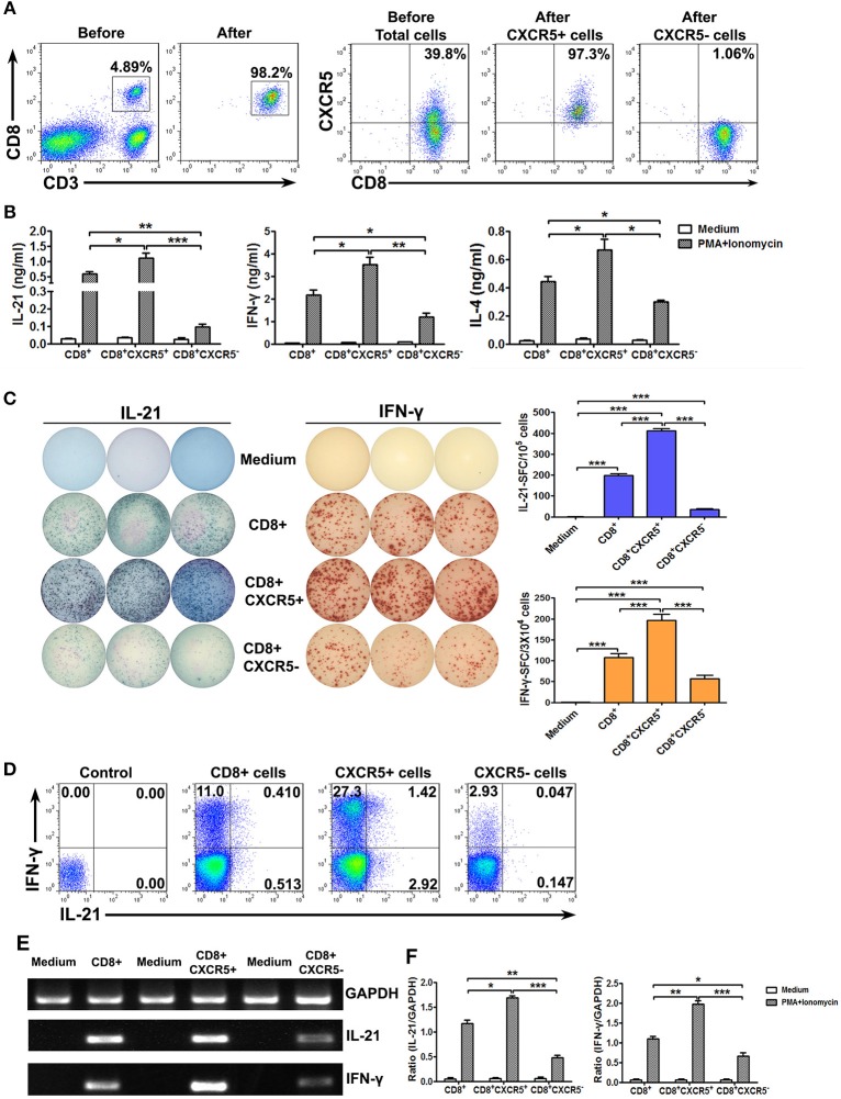
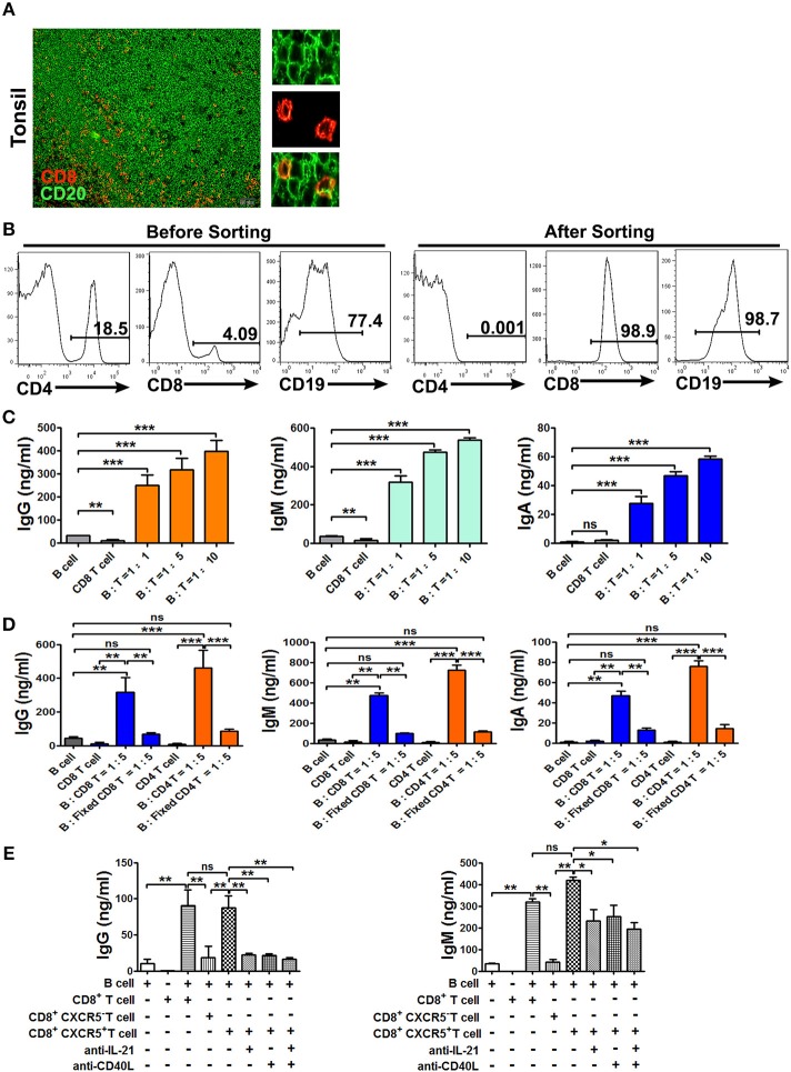
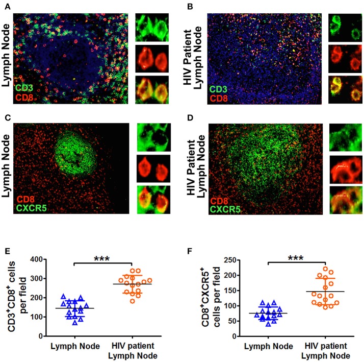
Recent studies indicated that CXCR5+CD8+ T cells in lymph nodes could eradicate virus-infected target cells. However, in the current study we found that a subset of CXCR5+CD8+ T cells in the germinal centers from human tonsils or lymph nodes are predominately memory cells that express CD45RO and CD27. The involvement of CXCR5+CD8+ T cells in humoral immune responses is suggested by their localization in B cell follicles and by the concomitant expression of costimulatory molecules, including CD40L and ICOS after activation. In addition, CXCR5+CD8+ memory T cells produced significantly higher levels of IL-21, IFN-γ, and IL-4 at mRNA and protein levels compared to CXCR5-CD8+ memory T cells, but IL-21-expressing CXCR5+CD8+ T cells did not express Granzyme B and perforin. When cocultured with sorted B cells, sorted CXCR5+CD8+ T cells promoted the production of antibodies compared to sorted CXCR5-CD8+ T cells. However, fixed CD8+ T cells failed to help B cells and the neutralyzing antibodies against IL-21 or CD40L inhibited the promoting effects of sorted CXCR5+CD8+ T cells on B cells for the production of antibodies. Finally, we found that in the germinal centers of lymph nodes from HIV-infected patients contained more CXCR5+CD8+ T cells compared to normal lymph nodes. Due to their versatile functional capacities, CXCR5+CD8+ T cells are promising candidate cells for immune therapies, particularly when CD4+ T cell help are limited.
Keywords: B cell; C-X-C chemokine receptor type 5; CD8 T cell; Follicular; Tfh-like cell.
Figures
Similar articles
-
CXCR5+ CCR7- CD8 T cells are early effector memory cells that infiltrate tonsil B cell follicles.Eur J Immunol. 2007 Dec;37(12):3352-62. doi: 10.1002/eji.200636746. Eur J Immunol. 2007. PMID: 18000950
-
CD25(+) Bcl6(low) T follicular helper cells provide help to maturing B cells in germinal centers of human tonsil.Eur J Immunol. 2015 Jan;45(1):298-308. doi: 10.1002/eji.201444911. Epub 2014 Oct 27. Eur J Immunol. 2015. PMID: 25263533 Free PMC article.
-
CXC chemokine receptor 5 expression defines follicular homing T cells with B cell helper function.J Exp Med. 2000 Dec 4;192(11):1553-62. doi: 10.1084/jem.192.11.1553. J Exp Med. 2000. PMID: 11104798 Free PMC article.
-
Human T follicular helper cells: development and subsets.Adv Exp Med Biol. 2013;785:87-94. doi: 10.1007/978-1-4614-6217-0_10. Adv Exp Med Biol. 2013. PMID: 23456841 Review.
-
T cell immune response within B-cell follicles.Adv Immunol. 2019;144:155-171. doi: 10.1016/bs.ai.2019.08.008. Epub 2019 Sep 16. Adv Immunol. 2019. PMID: 31699216 Review.
Cited by
-
CXCR4, CXCR5 and CD44 May Be Involved in Homing of Lymphoma Cells into the Eye in a Patient Derived Xenograft Homing Mouse Model for Primary Vitreoretinal Lymphoma.Int J Mol Sci. 2022 Oct 4;23(19):11757. doi: 10.3390/ijms231911757. Int J Mol Sci. 2022. PMID: 36233057 Free PMC article.
-
CD8 T-cell subsets: heterogeneity, functions, and therapeutic potential.Exp Mol Med. 2023 Nov;55(11):2287-2299. doi: 10.1038/s12276-023-01105-x. Epub 2023 Nov 1. Exp Mol Med. 2023. PMID: 37907738 Free PMC article. Review.
-
Complex human adenoid tissue-based ex vivo culture systems reveal anti-inflammatory drug effects on germinal center T and B cells.EBioMedicine. 2020 Mar;53:102684. doi: 10.1016/j.ebiom.2020.102684. Epub 2020 Feb 27. EBioMedicine. 2020. PMID: 32114393 Free PMC article.
-
Memory T Cells in Pregnancy.Front Immunol. 2019 Apr 2;10:625. doi: 10.3389/fimmu.2019.00625. eCollection 2019. Front Immunol. 2019. PMID: 31001255 Free PMC article. Review.
-
B Cell-mediated Humoral Immunity in Chronic Hepatitis B Infection.J Clin Transl Hepatol. 2021 Aug 28;9(4):592-597. doi: 10.14218/JCTH.2021.00051. Epub 2021 May 27. J Clin Transl Hepatol. 2021. PMID: 34447690 Free PMC article. Review.
References
Publication types
MeSH terms
Substances
LinkOut - more resources
Full Text Sources
Research Materials