. 2018 Oct 5:9:2314.
doi: 10.3389/fimmu.2018.02314. eCollection 2018.

Regulatory NLRs Control the RLR-Mediated Type I Interferon and Inflammatory Responses in Human Dendritic Cells

Affiliations

Regulatory NLRs Control the RLR-Mediated Type I Interferon and Inflammatory Responses in Human Dendritic Cells

Tünde Fekete et al. Front Immunol. .

Abstract

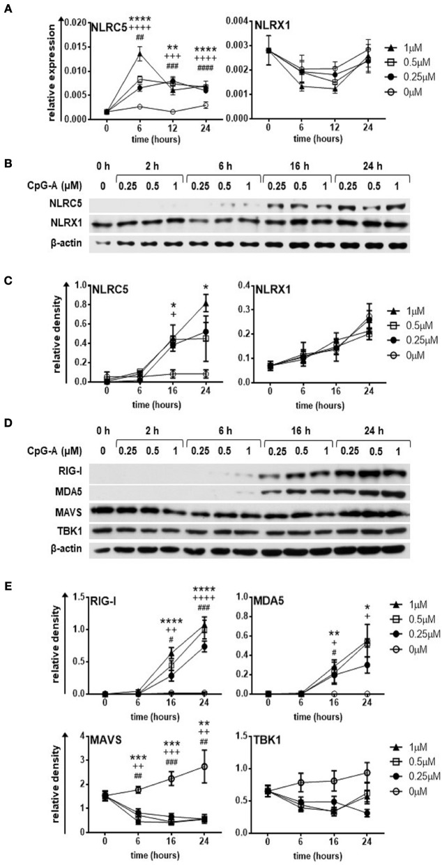
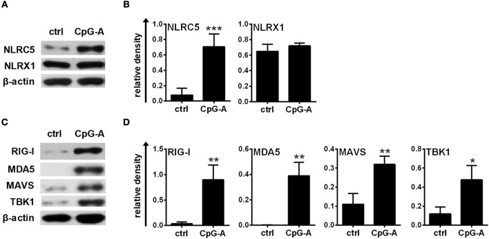
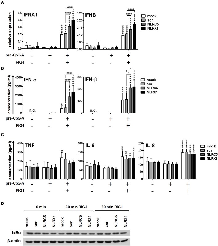
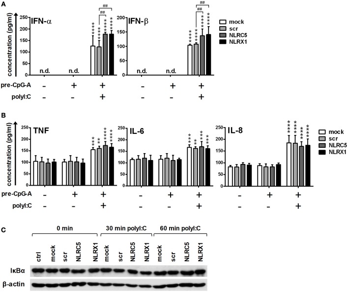
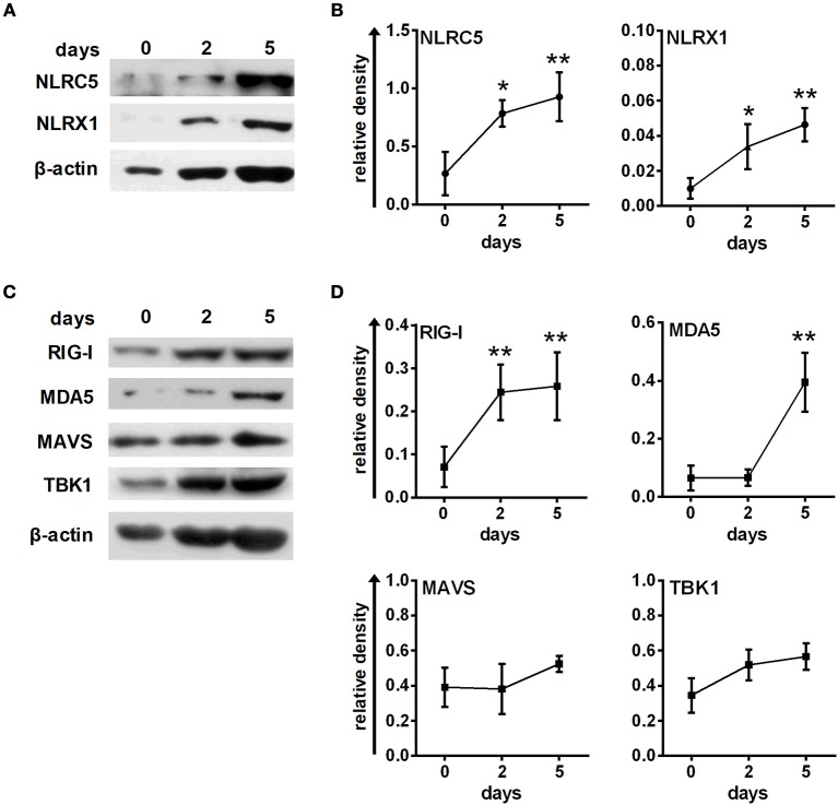
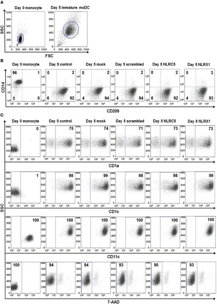
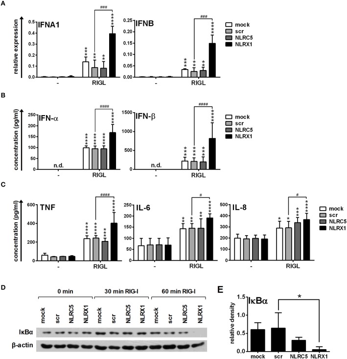
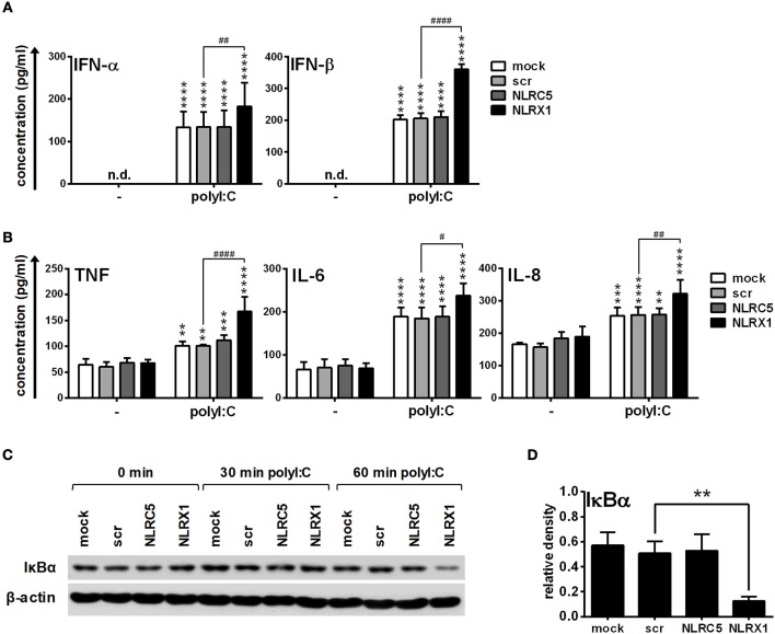
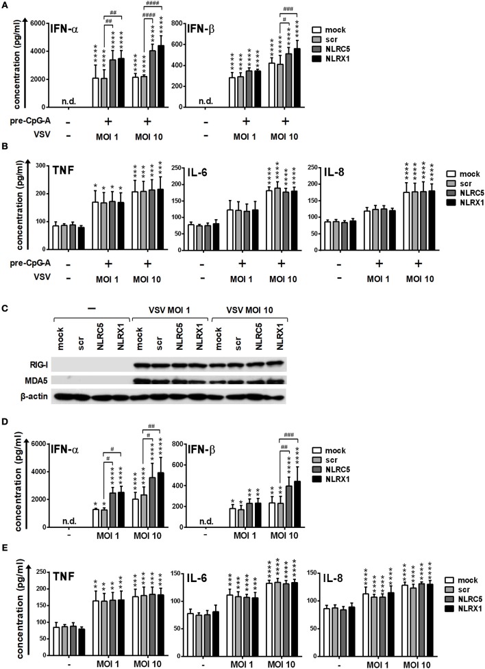
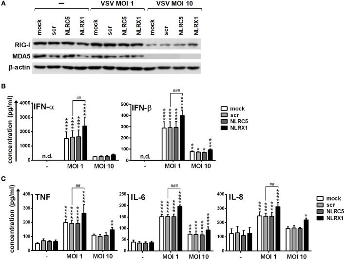
Unique members of the nucleotide-binding domain leucine-rich repeat (NLR) family have been found to regulate intracellular signaling pathways initiated by other families of pattern recognition receptors (PRR) such as Toll-like receptors (TLRs) and retinoic-acid inducible gene I (RIG-I)-like receptors (RLRs). Plasmacytoid dendritic cells (pDCs), the most powerful type I interferon (IFN) producing cells, preferentially employ endosomal TLRs to elicit antiviral IFN responses. By contrast, conventional DCs (cDCs) predominantly use cytosolic RLRs, which are constitutively expressed in them, to sense foreign nucleic acids. Previously we have reported that, though RIG-I is absent from resting pDCs, it is inducible upon TLR stimulation. In the recent study we investigated the regulatory ability of NLRs, namely NLRC5 and NLRX1 directly associated with the RLR-mediated signaling pathway in DC subtypes showing different RLR expression, particularly in pDCs, and monocyte-derived DCs (moDCs). Here we demonstrate that similarly to RLRs, NLRC5 is also inducible upon TLR9 stimulation, whereas NLRX1 is constitutively expressed in pDCs. Inhibition of NLRC5 and NLRX1 expression in pDCs augmented the RLR-stimulated expression of type I IFNs but did not affect the production of the pro-inflammatory cytokines TNF, IL-6, and the chemokine IL-8. Further we show that immature moDCs constantly express RLRs, NLRX1 and NLRC5 that are gradually upregulated during their differentiation. Similarly to pDCs, NLRX1 suppression increased the RLR-induced production of type I IFNs in moDCs. Interestingly, RLR stimulation of NLRX1-silenced moDCs leads to a significant increase in pro-inflammatory cytokine production and IκBα degradation, suggesting increased NF-κB activity. On the contrary, NLRC5 does not seem to have any effect on the RLR-mediated cytokine responses in moDCs. In summary, our results indicate that NLRX1 negatively regulates the RLR-mediated type I IFN production both in pDCs and moDCs. Further we show that NLRX1 inhibits pro-inflammatory cytokine secretion in moDCs but not in pDCs following RLR stimulation. Interestingly, NLRC5 suppresses the RLR-induced type I IFN secretion in pDCs but does not appear to have any regulatory function on the RLR pathway in moDCs. Collectively, our work demonstrates that RLR-mediated innate immune responses are primarily regulated by NLRX1 and partly controlled by NLRC5 in human DCs.

Keywords: NLR; RLR; antiviral; dendritic cell; interferon; regulate.


Figures

Figure 1
Figure 1
The expression of NLRC5, RIG-I and MDA5 but not that of NLRX1 is upregulated by CpG-A treatment in the human GEN2.2 pDC cell line. (A–E) GEN2.2 cells were treated with increasing concentration of CpG-A (0.25–1 μM) in a time dependent manner. The expression of NLRC5 and NLRX1 was measured at the mRNA level by Q-PCR (A) and at the protein level by western blotting (B,C). The changes in protein levels of RIG-I, MDA5, MAVS, and TBK1 were also analyzed after CpG-A treatments by western blotting (D,E). Representative blots are shown in (B,D). Data are shown as mean ± SD from 4 to 6 independent experiments in panels (A,C,E). Data were analyzed using one-way ANOVA followed by Bonferroni's post-hoc test. *p < 0.05, **p < 0.01, ***p < 0.001, ****p < 0.0001 1 μM CpG-A vs control; +p < 0.05, ++p < 0.01, +++p < 0.001, ++++p < 0.0001 0.5 μM CpG-A vs control; #p < 0.05, ##p < 0.01, ###p < 0.001, ####p < 0.0001 0.25 μM CpG-A vs control.
Figure 2
Figure 2
The expression of NLRC5, RIG-I, and MDA5 is inducible in primary human pDCs after CpG-A treatments. (A–D) Freshly isolated primary human pDCs were stimulated with 2.5 μM CpG-A for 16 h, thereafter the protein levels of NLRC5, NLRX1, RIG-I, MDA5, MAVS, and TBK1 were detected by western blotting. Representative blots are shown in (A,C). Data are shown as mean ± SD from 3 to 4 experiments in panels (B,D). (B,D) Statistical comparisons were performed using Student's t-test. *p < 0.05, **p < 0.01, ***p < 0.001.
Figure 3
Figure 3
The specific RIG-I agonist-induced type I IFN production is upregulated by NLRC5 or NLRX1 silencing while the NF-κB signaling pathway is not affected in GEN2.2 cells. (A–D) Cells were transfected with siRNAs specific for NLRC5, NLRX1 or scrambled (scr) siRNAs for 24 h then pre-treated with 0.25 μM CpG-A (pre-CpG-A) for 16 h to induce the cytosolic expression of RLRs. Following thorough washing steps cells were stimulated with the specific RIG-I agonist 5′ppp-dsRNA (RIGL, 1 μg/ml). The IFNA1 and IFNB mRNA expression levels were assessed by real-time PCR after 3 h (A) and IFN-α, IFN-β (B), TNF, IL-6, and IL-8 (C) protein levels were measured by ELISA after 6 (B) or 24 h (C). (D) Kinetics of IκBα degradation was determined by western blotting. (D) A representative blot is shown. (A–C) Data are represented as means ± SD of 3-5 individual experiments and analyzed using one-way ANOVA followed by Bonferroni's post-hoc test. *p < 0.05, **p < 0.01, ***p < 0.001, ****p < 0.0001 vs. pre-CpG-A-treated samples; #p < 0.05, ####p < 0.0001, n.d., not determined.
Figure 4
Figure 4
The RIG-I/MDA5 agonist-induced type I IFN production is upregulated by NLRC5 or NLRX1 silencing while the NF-κB signaling pathway is not affected in GEN2.2 cells. (A–C) Cells were transfected with siRNAs specific for NLRC5, NLRX1 or scrambled (scr) siRNAs for 24 h then pre-treated with 0.25 μM CpG-A (pre-CpG-A) for 16 h to induce the cytosolic expression of RLRs. Following thorough washing steps cells were stimulated with the RIG-I/MDA5 agonist polyI:C (1 μg/ml). The protein levels of IFN-α, IFN-β (A), TNF, IL-6, and IL-8 (B) were measured by ELISA after 6 (A) or 24 h (B). (C) Kinetics of IκBα degradation was determined by western blotting. (C) A representative blot is shown. Data are represented as means ± SD of 4 individual experiments and analyzed using one-way ANOVA followed by Bonferroni's post-hoc test. **p < 0.01, ***p < 0.0001, ****p < 0.0001 vs. pre-CpG-A-treated samples; ##p < 0.01, n.d., not determined.
Figure 5
Figure 5
Human immature moDCs constantly express NLRC5, NLRX1, RIG-I and MDA5. (A–D) Freshly isolated monocytes were seeded in 24-well plates and differentiated as described in the “Materials and Methods.” The protein levels of NLRC5, NLRX1, RIG-I, MDA5, MAVS, and TBK1 were measured by western blot. (A,C) Representative blots are shown. (B,D) Graphs represent the kinetics of protein expressions during moDC differentiation. Data are represented as mean ± SD of 3–5 individual experiments and were analyzed using one-way ANOVA followed by Bonferroni's post-hoc test. *p < 0.05, **p < 0.01 vs. day 0.
Figure 6
Figure 6
The silencing of NLRC5 or NLRX1 does not influence the differentiation process of human immature moDCs. (A–C) Freshly isolated monocytes were transfected with siRNAs specific for NLRC5, NLRX1, or scrambled (scr) siRNAs at day 0 and differentiated into immature moDCs. On day 5 of differentiation the phenotypic analysis of the cells were perfomed by flow cytometry. Cells were gated on forward vs. side scatter to exclude debris (A) and the expression levels of CD14 and CD209 (B) as well as CD1a, CD1c, and CD11c cell surface proteins and cell viability (C) were analyzed. (A–C) Representative dot blots are shown from 3 individual experiments. (B, C) Isotype controls antibodies were used to set gates for positive events and numbers indicate the percentage of positive cells. In case of 7AAD staining the numbers show the ratio of 7-AAD negative live cells.
Figure 7
Figure 7
NLRX1 but not NLRC5 affects the specific RIG-I agonist-induced type I IFN and pro-inflammatory responses in human moDCs. (A–E) moDCs transfected with the indicated siRNAs were stimulated with the RIG-I ligand 5′ppp-dsRNA (RIGL, 1 μg/ml). The mRNA expression levels of IFNA1 and IFNB were assessed by real-time PCR after 12 h (A) and IFN-α, IFN-β (B), TNF, IL-6, and IL-8 (C) protein levels were measured by ELISA after 24 h. (D,E) Kinetics of IκBα degradation was determined by western blotting. (D) A representative blot is shown. (E) Bar graphs show the relative density of IκBα measured at 60 min of stimulation. (A-C, E) Data are shown as mean ± SD from 4 independent experiments and analyzed using one-way ANOVA followed by Bonferroni's post-hoc test. *p < 0.05, **p < 0.01, ***p < 0.01 ****p < 0.0001 vs. untreated; #p < 0.05, ###p < 0.001, ####p < 0.0001, n.d., not determined.
Figure 8
Figure 8
NLRX1 but not NLRC5 controls the RIG-I/MDA5 agonist-induced type I IFN and pro-inflammatory responses in human moDCs. (A–D) moDCs transfected with the indicated siRNAs were stimulated with the RIG-I/MDA5 ligand polyI:C (1 μg/ml). The protein levels of IFN-α, IFN-β (A), TNF, IL-6, and IL-8 (B) were detected by ELISA after 24 h. (C,D) Kinetics of IκBα degradation was determined by western blotting. (C) A representative blot is shown. (D) Bar graphs show the relative density of IκBα measured at 60 min of stimulation. (A,B,D) Data are shown as mean ± SD from 4 independent experiments and analyzed using one-way ANOVA followed by Bonferroni's post-hoc test. **p < 0.01, ***p < 0.01 ****p < 0.0001 vs. untreated; #p < 0.05, ##p < 0.01, ####p < 0.0001, n.d., not determined.
Figure 9
Figure 9
Depletion of NLRC5 or NLRX1 enhances the type I IFN production of GEN2.2 cells but does not influence the NF-κB pathway activity in response to VSV infection. (A–E) Cells were transfected with siRNAs specific for NLRC5, NLRX1 or scrambled (scr) siRNAs for 24 h. (A,B) After silencing cells were pre-treated with 0.25 μM CpG-A (pre-CpG-A) for 16 h to induce the cytosolic expression of RLRs. Following thorough washing steps cells were infected with VSV at the indicated MOIs. The protein levels of IFN-α, IFN-β (A), TNF, IL-6, and IL-8 (B) were measured by ELISA after 18 h. (C–E) After silencing GEN2.2 cells were exposed to VSV at the indicated MOIs without CpG-A pre-treatment and the protein levels of RIG-I and MDA5 were detected by western blot at 24 h (C). Concentrations of IFN-α, IFN-β (D), TNF, IL-6, and IL-8 (E) were measured by ELISA from the supernatant of the VSV-infected cells. (C) A representative blot is shown. Data are represented as means ± SD of 4 individual experiments and analyzed using one-way ANOVA followed by Bonferroni's post-hoc test. *p < 0.05, **p < 0.01, ***p < 0.0001, ****p < 0.0001 vs. untreated; #p < 0.05, ##p < 0.01, ###p < 0.001, ####p < 0.0001, n.d., not determined.
Figure 10
Figure 10
NLRX1 but not NLRC5 affects the type I IFN and pro-inflammatory responses in VSV-infected human moDCs. (A–C) moDCs transfected with the indicated siRNAs were exposed to VSV at the indicated MOIs and after 18 h the protein levels of RIG-I and MDA5 (A) were analyzed by western blotting, and the concentrations of secreted IFN-α, IFN-β (B), TNF, IL-6, and IL-8 (C) were determined by ELISA. (A) A representative blot is shown. (B,C) Data are shown as mean ± SD from 4 independent experiments and analyzed using one-way ANOVA followed by Bonferroni's post-hoc test. *p < 0.05, **p < 0.01, ***p < 0.01 ****p < 0.0001 vs. untreated; ##p < 0.01, ###p < 0.001, n.d., not determined.

Similar articles

Cited by

References

    1. Thompson MR, Kaminski JJ, Kurt-Jones EA, Fitzgerald KA. Pattern recognition receptors and the innate immune response to viral infection. Viruses (2011) 3:920–40. 10.3390/v3060920 - DOI - PMC - PubMed
    1. Geginat J, Nizzoli G, Paroni M, Maglie S, Larghi P, Pascolo S, et al. . Immunity to Pathogens Taught by Specialized Human Dendritic Cell Subsets. Front Immunol. (2015) 6:527. 10.3389/fimmu.2015.00527 - DOI - PMC - PubMed
    1. Oviedo-Boyso J, Bravo-Patino A, Baizabal-Aguirre VM. Collaborative action of Toll-like and NOD-like receptors as modulators of the inflammatory response to pathogenic bacteria. Mediators Inflamm. (2014) 2014:432785. 10.1155/2014/432785 - DOI - PMC - PubMed
    1. Coutermarsh-Ott S, Eden K, Allen IC. Beyond the inflammasome: regulatory NOD-like receptor modulation of the host immune response following virus exposure. J Gen Virol. (2016) 97:825–38. 10.1099/jgv.0.000401 - DOI - PMC - PubMed
    1. Kawai T, Akira S. Toll-like receptors and their crosstalk with other innate receptors in infection and immunity. Immunity (2011) 34:637–50. 10.1016/j.immuni.2011.05.006 - DOI - PubMed

Publication types

LinkOut - more resources