. 2018 Oct;52(5):394-397.
doi: 10.1007/s13139-018-0536-7. Epub 2018 Aug 13.

Langerhans' Cell Histiocytosis Masquerading as Metastatic Papillary Thyroid Cancer on F-18 FDG PET/CT: Diagnostic Dilemma Solved by PET/CT-Guided Biopsy

Affiliations

Langerhans' Cell Histiocytosis Masquerading as Metastatic Papillary Thyroid Cancer on F-18 FDG PET/CT: Diagnostic Dilemma Solved by PET/CT-Guided Biopsy

Dharmender Malik et al. Nucl Med Mol Imaging. 2018 Oct.

Abstract

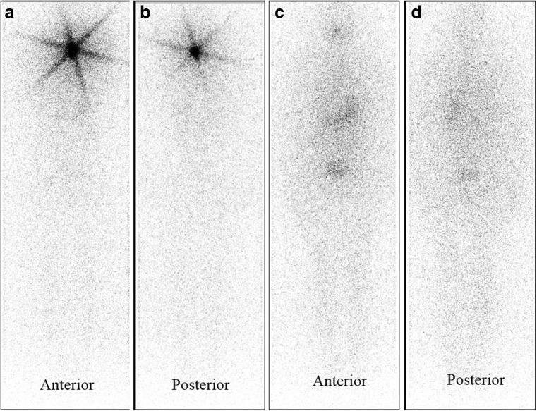
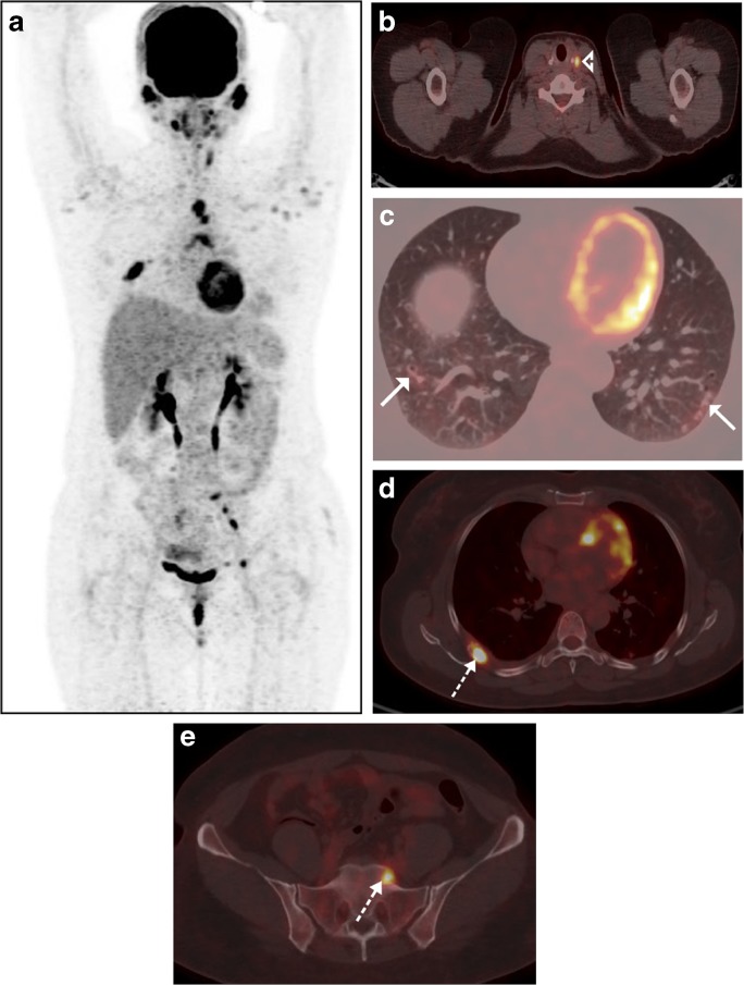
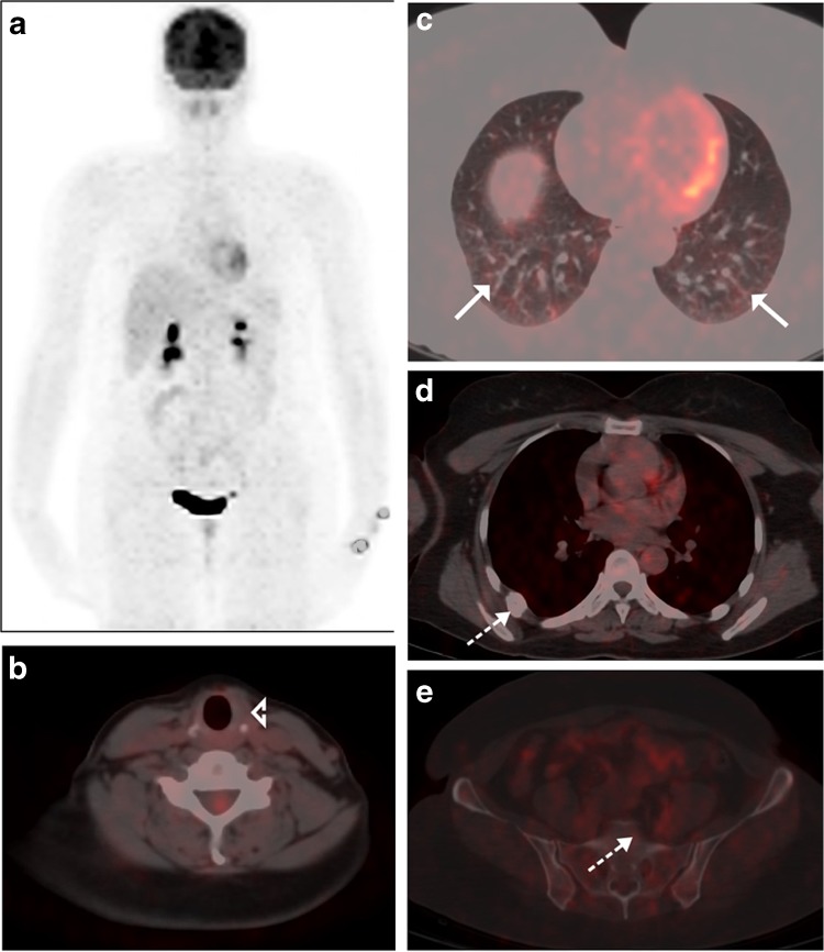
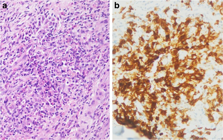
We present a case of papillary thyroid cancer (post-thyroidectomy status) on regular treatment with suppressive Levothyroxine therapy. On follow-up at 6 months after radioactive iodine ablation for remnant thyroid tissue, her thyroglobulin, and anti-thyroglobulin levels were 0.06 ng/ml and 670 IU/ml, respectively. Low-dose whole-body I-131 scan was negative. To look for the cause of isolated increased anti-thyroglobulin level, a whole-body 18F-FDG PET/CT was done which revealed multiple FDG-avid lytic skeletal lesions suggestive of metastases. For confirmation of diagnosis, 18F-FDG PET/CT-guided metabolic biopsy was done, which revealed Langerhans' cell histiocytosis on histopathological examination.

Keywords: 18F-FDG PET/CT; Anti-thyroglobulin level; Langerhans’ cell histiocytosis; PET/CT-guided biopsy; Papillary thyroid cancer.


Conflict of interest statement

Dharmender Malik, Rajender Kumar Basher, Bhagwant Rai Mittal, Gaurav Parkash, Amanjit Bal, and Anish Bhattacharya declare that they have no conflict of interest.All procedures performed in studies involving human participants were in accordance with the ethical standards of the institutional and/or national research committee and with the 1964 Helsinki declaration and its later amendments or comparable ethical standards. For this type of study formal consent is not required.The institutional review board of our institute approved this retrospective study, and the requirement to obtain informed consent was waived.

Figures

Fig. 1
Fig. 1
Forty-five-year-old female with left thyroid swelling (~ 5 cm) underwent total thyroidectomy and nodal exploration. Histopathology revealed papillary thyroid cancer (PTC) with no nodal involvement (classical variant, T3pNoMo). Post-therapy scan (a, b) acquired after 72 h of 50 mCi of I-131 iodine administration revealed remnant thyroid tissue in the thyroid bed. She was kept on follow-up with suppressive dose of levothyroxine therapy. On follow-up visit at 6 months, after levothyroxine withdrawal (serum TSH level 119 mIU/l), she had thyroglobulin level of 0.06 ng/ml but elevated anti-thyroglobulin antibody (anti-Tg Ab) titer (670 IU/ml; normal < 116) and a negative low-dose whole-body I-131 scan (c, d). 18F-FDG PET/CT was done with a suspicion of I-131-negative residual disease [1]
Fig. 2
Fig. 2
18F-FDG PET/CT maximum-intensity projection (MIP, a) revealed FDG-avid subcentimetric soft tissue lesion in thyroid bed (arrowhead, b), mediastinal lymph nodes, faintly FDG-avid subcentimetric nodules (some showing cavitation) in the lower lobe of both lung fields (arrow, c). Transaxial PET/CT images revealed FDG-avid lytic-expansile lesion in the right 6th rib (dotted arrow, d) and sacrum (dotted arrow, e) suggesting metastasis. 18F-FDG PET/CT-guided biopsy was performed from the right 6th rib lesion which revealed Langerhans’ cells histiocytosis (LCH; positive CD1a antigen on immunostaining). Reanalysis of initial thyroid pathology was consistent with concurrent LCH and PTC within the thyroid. The patient was started on chemotherapy for LCH
Fig. 3
Fig. 3
Follow-up 18F-FDG PET/CT (MIP, a) done after four cycles of chemotherapy revealed resolution of FDG-avid lesion in thyroid bed (b), lung nodules (c), and skeletal lesions (d, e) with appearance of sclerosis in right 6th rib suggesting favorable response to chemotherapy
Fig. 4
Fig. 4
Photomicrograph of bone biopsy specimen (a) showing sheets of Langerhans’ cells admixed with plasma cells and eosinophils (H&E, × 200), and CD1a immunostaining highlighting Langerhans’ cells (b). PTC accounts for more than 85 to 90% of all thyroid malignancies [2, 3]. It is seen more commonly in females between 3rd to 5th decades. At initial diagnosis, 1–3% has distant metastases while 7–23% will develop distant metastases during the follow-up period [–7]. Although, the distant metastases are quite uncommon in PTC, but lung and bone are the most common sites [8]. The BRAF V600E mutation is most common genetic mutation detected in patients with PTC and occurs in approximately 45% of patients [9]. BRAF V600E mutation is also found to be associated with LCH in ~ 50% of patients [10]. LCH in adults is a rare disorder occurring in 1–2 adults per million people with male predominance (2:1). It involves the bone, lung, skin, lymph nodes, and multiple sites and very rarely thyroid. Coexistence of LCH and PTC has been described in a few isolated case reports only; however, its significance is still unknown [11, 12]. LCH of the thyroid can both masquerade as a malignancy or coexist with other thyroid neoplasm because of the sharing of some common cytomorphologic features such as grooved nuclei, with PTC, which makes the pathological diagnosis even more difficult and may have increased anti-Tg Ab levels [13, 14]. The present case highlights the need of histopathology for distant skeletal lesions with increased anti-Tg Ab titer and negative iodine scintigraphy in PTC patients

References

    1. Qiu ZL, Wei WJ, Shen CT, Song HJ, Zhang XY, Sun ZK, et al. Diagnostic performance of 18F-FDG PET/CT in papillary thyroid carcinoma with negative 131I-WBS at first post-ablation, negative Tg and progressively increased TgAb level. Sci Rep. 2017;7:2849. doi: 10.1038/s41598-017-03001-7. - DOI - PMC - PubMed
    1. Fagin JA, Mitsiades N. Molecular pathology of thyroid cancer: diagnostic and clinical implications. Best Pract Res Clin Endocrinol Metab. 2008;22:955–969. doi: 10.1016/j.beem.2008.09.017. - DOI - PMC - PubMed
    1. Davies L, Welch HG. Increasing incidence of thyroid cancer in the United States, 1973-2002. JAMA. 2006;295:2164–2167. doi: 10.1001/jama.295.18.2164. - DOI - PubMed
    1. Mihailovic J, Stefanovic L, Malesevic M. Differentiated thyroid carcinoma with distant metastases: the probability of survival and its predicting factors. Cancer Biother Radiopharm. 2007;22:250–255. doi: 10.1089/cbr.2006.313. - DOI - PubMed
    1. Sugitani I, Fujimoto Y, Yamamoto N. Papillary thyroid carcinoma with distant metastases: survival predictors and the importance of local control. Surgery. 2008;143:35–42. doi: 10.1016/j.surg.2007.06.011. - DOI - PubMed

LinkOut - more resources