. 2018 Oct 15:4:40.
doi: 10.1038/s41420-018-0105-y. eCollection 2018.

Autophagy and unfolded protein response (UPR) regulate mammary gland involution by restraining apoptosis-driven irreversible changes

Affiliations

Autophagy and unfolded protein response (UPR) regulate mammary gland involution by restraining apoptosis-driven irreversible changes

Anni Wärri et al. Cell Death Discov. .

Erratum in

Abstract

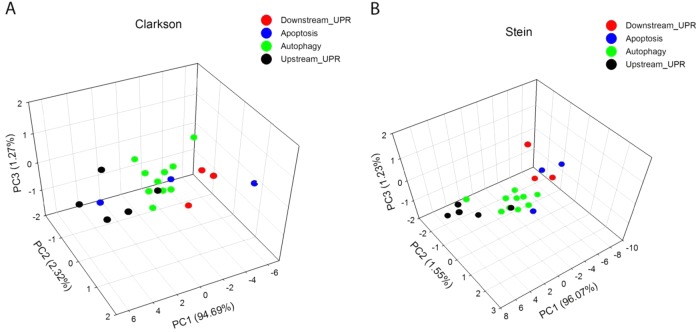
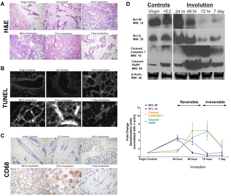
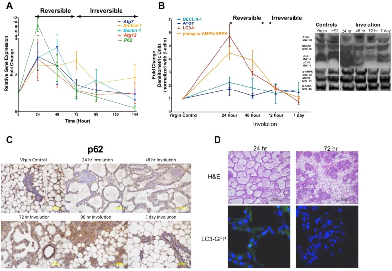
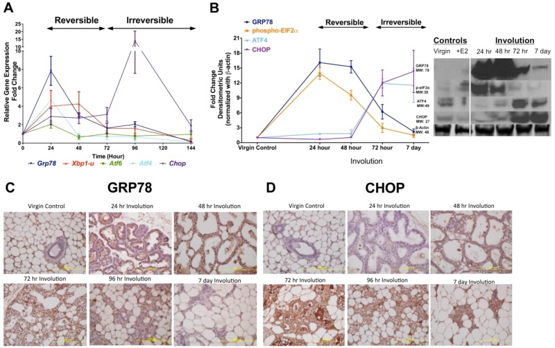
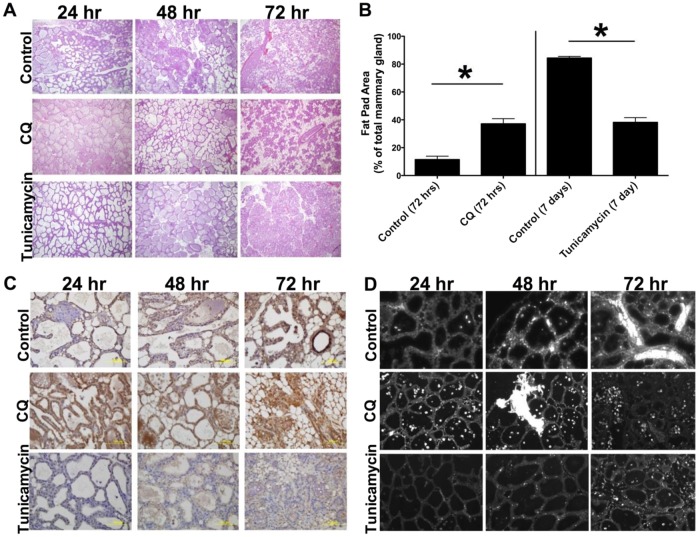
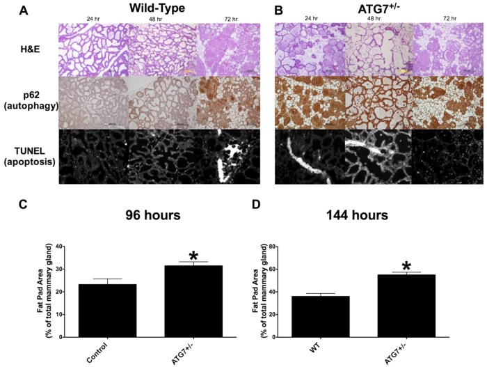
The postnatal mammary gland undergoes repeated cycles of proliferation and cell death, most notably when the fully differentiated (lactating) gland dedifferentiates to a prelactation state. Accumulation of milk proteins in the secretory epithelium creates the stress signal that triggers this process (involution). How this stress is perceived, and the cellular processes that are subsequently activated, remain unclear. We now report that Unfolded Protein Response (UPR), autophagy, and apoptosis related genes cluster separately during lactation and involution in the mouse mammary gland. Time-course experiments in rodents show that autophagy and UPR signaling are tightly co-regulated at the transition from reversible to irreversible involution. Inhibition of autophagy by chloroquine or genetic deletion of one ATG7 allele enhanced progression of mammary involution into the irreversible phase, as characterized by an early/precocious induction of apoptosis. These are the first preclinical in vivo data in support of a clinical trial testing an autophagy inhibitor for prevention of intraductal breast malignancy progression to invasive breast cancer. In marked contrast, stimulation of autophagy by low dose tunicamycin treatment reduced apoptosis and extended the reversible phase of involution by sustaining the secretory epithelium. Autophagy stimulators could be used short-term to promote lactation in women experiencing difficulties or irregularities in nursing. Taken together, these data indicate that UPR and autophagy play a key role in regulating the balance between cell survival and apoptosis during normal mammary gland regression.


Conflict of interest statement

Compliance with ethical standardsThe authors declare that they have no conflict of interest.

Figures

Fig. 1
Fig. 1. Principal component analysis (PCA) of gene expression microarray data sets.
Two published gene expression array data sets (A, B, ) were used. Data from these studies were derived from nonpooled samples. Multiple time-points of mouse mammary gland developmental stages, including lactation and involution, were analyzed. Datasets were acquired and analyzed as explained in Materials and methods. PCA analysis was performed on involution samples for a 21 gene profile representing apoptosis, autophagy, and UPR (Table 1; Supplementary Table S2) on each data set (a, b). A three-dimensional biplot shows the separation of apoptosis, autophagy, and UPR/downstream of UPR genes expressed at the different time points during mammary gland involution. Additional visual angles to show clustering of apoptosis genes better are presented in Supplementary Figure S2
Fig. 2
Fig. 2. Progression of mammary gland involution based on changes in tissue architecture, apoptosis, infiltrating magrophages, and the expression of apoptosis proteins.
Involution time course was created and samples collected as described in the Materials and methods. Mammary glands of virgin mice ± 17β-estradiol (E2) were used as negative controls; E2 treatment was used to block apoptosis. a Progression of involution was assessed based on tissue histology in the H&E stained slides and quantified in early involution (24–72 h) by grading of the H&E slides (shown in Supplementary Fig. S1) and in late involution (96 h, 7 d) by using ImageJ analysis of epithelial/adipose tissue area of the H&E stained slides (see Materials and methods); all slides were photographed using Olympus BX 61 microscope and 10× magnification (A–C). b Detection of apoptotic cells by TUNEL IHC. c Macrophage infiltration visualized by CD68 staining (IHC). d Expression of the known apoptosis markers and regulators BCL-W, BCL-XL, Cleaved caspase-7, Cleaved PARP proteins were measured by Western analysis. Results at each time point show average ± SD, n = 3
Fig. 3
Fig. 3. Evaluation of autophagy in the mammary gland involution.
a Expression of the autophagy genes Atg7, Atg12, Ambra-1, Beclin-1, and p62 was analyzed by qRT-PCR. Lactating mammary glands (involution 0 h) were used as controls; the expression of each gene at each time point is presented as fold-change relative to control. b Western analysis of the autophagy proteins BECLIN-1, ATG7, LC3-II, and pAMPK are shown. The horizontal black bars/arrows show the involution switch from reversible to irreversible stage, which has occurred by 72 h after forced weaning (A, B). The vertical bar indicates the involution 0 h (B, Western blot inset). Results at each time point show average ± SD, n = 3. c Autophagy marker, (downregulation of) p62 specific staining (IHC) at different time points during involution and in virgin control mammary glands. d H&E staining (upper panel) and autophagy specific LC3-GFP punctate formation (lower panel) in identical involution samples from LC3-GFP transgenic mice. All slides were photographed using Olympus BX 61 microscope and 10× magnification (C, D)
Fig. 4
Fig. 4. Evaluation of UPR in the mammary gland involution.
a Expression of UPR genes Grp78, Atf4, Atf6, Xbp1 (unspliced form), and Chop/Ddit3 was analyzed by qRT-PCR. Lactating mammary gland (involution 0 h) was used as a control, and the expression of each gene at each time point is presented as fold-change relative to control. The horizontal black bar/arrow shows the involution switch from reversible to irreversible stage, which has occurred by 72 h after forced weaning (A, B). b Western analysis of UPR proteins GRP78, ATF4, ATF6, XBP1 (unspliced form), phospho-eIF2a, and CHOP/DDIT3. The vertical bar indicates the involution 0 h (B, Western blot inset). Results at each time point show average ± SD, n = 3. c GRP78 and d CHOP/DDIT3-specific staining (IHC) at different time points during involution and in virgin control mammary glands. All slides were photographed using Olympus BX 61 microscope and 10× magnification (C, D)
Fig. 5
Fig. 5. Drug interventions inhibiting and stimulating autophagy enhanced and delayed involution, respectively.
At the time of forced weaning (involution 0 h) drug treatments were started to inhibit (with low dose chloroquine [CQ], middle panels) and stimulate (with low dose tunicamycin [Tm], bottom panels) autophagy, as described in the Materials and methods. Time-course involution samples were collected as in Figs. 2–4. a, b Histology and quantification of involution progress by ImageJ analysis of epithelial/fat pad area of the H&E stained mammary gland slides, as described in Materials and methods. Average ± SD are shown, n = 3. Tm vs. vehicle control: Student’s t test, P ≤ 0.001 (96 h); Mann–Whitney rank sum test P ≤ 0.001 (7d). CQ vs. vehicle control: Mann–Whitney rank sum test P ≤ 0.001 (72 h). c Representative slides of (c) autophagy marker (downregulation of) p62 specific staining (IHC), and d apoptosis marker TUNEL IHC are shown
Fig. 6
Fig. 6. Enhanced involution in autophagy gene deficient mice.
An involution time course was created using wild type (Atg7+/+) and Atg7 heterozygous (Atg7+/−) mice. Samples were collected as in Figs. 2–4 and as described in the Materials and methods. a, b H&E staining (top panels), p62 IHC (middle panels), and TUNEL IHC (bottom panels) of mammary glands of Atg7+/+ and Atg7+/− mice after 24 h, 48 h, and 72 h of involution. c, d Quantification of epithelial/adipose tissue area of the H&E stained slides as in Fig. 2, and as described in the Materials and methods, and in late involution (96 h, 7 d) by using ImageJ analysis, as described in the Materials and methods. Average ± SD are shown, n = 3–5. Student’s t test, P = 0.026 (96 h), P = 0.001 (7 d)

References

    1. Jaggi R, Marti A, Guo K, Feng Z, Friis RR. Regulation of a physiological apoptosis: mouse mammary involution. J. Dairy Sci. 1996;79:1074–1084. doi: 10.3168/jds.S0022-0302(96)76461-5. - DOI - PubMed
    1. Stein T, Salomonis N, Gusterson BA. Mammary gland involution as a multi-step process. J. Mammary Gland Biol. Neoplasia. 2007;12:25–35. doi: 10.1007/s10911-007-9035-7. - DOI - PubMed
    1. McDaniel SM, et al. Remodeling of the mammary microenvironment after lactation promotes breast tumor cell metastasis. Am. J. Pathol. 2006;168:608–620. doi: 10.2353/ajpath.2006.050677. - DOI - PMC - PubMed
    1. Lyons TR, et al. Postpartum mammary gland involution drives progression of ductal carcinoma in situ through collagen and COX-2. Nat. Med. 2011;17:1109–1115. doi: 10.1038/nm.2416. - DOI - PMC - PubMed
    1. Schedin P, O’Brien J, Rudolph M, Stein T, Borges V. Microenvironment of the involuting mammary gland mediates mammary cancer progression. J. Mammary Gland Biol. Neoplasia. 2007;12:71–82. doi: 10.1007/s10911-007-9039-3. - DOI - PubMed

LinkOut - more resources