Review
. 2018 Aug;10(Suppl 22):S2611-S2627.
doi: 10.21037/jtd.2018.05.86.

Management of incidental lung nodules <8 mm in diameter

Affiliations
Review

Management of incidental lung nodules <8 mm in diameter

Marcelo Sánchez et al. J Thorac Dis. 2018 Aug.

Abstract

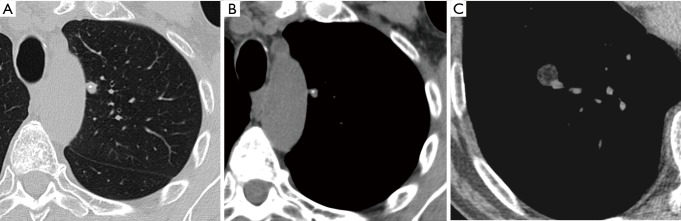
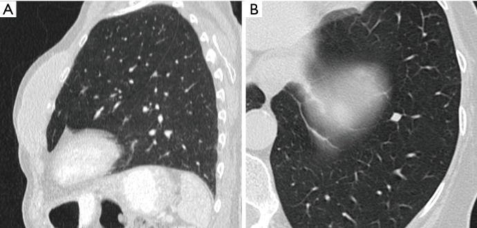
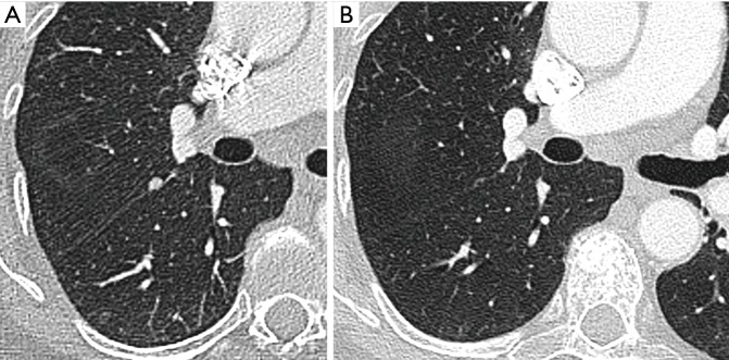
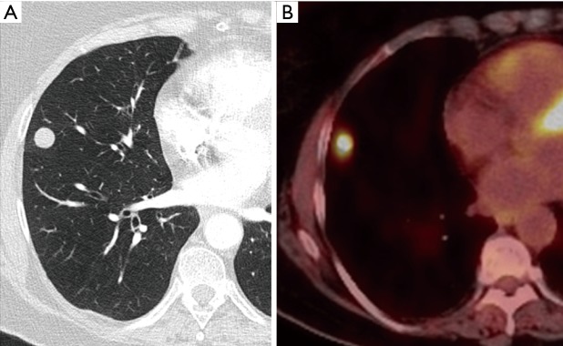
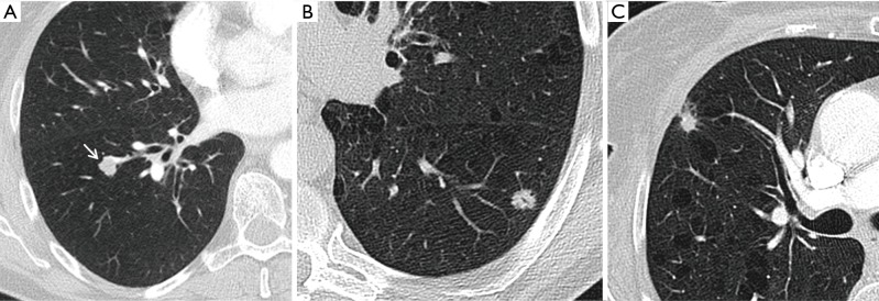
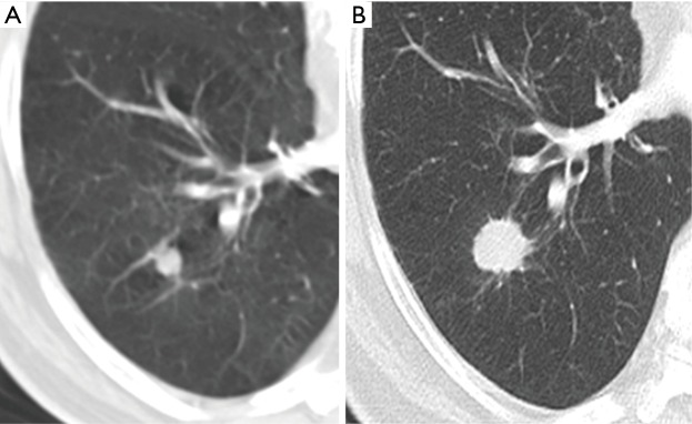
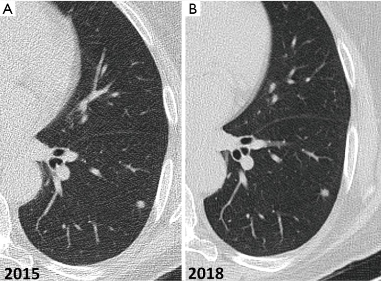
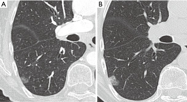
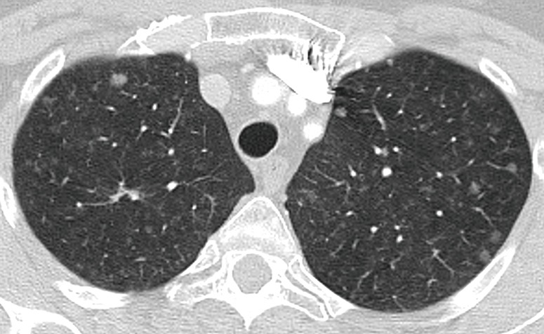
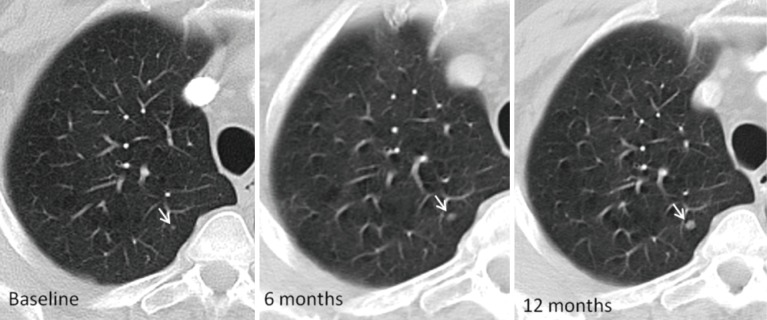
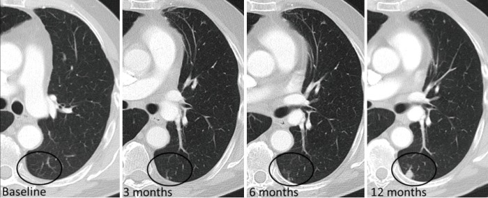
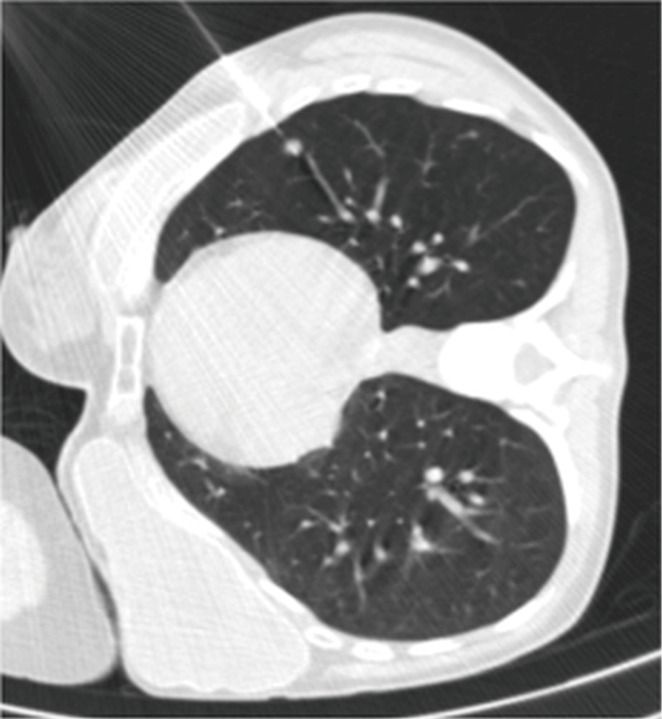
Due to the increase of incidentally detected pulmonary nodules and the information obtained from several screening programs, updated guidelines with new recommendations for the management of small pulmonary nodules have been proposed. These international guidelines coincide in proposing periodic follow-up for small nodules, less than 8 mm of diameter. Fleischner and British Thoracic Society guidelines are the most recent and popular guidelines for incidental pulmonary nodules management. They have specific recommendations according to nodule characteristics (density and size) and cancer risk of the patient. Both guidelines separate recommendations for solid and subsolid nodules. Predictive risk models have been developed to improve the nodule management. In certain cases follow up may not be the best option. We discuss the scenarios and options to achieve a histologic diagnosis of these tiny pulmonary nodules.

Keywords: Pulmonary nodules; computed tomography; lung cancer; management.


Conflict of interest statement

Conflicts of Interest: The authors have no conflicts of interest to declare.

Figures

Figure 1
Figure 1
CT images show (A) lung window and (B) soft-tissue window thin transverse sections of a small solid nodule with internal calcification, typical of a healed granuloma. (C) Lung window transverse CT section shows a small solid nodule with fat content, consistent with a hamartoma. No further CT follow-up is recommended.
Figure 2
Figure 2
Intrapulmonary lymph node characteristics. (A) Sagittal CT section shows a typical perifissural lymph nodule with a triangular shape and defined borders; (B) transverse CT section shows a solid nodule in the left lower lobe with a trapezoidal shaped suggestive of intrapulmonary lymph nodes.
Figure 3
Figure 3
Transverse CT image shows a perifissural nodule with spherical shape and irregular margins in an oncologic patient (A), the nodule practically disappeared in the follow-up CT (B).
Figure 4
Figure 4
Transverse CT sections of a solid nodule with well-defined and regular margins in the right middle lobe with the corresponding image of PET/CT showing increased FDG uptake, suspicious of malignancy.
Figure 5
Figure 5
Pulmonary nodule, CT baseline and follow up. (A) Transverse CT image shows a part-solid nodule located in the left lower lobe with a central 8 mm solid component; (B) follow-up CT image after 3 months shows complete resolution, consistent with a benign etiology.
Figure 6
Figure 6
Multiple small pulmonary nodules in the right lung detected with the maximum intensity projection (MIP) reformation that increase the sensitivity of detection of small nodules, particularly in the central lung.
Figure 7
Figure 7
Small solid pulmonary nodule measured in the long and perpendicular short axis diameters to obtain the mean diameter, measured in the axial plane.
Figure 8
Figure 8
Pulmonary nodule. Volumetric evaluation. (A) Volumetric nodule measurement in a small nodule shows that small increase in diameter (from 5.7 to 6.3 mm large diameter) corresponds to a doubling of overall volume in the 6 months follow-up CT (B). The nodule increased from 33 to 63 mm3, greater than 25% and corresponds to a VDT of 252 days.
Figure 9
Figure 9
Radiologic features of pulmonary nodules. (A) Transverse CT section of a 8 mm solid pulmonary nodule with irregular margins (arrow); (B) transverse CT section shows a spiculated predominantly solid nodule with small air bronchiologram; (C) transverse CT sections show a small nodule with spiculated margins and pleural retraction. All three pulmonary nodules, regardless of its size, have radiological features of malignancy.
Figure 10
Figure 10
Solid pulmonary nodule growth. (A) Transverse CT section shows a small solid pulmonary nodule in the right lower lobe; (B) follow-up CT image after 6 months shows increase in the size of the nodule with spiculated borders. Surgery revealed invasive adenocarcinoma.
Figure 11
Figure 11
CT images showing a 6 mm solid pulmonary nodule in the left lower lobe, stable for 3 years. Solid lesions without changes during at least 2 years are considered benign lesions.
Figure 12
Figure 12
Subsolid pulmonary nodule growth. (A) Transverse 1.5 mm CT section shows a pure ground-glass nodule in the right lower lobe; (B) CT obtained 3 years after shows a subtle increase in the size with the appearance of a 5 mm solid component, highly suspicious of adenocarcinoma. Patient underwent to surgery and pathology showed lepidic growth with a small central area of invasion confirming a minimally invasive adenocarcinoma. In subsolid nodules a longer follow-up period is required because many of them are indolent or slow growing neoplasms.
Figure 13
Figure 13
Transverse thin CT section of the upper lobes shows multiple and bilateral subsolid nodules of variable size. A nodule of greater size and density is identified in the right upper lobe (arrow). In the context of multiple pulmonary nodules the recommendations is to assess the risk based on that of the largest nodule. Follow-up CT is recommended in 3–6 months.
Figure 14
Figure 14
Transverse CT sections through the right upper lobe in a oncologic patient shows increase in size of a micronodule (arrow) during 12 months CT follow-up, suggestive of malignancy, regardless of size.
Figure 15
Figure 15
Transverse CT sections show growth of a new nodule (circle area) in the CT follow-up in a patient with melanoma, suspicious of metastases. In oncologic patients new or incident nodules have higher risk of malignancy than baseline nodules.
Figure 16
Figure 16
Transverse CT section in right lateral decubitus shows a 7 mm nodule located in the left lower lobe in a patient with history of osteosarcoma and breast tumor. A18G core needle biopsy was performed with the result of metastatic breast cancer.
Figure 17
Figure 17
Transverse chest CT showing a pulmonary nodule in the right middle lobe with the hook wire located in the nodule. The patient underwent video-assisted thoracoscopic surgery (VATS) on the same day after placement.
Figure 18
Figure 18
Images showing the radio-guided surgery procedure. Under CT guidance, (A) a 7 mm pulmonary nodule located in the left upper lobe is identified and (B) a needle is inserted to reach the nodule; (C) a radiotracer (solution of 99mtechnetium (99mTc) macro-aggregates albumin diluted with iodized contrast medium) is injected. After injection, CT is performed to confirm precise staining, showing peritumoral hemorrhage after the injection of the radiotracer (D); (E) a SPECT-CT is performed showing uptake inside the pulmonary nodule; (F) SPECT-CT 3D reconstructions showing activity inside the nodule.

Similar articles

Cited by

References

    1. Swensen SJ. The probability of malignancy in solitary pulmonary nodules. Arch Intern Med 1997;157:849. 10.1001/archinte.1997.00440290031002 - DOI - PubMed
    1. Aberle DR, Adams AM, Berg CD, et al. Reduced lung-cancer mortality with low-dose computed tomographic screening. N Engl J Med 2011;365:395-409. 10.1056/NEJMoa1102873 - DOI - PMC - PubMed
    1. MacMahon H, Naidich DP, Goo JM, et al. Guidelines for management of incidental pulmonary nodules detected on CT Images: From the Fleischner Society 2017. Radiology 2017;284:228-43. 10.1148/radiol.2017161659 - DOI - PubMed
    1. Callister MEJ, Baldwin DR, Akram AR, et al. BTS guidelines for the investigation and management of pulmonary nodules. Thorax 2015;70 Suppl 2:ii1-54. 10.1136/thoraxjnl-2015-207168 - DOI - PubMed
    1. Rami-Porta R, Bolejack V, Crowley J, et al. The IASLC Lung Cancer Staging Project: Proposals for the Revisions of the T Descriptors in the Forthcoming Eighth Edition of the TNM Classification for Lung Cancer. J Thorac Oncol 2015;10:990-1003. - PubMed