. 2018 Aug;10(Suppl 22):S2649-S2655.
doi: 10.21037/jtd.2018.03.132.

Transcervical videomediastino-thoracoscopy

Affiliations

Transcervical videomediastino-thoracoscopy

Josep Belda-Sanchis et al. J Thorac Dis. 2018 Aug.

Abstract

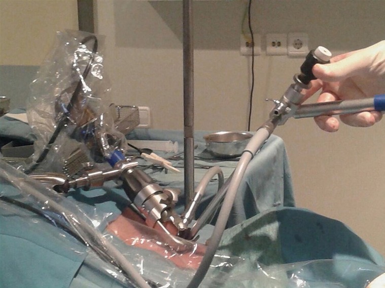
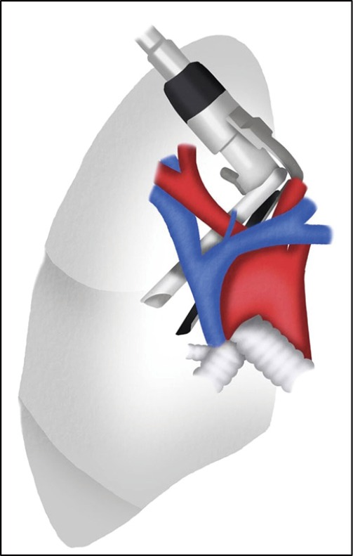
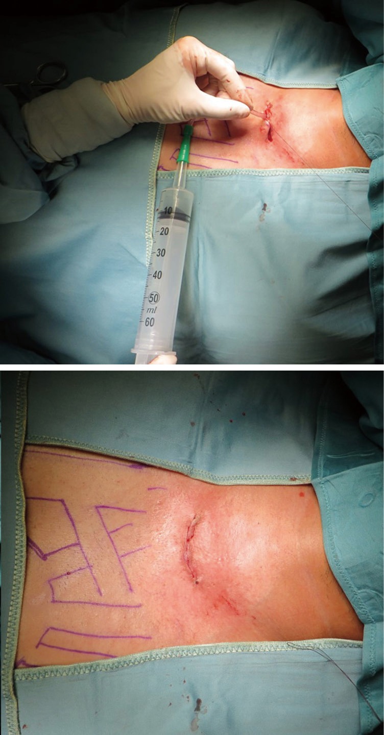
Although technical advances in non-invasive and minimally invasive approaches to lung and pleural cancer diagnosis and staging have become more widely available and accurate, surgical techniques remain the gold standard in assessing the extent of loco-regional involvement. Precise surgical staging of lung or pleural tumours is pivotal in the selection of surgical candidates and for predicting survival. In some patients, both mediastinal and pleural exploration may be needed for many different reasons. Transcervical videomediastino-thoracoscopy (VMT) combines simultaneously the exploration of both the mediastinum and the pleural cavities through a single cervical incision, allowing for biopsies or sampling of the mediastinal lymph nodes, lymphadenectomy and pleuropulmonary assessment (mainly pleural effusions, tumour involvement of the visceral and parietal pleura and pulmonary nodules). Thoracic surgeons should be aware of this combined surgical approach and completely familiar with classical indications and technical details of the transcervical approach to the mediastinum and thoracoscopic exploration of the pleural cavities.

Keywords: Staging; lung cancer; thoracoscopy; videomediastinoscopy.


Conflict of interest statement

Conflicts of Interest: The authors have no conflicts of interest to declare.

Figures

Figure 1
Figure 1
Patient’s head hyperextended. A head ring provides stability during surgery.
Figure 2
Figure 2
U-shaped retaining arm with special clamping device used to fix and stabilize the video mediastinoscope (Richard Wolf, Knittlingen, Germany).
Figure 3
Figure 3
Set of specific instruments required for videomediastinoscopy: (A) dissection-suction-coagulation cannula; (B) glass tube for test puncture; (C) grasping forceps to retrieve inserted gauzes; (D) biopsy forceps; (E) grasper; (F) Linder-Dahan spreadable blade video mediastinoscope (Richard Wolf, Knittlingen, Germany).
Figure 4
Figure 4
A bayonet thoracoscope with a working channel inserted through the video mediastinoscope.
Figure 5
Figure 5
Transcervical access to the right pleural cavity.
Figure 6
Figure 6
The SVC pulled up to facilitate the exposition of the RMP and pleurotomy. AV, azygos vein. AV, azygos vein; SVC, superior vena cava; RMP, right mediastinal pleura.
Figure 7
Figure 7
The sequence of figures shows the main steps of the right transcervical videomediastino-thoracoscopy. (A) RMP exposed; (B) by grasping and pulling the mediastinal pleura; (C,D) the pleurotomy can be performed using endoscopic scissors. AV, azygos vein; RMP, right mediastinal pleura.
Figure 8
Figure 8
Video mediastinoscope inserted between the innominate artery and the left common carotid artery at their origin in the aortic arch (A) and advanced in front (B) or behind (C) the left innominate vein.
Figure 9
Figure 9
Transcervical access to the left pleural cavity in front of the LIV. Exposition of the LMP at the level of the aorto-pulmonary window. LIV, left innominate vein; LMP, left mediastinal pleura.
Figure 10
Figure 10
Thoracoscopic view of the right pleurotomy through the working channel of the video mediastinoscope. RMP, right mediastinal pleura.
Figure 11
Figure 11
View of the small drainage tube introduced into the pleural cavity. SVC, superior vena cava; RMP, right mediastinal pleura; AV, azygos vein.
Figure 12
Figure 12
In this case, a nasogastric tube was introduced into the pleural cavity through the opened mediastinal pleura and exteriorized through the cervicotomy. A 50 mL syringe was connected directly to the nasogastric tube and constant negative pressure is applied to the plunger of the syringe to aspirate air from the chest cavity.

References

    1. Deslauriers J, Beaulieu M, Dufour C, et al. Mediastinopleuroscopy: a new approach to the diagnosis of intrathoracic diseases. Ann Thorac Surg 1976;22:265-9. 10.1016/S0003-4975(10)64914-4 - DOI - PubMed
    1. Rami-Porta R, Call S, Serra-Mitjans M. Mediastinoscopy. In: Zieliński M, Rami-Porta R. editors. The Transcervical Approach in Thoracic Surgery. Heidelberg: Springer-Verlag, 2014:9-27.
    1. Witte B, Wolf M, Hürtgen M. Video-assisted mediastinoscopic lymphadenectomy (VAMLA). In: Zieliński M, Rami-Porta R. editors. The Transcervical Approach in Thoracic Surgery. Heidelberg: Springer-Verlag, 2014:89-99.
    1. Hürtgen M, Friedel G, Toomes H, et al. Radical video-assisted mediastinoscopic lymphadenectomy (VAMLA)--technique and first results. Eur J Cardiothorac Surg 2002;21:348-51. 10.1016/S1010-7940(01)01125-3 - DOI - PubMed
    1. Witte B, Hürtgen M. Video-assisted mediastinoscopic lymphadenectomy (VAMLA). J Thorac Oncol 2007;2:367-9. 10.1097/01.JTO.0000263725.89512.d7 - DOI - PubMed