. 2018 Oct 5:8:352.
doi: 10.3389/fcimb.2018.00352. eCollection 2018.

CSGID Solves Structures and Identifies Phenotypes for Five Enzymes in Toxoplasma gondii

Affiliations

CSGID Solves Structures and Identifies Phenotypes for Five Enzymes in Toxoplasma gondii

Joseph D Lykins et al. Front Cell Infect Microbiol. .

Abstract

Toxoplasma gondii, an Apicomplexan parasite, causes significant morbidity and mortality, including severe disease in immunocompromised hosts and devastating congenital disease, with no effective treatment for the bradyzoite stage. To address this, we used the Tropical Disease Research database, crystallography, molecular modeling, and antisense to identify and characterize a range of potential therapeutic targets for toxoplasmosis. Phosphoglycerate mutase II (PGMII), nucleoside diphosphate kinase (NDK), ribulose phosphate 3-epimerase (RPE), ribose-5-phosphate isomerase (RPI), and ornithine aminotransferase (OAT) were structurally characterized. Crystallography revealed insights into the overall structure, protein oligomeric states and molecular details of active sites important for ligand recognition. Literature and molecular modeling suggested potential inhibitors and druggability. The targets were further studied with vivoPMO to interrupt enzyme synthesis, identifying the targets as potentially important to parasitic replication and, therefore, of therapeutic interest. Targeted vivoPMO resulted in statistically significant perturbation of parasite replication without concomitant host cell toxicity, consistent with a previous CRISPR/Cas9 screen showing PGM, RPE, and RPI contribute to parasite fitness. PGM, RPE, and RPI have the greatest promise for affecting replication in tachyzoites. These targets are shared between other medically important parasites and may have wider therapeutic potential.

Keywords: PPMO; Toxoplasma gondii; crystallography; nucleoside diphoshate kinase; ornithine aminotransferase; phosphoglycerate mutase; ribose-5-phosphate isomerase; ribulose-3-phosphate epimerase.


Figures

Figure 1
Figure 1
The Toxoplasma Structural Genomics Pipeline identified promising targets with good predicted druggability (Phase III) and outstanding interest to the Toxoplasma research community (Phase I, II). Toxoplasma proteins were selected for Phase I from published work up to 2009 and communicated with the research laboratories. Phase II included proteins from published and unpublished studies suggested by international researchers at the end of 2011. Phase III candidates were selected from the Tropical Diseases Research Database (TDRT). Proteins judged very unlikely to crystallize based on analysis by XtalPred were eliminated (Slabinski et al., 2007). Several structures solved have already been described (Ruan et al., ; Tonkin et al., ; Dubey et al., 2017). This crystallography pipeline remains available for production and solution of structures of proteins for scientists in the Toxoplasma research community.
Figure 2
Figure 2
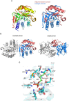
Crystal structure of TgPGM (A). Ribbon representation of TgPGM monomer (left side) colored blue (N-terminus) to red (C-terminus) and tetramer of TgPGM (right side) (B). Domain structure of TgPGM [red—an α/β/α-sandwich domain (residues 12–107 and 169–257) and blue—a domain without any defined folding motif (residues 108–168)] (C). Pairwise structural alignment of TgPGM (gray) and BpPGM (cyan) showing active site with residues of TgPGM shown in sticks and labeled in one-letter code. Equivalent residues of BpPGM are displayed. BpPGM binds (2R)-2,3-diphosphoglyceric acid, DG2, in the active site.
Figure 3
Figure 3
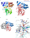
Crystal structure of TgNDK (A). Ribbon representation of TgNDK monomer (left side) colored blue (N-terminus) to red (C-terminus) and trimeric and hexameric assemblies of TgNDK (middle and right side) (B). Pairwise structural alignment of TgPGM (gray; SO4 is shown in sticks) and human NM23-H2 transcription factor [cyan (chain E with bound 2′-deoxyguanosine-5′-monophosphate (DG) and 2′-deoxyadenosine-5′-monophosphate (DA)] and magenta [chain B with bound 2′-deoxyguanosine-5′-monophosphate (DG)] showing active site with residues of TgNDK shown in sticks and labeled in one-letter code. Equivalent residues of human NM23-H2 transcription factor are displayed.
Figure 4
Figure 4
Crystal structure of TgRPE (A). Ribbon representation of TgRPE monomer (left side) colored blue (N-terminus) to red (C-terminus) and TgRPE dimer (right side) (B). Pairwise structural alignment of TgRPE (gray; SO4 is shown in sticks and Zn2+ as green sphere) and human RPE with bound Fe2+ (yellow ribbon), 5-O-phosphono-D-xylulose (5SR; magenta ribbon), and ribulose-5-phosphate (5RP; cyan ribbon) showing active site with residues of TgRPE are shown in sticks and labeled in one-letter code. Equivalent residues of human RPE are displayed.
Figure 5
Figure 5
Crystal structure of TgRPI (A). Ribbon representation of TgRPI monomer (left side) colored blue (N-terminus) to red (C-terminus). Catalytic domain and oligomerization domain of TgRPI with bound D-malate (MLT; sticks) and chloride ion (Cl; green sphere) are shown on the right. (B). Unstable and stable TgRPI dimers according to PISA prediction (C). Pairwise structural alignment of TgRPI (gray; MLT is shown in sticks and Cl as green sphere) and TtRPI (cyan ribbon) with bound arabinose 5-phosphate (A5P; sticks) showing active site with residues of TgRPI are shown in sticks and labeled in one-letter code. Equivalent residues of TtRPI are displayed. Water molecules are shown as small red spheres.
Figure 6
Figure 6
Crystal structure of TgOAT (A). Ribbon representation of TgOAT monomer (left side) colored blue (N-terminus) to red (C-terminus). Catalytic and N/C-termini domains with bound PLP are shown on the right (B). Dimer of TgOAT (C). Pairwise structural alignment of TgOAT (gray; PLP is shown in sticks) and hOAT with bound PLP (cyan ribbon; PDB 1oat). Residues of TgOAT are shown in sticks and labeled in one-letter code. Equivalent lysine residues of hOAT are displayed. Chain identifier in parentheses.
Figure 7
Figure 7
(A) Schematic of Interaction Between vivoPMO and RNA. (B) Sequences for PMO. Sequences for all enzymes were designed to be complementary to regions overlapping splice sites so as to prevent appropriate interaction between mRNA and the spliceosome predicted to lead to the production of a non-functional protein product.
Figure 8
Figure 8
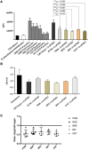
(A) YFP/vivoPMO Efficacy Assay at 10 μM. Targeted vivoPMOs show statistically significant knockdown relative to off-target and to a standardized parasite concentration as determined by student t-test. (B) Wst-1 Cell Viability/ vivoPMO Toxicity Assay at 10 μM. Targeted vivoPMOs do not show statistically significantly (p < 0.05 by student T-test) different levels of optical density relative to off-target vivoPMO or uninfected fibroblasts at 10 μM concentrations. (C) Ratio of On- to Off-Target vivoPMO for Replicate Experiments Using 10 μM of Targeted vivoPMO. The data presented here represent five replicate experiments. Lower individual values suggest a larger effect of the morpholino.
Figure 9
Figure 9
Immunofluorescence Assay. This series of images depict the localization of nucleoside diphosphate kinase, one of the molecular targets herein discussed. Note the concentration of red fluorescence at the periphery of the parasite. This is consistent with the localization of secreted proteins like Gra1, suggesting that NDK could be secreted by the parasite. This has precedents in other pathogens, including M. tuberculosis and Leishmania.
Figure 10
Figure 10
Summary figure showing the T. gondii drug discovery pipeline as described herein. The TDR Database provided important initial insights into potentially important parasite proteins (Agüero et al., ; Magariños et al., 2012). Identification of proteins amenable to crystallographic analysis, solution of their structures, abrogation with vivoPMO and confirmation of essentiality by querying the CRISPR/Cas9 database completed the pathway, identifying enriched targets with improved therapeutic potential.

References

    1. Agüero F., Al-Lazikani B., Aslett M., Berriman M., Buckner F. S., Campbell R. K., et al. (2008). Genomic-scale prioritization of drug targets: the TDR Targets database. Nat. Rev. Drug Discov. 7, 900–907. 10.1038/nrd2684 - DOI - PMC - PubMed
    1. Akana J., Fedorov A. A., Fedorov E., Novak W. R. P., Babbitt P. C., Almo S. C., et al. (2006). D-Ribulose 5-phosphate 3-epimerase: functional and structural relationships to members of the ribulose-phosphate binding (beta/alpha)8-barrel superfamily. Biochemistry 45, 2493–2503. 10.1021/bi052474m - DOI - PubMed
    1. Ananvoranich S., Al Rayes M., Al Riyahi A., Wang X. (2006). RNA silencing of glycolysis pathway in Toxoplasma gondii. J. Eukaryot. Microbiol. 53(Suppl. 1), S162–S163. 10.1111/j.1550-7408.2006.00216.x - DOI - PubMed
    1. Anderson W. F. (2009). Structural genomics and drug discovery for infectious diseases. Infect. Disord. 9, 507–517. 10.2174/187152609789105713 - DOI - PMC - PubMed
    1. Aslanidis C., de Jong P. J. (1990). Ligation-independent cloning of PCR products (LIC-PCR). Nucleic Acids Res. 18, 6069–6074. 10.1093/nar/18.20.6069 - DOI - PMC - PubMed

Publication types

LinkOut - more resources