. 2019 Aug;45(5):459-475.
doi: 10.1111/nan.12525. Epub 2018 Nov 23.

Rapidly progressive amyotrophic lateral sclerosis is associated with microglial reactivity and small heat shock protein expression in reactive astrocytes

Affiliations

Rapidly progressive amyotrophic lateral sclerosis is associated with microglial reactivity and small heat shock protein expression in reactive astrocytes

R P Gorter et al. Neuropathol Appl Neurobiol. 2019 Aug.

Abstract

Aims: Amyotrophic lateral sclerosis (ALS) is a chronic neurodegenerative disease characterized by progressive loss of motor neurons, muscle weakness, spasticity, paralysis and death usually within 2-5 years of onset. Neuroinflammation is a hallmark of ALS pathology characterized by activation of glial cells, which respond by upregulating small heat shock proteins (HSPBs), but the exact underlying pathological mechanisms are still largely unknown. Here, we investigated the association between ALS disease duration, lower motor neuron loss, TARDNA-binding protein 43 (TDP-43) pathology, neuroinflammation and HSPB expression.

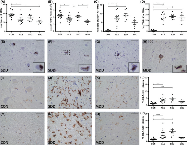
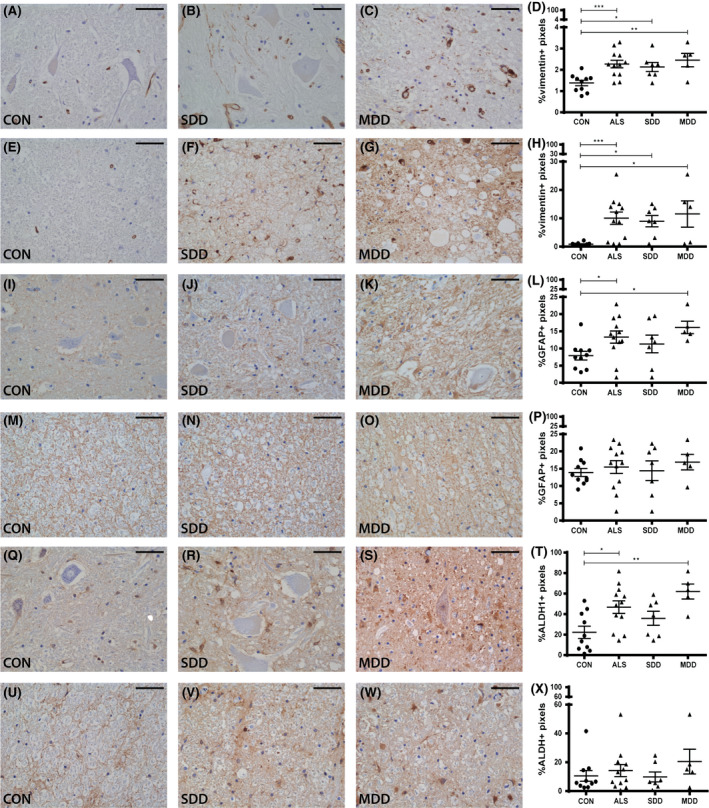
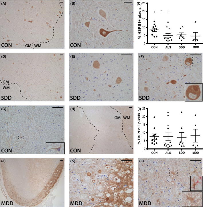
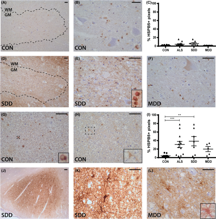
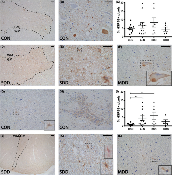
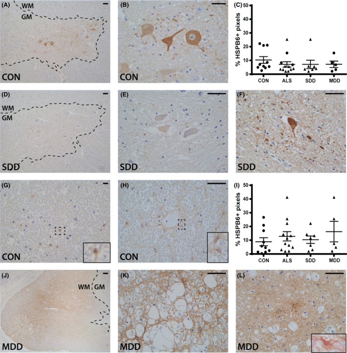
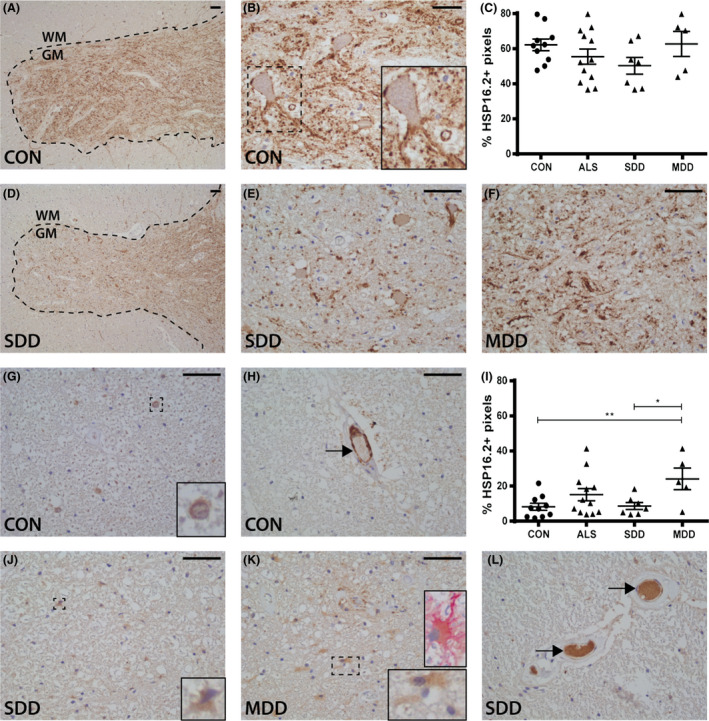
Methods: With immunohistochemistry, we examined HSPB1, HSPB5, HSPB6, HSPB8 and HSP16.2 expression in cervical, thoracic and sacral spinal cord regions in 12 ALS cases, seven with short disease duration (SDD), five with moderate disease duration (MDD), and ten age-matched controls. Expression was quantified using ImageJ to examine HSP expression, motor neuron numbers, microglial and astrocyte density and phosphorylated TDP-43 (pTDP-43+) inclusions.

Results: SDD was associated with elevated HSPB5 and 8 expression in lateral tract astrocytes, while HSP16.2 expression was increased in astrocytes in MDD cases. SDD cases had higher numbers of motor neurons and microglial activation than MDD cases, but similar levels of motor neurons with pTDP-43+ inclusions.

Conclusions: Increased expression of several HSPBs in lateral column astrocytes suggests that astrocytes play a role in the pathogenesis of ALS. SDD is associated with increased microgliosis, HSPB5 and 8 expression in astrocytes, and only minor changes in motor neuron loss. This suggests that the interaction between motor neurons, microglia and astrocytes determines neuronal fate and functional decline in ALS.

Keywords: HSPB; TDP-43 pathology; amyotrophic lateral sclerosis; astrocytes; inflammation; small heat shock proteins.


Figures

Figure 1
Figure 1
Motor neurons, pTDP‐43 inclusions and microglial activation in ALS spinal cord. Quantification of (A) motor neuron counts, (B) ventral horn surface, (C) number of pTDP‐43+ inclusions and (D) percentage of pTDP‐43+ inclusion containing motor neurons in controls and ALS patients (subgroups: SDD and MDD). pTDP‐43+ motor neurons in ALS patients with (E,F) SDD and (G,H) MDD. HLA‐DR expression in (IK) ventral horns and (MO) lateral tracts of controls and SDD and MDD patients. Quantification of HLA‐DR+ pixels in (L) ventral horns and (P) lateral tracts of controls and ALS patients (subgroups: SDD and MDD). Data points represent the mean value for each patient. Data are shown as mean ± SEM. Significance was analysed between ALS patients (n = 12) and controls (n = 10) with Student's t‐test or Mann–Whitney U‐test. ALS patients SDD (n = 7) and MDD (n = 5) were compared to controls (n = 10) using anova and Tukey's post‐test or Kruskal–Wallis H test and Dunn's post hoc multiple comparisons test. Significant data are presented (****= <0.0001, ***= <0.001, **<0.01, *= <0.05). Scale bar in all pictures = 50 μm. Inserts are digitally enlarged. SDD, short disease duration; MDD, moderate disease duration; ALS, amyotrophic lateral sclerosis; HLA‐DR, human leucocyte antigen‐D related; TDP, TARDNA‐binding protein; pTDP‐43, phosphorylated TDP.
Figure 2
Figure 2
Astrocyte reactivity in ALS spinal cord. Expression (AC) and quantification (D) of vimentin+ pixels in ventral horns of controls and ALS patients (subgroups: SDD and MDD). Expression (EG) and quantification (H) of vimentin+ pixels in lateral columns of controls and ALS patients (subgroups: SDD and MDD). Expression (IK) and quantification (L) of GFAP+ pixels in ventral horns of controls and ALS patients (subgroups: SDD and MDD). Expression (MO) and quantification (P) of GFAP+ pixels in lateral columns of controls and ALS patients (subgroups: SDD and MDD). Expression (QS) and quantification (T) of ALDH1+ pixels in ventral horns of controls and ALS patients (subgroups: SDD and MDD). Expression (UW) and quantification (X) of ALDH1+ pixels in lateral columns of controls and ALS patients (subgroups: SDD and MDD). Data points represent the mean value for each patient. Data are shown as mean ± SEM. Significance was analysed between ALS patients (n = 12) and controls (n = 10) with Student's t‐test or Mann–Whitney U‐test. ALS patients with short disease duration (SDD; n = 7) and moderate disease duration (MDD; n = 5) were compared to controls (n = 10) using anova and Tukey's post‐test or Kruskal–Wallis H test and Dunn's post hoc multiple comparisons test. Significant data are presented (****= <0.0001, ***= <0.001, **<0.01, *= <0.05). Scale bar in all pictures = 50 μm. SDD, short disease duration; MDD, moderate disease duration; ALS, amyotrophic lateral sclerosis; GFAP, glial fibrillary acid protein; ALDH1, aldehyde dehydrogenase 1.
Figure 3
Figure 3
HSPB1 expression in ALS spinal cord. HSPB1 expression in (A,B,DF) ventral horns and (G,H,JL) lateral tracts of controls and ALS patients (subgroups: SDD and MDD) with (L) an insert of a vimentin+ (pink) and HSPB1+ (brown) astrocyte. Grey matter is delineated with a dotted line. Quantification of HSPB1+ pixels in (C) ventral horns and (I) lateral columns of controls and ALS patients (subgroups: SDD and MDD). Data points represent the mean value for each patient. Data are shown as mean ± SEM. Significance was analysed between ALS patients (n = 12) and controls (n = 10) with Student's t‐test or Mann–Whitney U‐test. ALS patients with SDD (n = 7) and MDD (n = 5) were compared to controls (n = 10) using anova and Tukey's post‐test or Kruskal–Wallis H test and Dunn's post hoc multiple comparisons test. Significant data are presented (****P = <0.0001, ***P = <0.001, **<0.01, *P = <0.05). Scale bar in all pictures = 50 μm. Inserts are digitally enlarged. SDD, short disease duration; MDD, moderate disease duration; ALS, amyotrophic lateral sclerosis; HSPB, heat shock protein B.
Figure 4
Figure 4
HSPB5 expression in ALS spinal cord. HSPB5 expression in (A,B,DF) ventral horns and (G,H,JL) lateral tracts of controls and ALS patients (subgroups: SDD and MDD) with inserts of (E) oligodendrocyte transcription factor 2 (olig2+) (pink) and HSPB5+ (brown) oligodendrocytes and (L) a vimentin+ (pink) and HSPB5+ (brown) astrocyte. Grey matter is delineated with a dotted line. Quantification of HSPB5+ pixels in (C) ventral horns and (I) lateral columns of controls and ALS patients (subgroups: SDD and MDD). Data points represent the mean value for each patient. Data are shown as mean ± SEM. Significance was analysed between ALS patients (n = 12) and controls (n = 10) with Student's t‐test or Mann–Whitney U‐test. ALS patients with SDD (n = 7) and MDD (n = 5) were compared to controls (n = 10) using anova and Tukey's post‐test or Kruskal–Wallis H test and Dunn's post hoc multiple comparisons test. Significant data are presented (****= <0.0001, ***= <0.001, **<0.01, *= <0.05). Scale bar in all pictures = 50 μm. Inserts are digitally enlarged. SDD, short disease duration; MDD, moderate disease duration; ALS, amyotrophic lateral sclerosis; HSPB, heat shock protein B.
Figure 5
Figure 5
HSPB8 expression in ALS spinal cord. HSPB8 expression in (A,B,DF) ventral horns and (G,H,JL) lateral tracts of controls and ALS patients (subgroups: SDD and MDD) with (K) an insert of a vimentin+ (pink) and HSPB8+ (brown) astrocyte. Grey matter is delineated with a dotted line. Quantification of HSPB8+ pixels in (C) ventral horns and (I) lateral columns of controls and ALS patients (subgroups: SDD and MDD). Data points represent the mean value for each patient. Data are shown as mean ± SEM. Significance was analysed between ALS patients (n = 12) and controls (n = 10) with Student's t‐test or Mann–Whitney U‐test. ALS patients with SDD (n = 7) and MDD (n = 5) were compared to controls (n = 10) using anova and Tukey's post‐test or Kruskal–Wallis H test and Dunn's post hoc multiple comparisons test. Significant data are presented (****= <0.0001, ***= <0.001, **<0.01, *= <0.05). Scale bar in all pictures = 50 μm. Inserts are digitally enlarged. SDD, short disease duration; MDD, moderate disease duration; ALS, amyotrophic lateral sclerosis; HSPB, heat shock protein B.
Figure 6
Figure 6
HSPB6 expression in ALS spinal cord. HSPB6 expression in (A,B,DF) ventral horns and (G,H,JL) lateral tracts of controls and ALS patients (subgroups: SDD and MDD) with (L) an insert of a vimentin+ (pink) and HSPB6+ (brown) astrocyte. Grey matter is delineated with a dotted line. Quantification of HSPB6+ pixels in (C) ventral horns and (I) lateral columns of controls and ALS patients (subgroups: SDD and MDD). Data points represent the mean value for each patient. Data are shown as mean ± SEM. Significance was analysed between ALS patients (n = 12) and controls (n = 10) with Student's t‐test or Mann–Whitney U‐test. ALS patients with SDD (n = 7) and MDD (n = 5) were compared to controls (n = 10) using anova and Tukey's post‐test or Kruskal–Wallis H test and Dunn's post hoc multiple comparisons test. Scale bar in all pictures = 50 μm. Inserts are digitally enlarged. SDD, short disease duration; MDD, moderate disease duration; ALS, amyotrophic lateral sclerosis; HSPB, heat shock protein B.
Figure 7
Figure 7
HSP16.2 expression in ALS spinal cord. HSP16.2 expression in (A,B,DF) ventral horns and (G,H,JL) lateral tracts of controls and ALS patients (subgroups: SDD and MDD) with (K) an insert of a vimentin+ (pink) and HSP16.2+ (brown) astrocyte. HSP16.2+ blood vessels are indicated with arrows (H,L). Grey matter is delineated with a dotted line. Quantification of HSP16.2+ pixels in (C) ventral horns and (I) lateral columns of controls and ALS patients (subgroups: SDD and MDD). Data points represent the mean value for each patient. Data are shown as mean ± SEM. Significance was analysed between ALS patients (n = 12) and controls (n = 10) with Student's t‐test or Mann–Whitney U‐test. ALS patients with SDD (n = 7) and MDD (n = 5) were compared to controls (n = 10) using anova and Tukey's post‐test or Kruskal–Wallis H test and Dunn's post hoc multiple comparisons test. Significant data are presented (****= <0.0001, ***= <0.001, **<0.01, *= <0.05). Scale bar in all pictures = 50 μm. Inserts are digitally enlarged. SDD, short disease duration; MDD, moderate disease duration; ALS, amyotrophic lateral sclerosis; HSP heat shock protein.

Similar articles

Cited by

References

    1. van Es MA, Hardiman O, Chio A, Al‐Chalabi A, Pasterkamp RJ, Veldink JH, et al Amyotrophic lateral sclerosis. Lancet 2017; 390: 2084–98 - PubMed
    1. Hoppitt T, Pall H, Calvert M, Gill P, Yao G, Ramsay J, et al A systematic review of the incidence and prevalence of long‐term neurological conditions in the UK. Neuroepidemiology 2011; 36: 19–28 - PMC - PubMed
    1. Berlowitz DJ, Howard ME, Fiore JFJ, Vander Hoorn S, O'Donoghue FJ, Westlake J, et al Identifying who will benefit from non‐invasive ventilation in amyotrophic lateral sclerosis/motor neurone disease in a clinical cohort. J Neurol Neurosurg Psychiatry 2016; 87: 280–6 - PubMed
    1. Kalmar B, Greensmith L. Cellular chaperones as therapeutic targets in ALS to restore protein homeostasis and improve cellular function. Front Mol Neurosci 2017; 10: 251 - PMC - PubMed
    1. Webster CP, Smith EF, Shaw PJ, De Vos KJ. Protein homeostasis in amyotrophic lateral sclerosis: therapeutic opportunities? Front Mol Neurosci 2017; 10: 123 - PMC - PubMed

Substances