

Thalamic afferents to prefrontal cortices from ventral motor nuclei in decision-making
- PMID: 30346073
- PMCID: PMC6587977
- DOI: 10.1111/ejn.14215
Thalamic afferents to prefrontal cortices from ventral motor nuclei in decision-making
Abstract
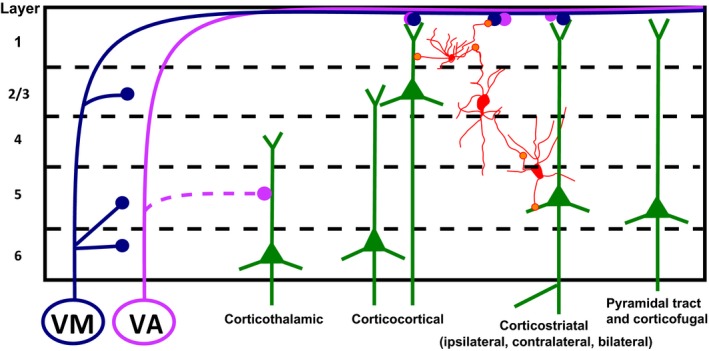
The focus of this literature review is on the three interacting brain areas that participate in decision-making: basal ganglia, ventral motor thalamic nuclei, and medial prefrontal cortex, with an emphasis on the participation of the ventromedial and ventral anterior motor thalamic nuclei in prefrontal cortical function. Apart from a defining input from the mediodorsal thalamus, the prefrontal cortex receives inputs from ventral motor thalamic nuclei that combine to mediate typical prefrontal functions such as associative learning, action selection, and decision-making. Motor, somatosensory and medial prefrontal cortices are mainly contacted in layer 1 by the ventral motor thalamic nuclei and in layer 3 by thalamocortical input from mediodorsal thalamus. We will review anatomical, electrophysiological, and behavioral evidence for the proposed participation of ventral motor thalamic nuclei and medial prefrontal cortex in rat and mouse motor decision-making.
Keywords: action selection; basal ganglia; cognitive functions; rodents.
© 2018 The Authors. European Journal of Neuroscience published by Federation of European Neuroscience Societies and John Wiley & Sons Ltd.
Conflict of interest statement
The authors declare no conflict of interest, financial, or otherwise.
Figures

References
-
- Aceves, J.J. , Rueda‐Orozco, P.E. , Hernandez‐Martinez, R. , Galarraga, E. & Bargas, J. (2011) Bidirectional plasticity in striatonigral synapses: a switch to balance direct and indirect basal ganglia pathways. Learn. Mem., 18, 764–773. - PubMed
-
- Afifi, A.K. (2003) The basal ganglia: a neural network with more than motor function. Semin. Peadiatr. Neurol., 10, 3–10. - PubMed
-
- Albin, R.L. , Young, A.B. & Penney, J.B. (1989) The functional anatomy of basal ganglia disorders. Trends Neurosci., 12, 366–375. - PubMed
-
- Alcaraz, F. , Marchand, A.R. , Courtand, G. , Coutureau, E. & Wolff, M. (2016) Parallel inputs from the mediodorsal thalamus to the prefrontal cortex in the rat. Eur. J. Neuorsci., 44, 1972–1986. - PubMed
-
- Arbuthnott, G.W. , MacLeod, N.K. , Maxwell, D.J. & Wright, A.K. (1990) Distribution and synaptic contacts of the cortical terminals arising from neurons in the rat ventromedial thalamic nucleus. Neuroscience, 38, 47–60. - PubMed
Publication types
MeSH terms
Grants and funding
LinkOut - more resources
Full Text Sources
