. 2019 Feb 1;167(2):581-592.
doi: 10.1093/toxsci/kfy263.

Constitutive Activation of the Human Aryl Hydrocarbon Receptor in Mice Promotes Hepatocarcinogenesis Independent of Its Coactivator Gadd45b

Affiliations

Constitutive Activation of the Human Aryl Hydrocarbon Receptor in Mice Promotes Hepatocarcinogenesis Independent of Its Coactivator Gadd45b

Peipei Lu et al. Toxicol Sci. .

Abstract

2,3,7,8-tetrachlorodibenzo-p-dioxin (TCDD), or dioxin, is a potent liver cancer promoter through its sustained activation of the aryl hydrocarbon receptor (Ahr) in rodents. However, the carcinogenic effect of TCDD and AHR in humans has been controversial. It has been suggested that the inter-species difference in the carcinogenic activity of AhR is largely due to different ligand affinity in that TCDD has a 10-fold lower affinity for the human AHR compared with the mouse Ahr. It remains unclear whether the activation of human AHR is sufficient to promote hepatocellular carcinogenesis. The goal of this study is to clarify whether activation of human AHR can promote hepatocarcinogenesis. Here we reported the oncogenic activity of human AHR in promoting hepatocellular carcinogenesis. Constitutive activation of the human AHR in transgenic mice was as efficient as its mouse counterpart in promoting diethylnitrosamine (DEN)-initiated hepatocellular carcinogenesis. The growth arrest and DNA damage-inducible gene 45 β (Gadd45b), a signaling molecule inducible by external stress and UV irradiation, is highly induced upon AHR activation. Further analysis revealed that Gadd45b is a novel AHR target gene and a transcriptional coactivator of AHR. Interestingly, ablation of Gadd45b in mice did not abolish the tumor promoting effects of the human AHR. Collectively, our findings suggested that constitutive activation of human AHR was sufficient to promote hepatocarcinogenesis.


Figures

Figure 1.
Figure 1.
Transgenic activation of human AHR promotes DEN-initiated hepatocarcinogenesis. A, Schematic presentation of the FABP-tTA/TetRE-CA-AHR or FABP-tTA/TetRE-CA-Ahr transgenic system. PCMV, minimal cytomegalovirus promoter. B, Mice were i.p. injected with a single dose of 90 mg/kg DEN at 6-week old. Shown are the representative gross appearances of the mouse livers at 9 months after the DEN challenge. C, Representative liver tumor histology by H&E staining (100×) with dotted lines denoting the tumor nodule areas (top), and H&E staining (400×) with arrows indicating cells that are undergoing mitosis (bottom). D, Immunohistochemical staining (400×) of the proliferation marker Ki67 in liver tissues from tumor-surrounding/nontumor (N) and tumor (T) areas in CA-Ahr and CA-AHR mice. Arrows indicate positive staining. n = 5–7 for each group.
Figure 2.
Figure 2.
Activation of human AHR increases inflammation and impairs liver function upon the DEN treatment. Mice were i.p. injected with a single dose of 90 mg/kg DEN at 6-week old and sacrificed after 9 months. Liver and serum samples were collected at the end of the experiment. A, Hepatic mRNA expression of Il-6 and Tnf-α. B, Serum levels of ALT and AST. n = 5–7 for each group. *p < .05; **p < .01, all compared with WT. Results are presented as means ± standard deviation (SD).
Figure 3.
Figure 3.
AHR activation induces the expression of Gadd45b. Mice were i.p. injected with a single dose of 90 mg/kg DEN at 6-week old and sacrificed after 9 months. Tumor bearing liver tissues and the surrounding nontumor tissues were collected. A, Hepatic mRNA expressions of Gadd45b, c-Myc, and cell cycle-related genes in liver tissues from tumor-surrounding/nontumor (N) and tumor (T) areas in CA-Ahr mice, CA-AHR mice, and WT mice were measured by real-time PCR. B, The protein expression of Gadd45b in liver tissues from tumor-surrounding/nontumor (N) and tumor (T) areas in CA-AHR mice and WT mice was analyzed by immunohistochemistry. Arrows in (B) indicate positive staining. n = 5–7 for each group. *p < .05; **p < .01, all compared with WT. Results are presented as means ± standard deviation (SD).
Figure 4.
Figure 4.
Gadd45b is an AHR target gene. A, The hepatic mRNA expressions of Gadd45b, c-Myc, Cyclin D1, and Cdk1 was measured in naïve 6-week old WT and CA-AHR mice. n = 5 for each group. B, Eight-week old WT and Ahr−/− male mice were i.p. injected with TCDD (10 μg/kg) or corn oil for four consecutive days before tissue harvesting. The hepatic gene expression was measured by real-time PCR. n = 5 for each group. C, Human primary hepatocytes were treated with DMSO or 3-MC (4 μM) for 24 hours before harvesting. The expression of CYP1A2 was included as a positive control. D, Schematic representation of the mouse Gadd45b gene promoter and the positions of putative DREs. E, The sequence of Gadd45b DRE1 and the mutant variant (top) and EMSA results (bottom). Arrows indicate the specific shift bands. F, ChIP assay to show the recruitment of CA-AHR onto the Gadd45b gene promoters. WT CD-1 mouse (n = 4 for each group) livers were hydrodynamically transfected with pCMX-Flag empty construct or pCMX-Flag-CA-AHR expressing construct. The anti-Flag antibody was used for ChiP analysis. G, Activation of the Gadd45b gene promoter reporter gene by AHR in the presence of 3-MC. CV1 cells were cotransfected in triplicate with indicated reporters and receptors. Transfected cells were treated with vehicle DMSO or 3-MC (2 μM) for 24 h before luciferase assay. *p < .05; **p < .01, compared with the vehicle (DMSO) control. Results are presented as means ± standard deviation (SD).
Figure 5.
Figure 5.
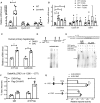
Gadd45b functions as an AHR coactivator. A, Contransfection of Gadd45b increases the transcriptional activity of AHR. Huh7 cells were cotransfected in triplicate with Flag-Gadd45b, pCMX-AHR, and the AHR responsive pGud-luciferase reporter gene. The transfected cells were subsequently treated with DMSO or 3-MC (2 μM) for 24 h before the luciferase assay. B, CD-1 male mice (n = 5 for each group) were hydrodynamically transfected with the pCMX-Flag empty vector or pCMX-Flag-Gadd45b vector before being treated with vehicle or TCDD (10 μg/kg) for 24 h. The hepatic gene expression was measured by real-time PCR. C, Hepatic gene expression was measured in WT and Gadd45b−/− mice (n = 5 for each group) treated with vehicle corn oil or TCDD (10 μg/kg) for 24 h. D, Co-immunoprecipitation to assess the interaction between AHR and Gadd45b. HA-AHR and Flag-Gadd45b plasmids (0.4 μg/well for each) were cotransfected in 6-well 293T cells, and cell lysates from 3 wells were combined for co-IP with the anti-HA antibody. The resulting proteins were subjected to Western blotting and blotted with both anti-Flag and anti-HA antibodies. Arrows indicate specific bands of expected sizes. E, Mammalian 2-hybrid assay to map the Ahr binding domain on Gadd45b. Shown are the schematic representations of the full-length Gadd45b and its deletion mutants (top), Western blotting to confirm the expression of Gal4-Gadd45b constructs using an antibody against Gal4-DBD (middle), and mammalian two-hybrid luciferase reporter assay to show the interaction between Gal4-Gadd45b or its deletion mutants with Ahr-VP (bottom). *p < .05; **p < .01. The comparisons are labeled. Results are presented as means ± standard deviation (SD).
Figure 6.
Figure 6.
Gadd45b is not required for AHR-promoted hepatocellular carcinogenesis. A, Representative gross appearance of livers of male and female CA-AHR-Gadd45b−/− mice 9 months after the DEN injection. B, H&E staining of liver sections of DEN-treated male CA-AHR-Gadd45b−/− mice. Dashed line denotes the nodule area (top). Dotted line denotes abnormal liver cell plates that are 3+ cells thick (middle). Arrowheads indicate apoptotic cells (bottom). C, Immunohistochemical staining of Ki67 and PCNA in DEN-treated male CA-AHR and CA-AHR-Gadd45b−/− mice. Arrows indicate positive staining. Magnification, 400×. D, The hepatic mRNA expression of Il-6 and Tnf-α from tumor-surrounding (N) and tumor (T) areas in male CA-AHR mice and CA-AHR-Gadd45b−/− mice. n = 8–15 for each group. E, The serum levels of ALT and AST in male CA-AHR mice and CA-AHR-Gadd45b−/− mice. n = 8–15 for each group. Results are presented as means ± standard deviation (SD).

Similar articles

Cited by

References

    1. Balkwill F., Mantovani A. (2001). Inflammation and cancer: Back to Virchow? Lancet 357, 539–545. - PubMed
    1. Cole P., Trichopoulos D., Pastides H., Starr T., Mandel J. S. (2003). Dioxin and cancer: A critical review. Regul. Toxicol. Pharmacol. 38, 378–388. - PubMed
    1. Deshpande A., Sicinski P., Hinds P. W. (2005). Cyclins and cdks in development and cancer: A perspective. Oncogene 24, 2909–2915. - PubMed
    1. Ema M., Ohe N., Suzuki M., Mimura J., Sogawa K., Ikawa S., Fujii-Kuriyama Y. (1994). Dioxin binding activities of polymorphic forms of mouse and human arylhydrocarbon receptors. J. Biol. Chem. 269, 27337–27343. - PubMed
    1. Flaveny C. A., Perdew G. H. (2009). Transgenic humanized AHR mouse reveals differences between human and mouse AHR ligand selectivity. Mol. Cell. Pharmacol. 1, 119–123. - PMC - PubMed

Publication types

MeSH terms

Substances