. 2018 Nov 14;18(11):7021-7029.
doi: 10.1021/acs.nanolett.8b02989. Epub 2018 Oct 23.

Nanothermometry Reveals Calcium-Induced Remodeling of Myosin

Affiliations

Nanothermometry Reveals Calcium-Induced Remodeling of Myosin

Eric R Kuhn et al. Nano Lett. .

Abstract

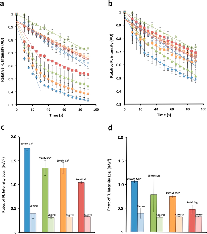
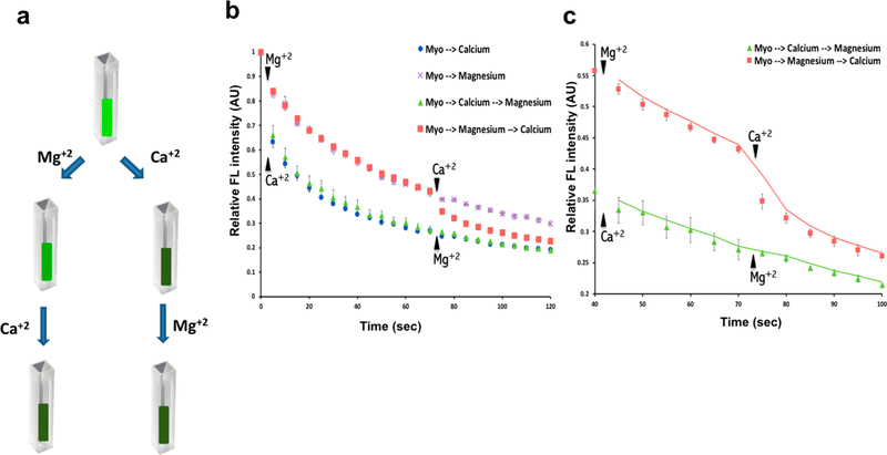
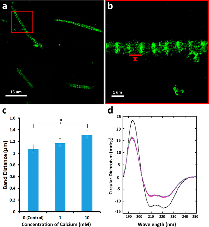
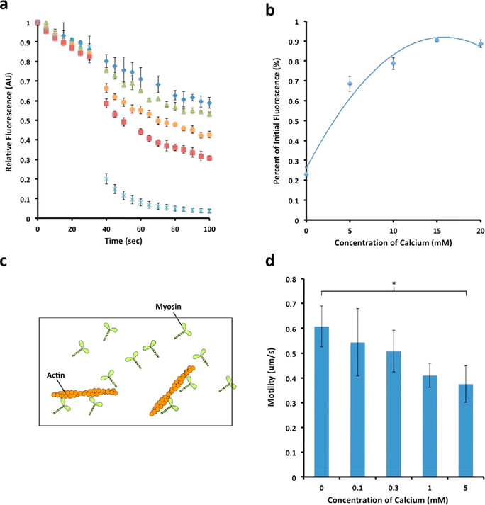
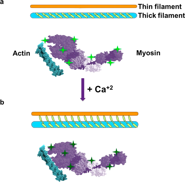
Ions greatly influence protein structure-function and are critical to health and disease. A 10, 000-fold higher calcium in the sarcoplasmic reticulum (SR) of muscle suggests elevated calcium levels near active calcium channels at the SR membrane and the impact of localized high calcium on the structure-function of the motor protein myosin. In the current study, combined quantum dot (QD)-based nanothermometry and circular dichroism (CD) spectroscopy enabled detection of previously unknown enthalpy changes and associated structural remodeling of myosin, impacting its function following exposure to elevated calcium. Cadmium telluride QDs adhere to myosin, function as thermal sensors, and reveal that exposure of myosin to calcium is exothermic, resulting in lowering of enthalpy, a decrease in alpha helical content measured using CD spectroscopy, and the consequent increase in motor efficiency. Isolated muscle fibers subjected to elevated levels of calcium further demonstrate fiber lengthening and decreased motility of actin filaments on myosin-functionalized substrates. Our results, in addition to providing new insights into our understanding of muscle structure-function, establish a novel approach to understand the enthalpy of protein-ion interactions and the accompanying structural changes that may occur within the protein molecule.

Keywords: Quantum dot thermometry; calcium; molecular motor myosin; muscle function.


Conflict of interest statement

The authors declare no competing financial interest.

Figures

Figure 1.
Figure 1.
Cadmium telluride quantum dots adhered to myosin molecules function as nanoscale thermometers, enabling the detection of heat loss from the motor protein on binding to calcium or magnesium. Binding of calcium to bovine cardiac (BC) myosin lowers the enthalpy of the molecule to a greater degree than magnesium. (a–d) Binding of calcium to bovine cardiac (BC) myosin lowers the energy state of the motor protein greater than magnesium. (a) Cadmium telluride quantum dots associated with the myosin molecule and used as molecular thermometers are able to detect mK changes in temperature of the myosin molecule. Changes in temperature are reflected as a change in fluorescence. The greater the heat loss, the lower the fluorescence. Calcium concentrations used were 5 mM (solid red squares), 10 mM (solid green triangles), 15 mM (solid orange circles), and 20 mM (solid blue diamonds). Color coded corresponding controls were empty symbols. This calcium and magnesium dependent drop in fluorescence is a measure of heat release. Real-time fluorimetry demonstrates the extent and rate of this heat loss. (b) There is similarly a magnesium dependent drop in fluorescence, however, to a lesser extent than calcium. (c, d) Rate of fluorescence loss in the first 30 s following addition of calcium or magnesium. (c) Note that the rate of heat release in the presence of increasing calcium concentration is 30% greater than is observed with (d) magnesium. (Data presented is MEAN ± SEM, n = 3, *p < 0.005). Similar study using rabbit skeletal (RS) myosin is presented in Figure 1S (Supporting Information).
Figure 2.
Figure 2.
Calcium and magnesium share the same binding sites in the myosin molecule. (a) Schematic drawing of the experiment showing that myosin pre-exposed to 10 mM calcium demonstrate little change in the enthalpy following exposure to 10 mM magnesium. In contrast, myosin preexposed to 10 mM magnesium demonstrate a near 10% drop in the enthalpy following exposure to 10 mM calcium. (b) Spectrofluorimetric measurements of time-dependent loss in fluorescence when myosin pre-exposed to 10 mM magnesium is followed by exposure to 10 mM calcium. Little change in enthalpy is observed when myosin pre-exposed to 10 mM calcium is followed by exposure to 10 mM magnesium, demonstrating that the loss of enthalpy of myosin is not additive when the motor protein is first exposed to calcium followed by magnesium addition (data presented is MEAN ± SEM, n = 3). (c) Section of Figure 2b has been rescaled for clarity. These results suggest that calcium and magnesium likely share the same binding sites on myosin and that calcium has a greater binding affinity to myosin than magnesium.
Figure 3.
Figure 3.
Myosin undergoes structural changes in the presence of elevated calcium. (a, b) Mouse muscle fibers exposed to calcium results in approximately a 10% increase in the distance between the A bands (data presented are MEAN ± SEM, n = 5). (c) Dose-dependent increase in A band distances (*p < 0.005). (d) Circular dichroism spectroscopy of BC myosin demonstrates unwinding of the α helix (Supporting Information, Table 1S). Calcium concentrations used are 0 mM (black), 5 mM (blue), 10 mM (green), and 15 mM (red). An average of 30 scans were collected in total for each sample from triplicate preparations.
Figure 4.
Figure 4.
Calcium-dependent increase in myosin efficiency and its impact on motility. (a) Molecular motor myosin consumes energy in the form of ATP for mechanical work. The greater the heat loss in the hydrolysis of ATP by myosin, less is available for mechanical work and hence lower the efficiency (data presented are MEAN ± SEM, n = 4). Note the lower heat loss at higher calcium concentrations, hence greater efficiency. (b) Plot of panel a showing calcium dose dependence retention of fluorescence, hence less heat loss at increased calcium concentrations. (c) Schematic diagram of a motility assay setup. Myosin attached to the surface of the coverslip is used to observe and measure motility of fluorescently labeled actin filaments. (d) Myosin motility assay demonstrates decreased motility with increased calcium (see Supporting Information, Table 3S), suggesting an energy-speed-precision trade-off as previously reported (data presented are MEAN ± SEM, n = 4, *p < 0.02).
Figure 5.
Figure 5.
Schematic diagram showing myosin heads in the thick filament unwind and loses enthalpy in the presence of calcium, resulting in increased interactions with the thin filament. (a) Thick and thin filaments before calcium addition. Note the bright green fluorescing QDs (bright green stars). (b) Calcium addition results in the loss of alpha helical content of myosin molecules demonstrated using CD and the consequent release of heat and therefore loss of QD fluorescence (dim green stars). Loss in alpha helical content of the myosin molecule in the presence of calcium could result in a radial increase in size of the molecule, enabling myosin heads greater probability to interaction with actin in the thin filament.

References

    1. Reconditi M Motion of myosin head domains during activation and force development in skeletal muscle. Proc. Natl. Acad. Sci. U. S. A 2011, 108, 7236–7240. - PMC - PubMed
    1. Gordon AM; Homsher E; Regnier M Regulation of contraction in striated muscle. Physiol. Rev 2000, 80, 853–924. - PubMed
    1. Linari M; et al. Force generation by skeletal muscle is controlled by mechanosensing in myosin filaments. Nature 2015, 528, 276–279. - PubMed
    1. Toyoshima C How Ca2+-ATPase pumps ions across the sarcoplasmic reticulum membrane. Biochim. Biophys. Acta, Mol. Cell Res 2009, 1793 (6), 941. - PubMed
    1. Huxley HE; Brown W The low-angle x-ray diagram of vertebrate striated muscle and its behaviour during contraction and rigor. J. Mol. Biol 1967, 30, 383–434. - PubMed

Publication types