. 2018 Oct 22;13(10):e0206130.
doi: 10.1371/journal.pone.0206130. eCollection 2018.

Bone marrow derived-mesenchymal stem cells downregulate IL17A dependent IL6/STAT3 signaling pathway in CCl4-induced rat liver fibrosis

Affiliations

Bone marrow derived-mesenchymal stem cells downregulate IL17A dependent IL6/STAT3 signaling pathway in CCl4-induced rat liver fibrosis

Shimaa Farouk et al. PLoS One. .

Abstract

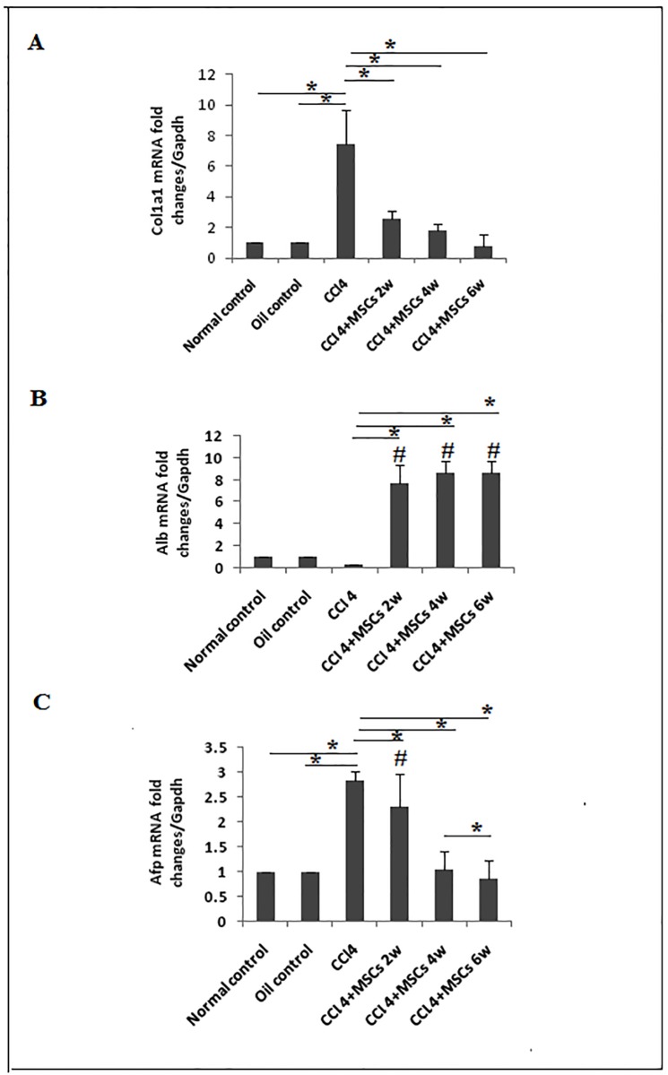
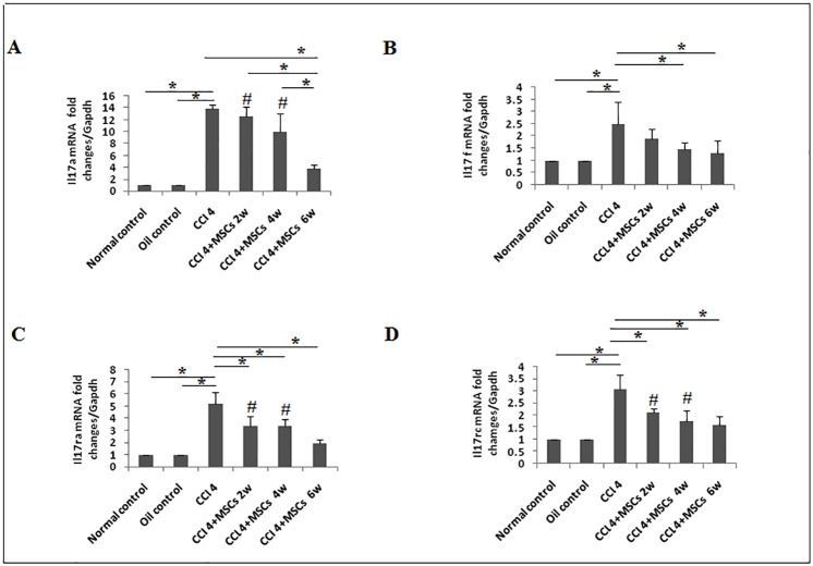
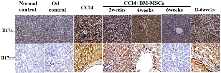
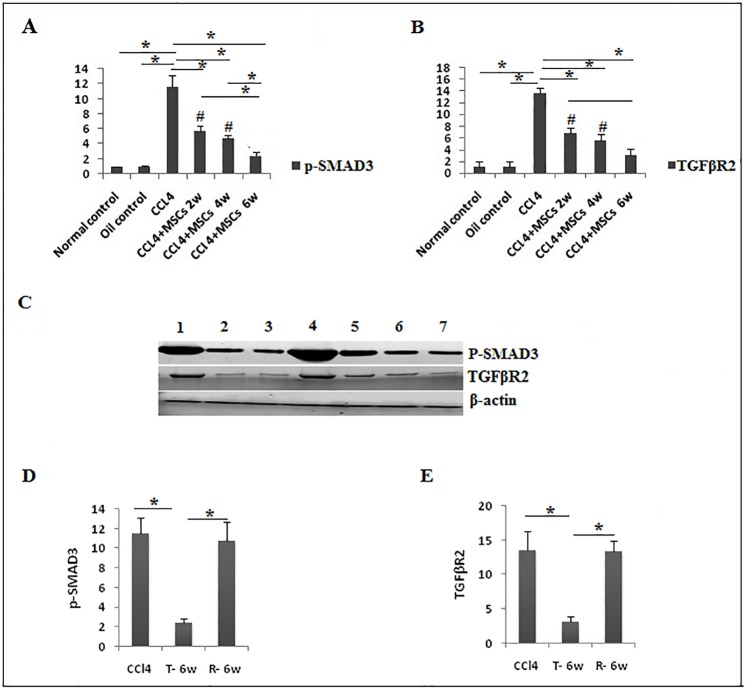
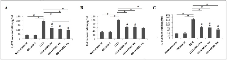
Therapeutic potential of bone marrow-derived mesenchymal stem cells (BM-MSCs) has been reported in several animal models of liver fibrosis. Interleukin (IL) 17A, IL6 and Stat3 have been described to play crucial roles in chronic liver injury. However, the modulatory effect of MSCs on these markers was controversial in different diseases. BM-MSCs might activate the IL6/STAT3 signaling pathway and promote cell invasion in hepatocellular carcinoma, but the immunomodulatory role of BM-MSCs on IL17A/IL6/STAT3 was not fully elucidated in liver fibrosis. In the present study, we evaluated the capacity of the BM-MSCs in the modulation of cytokines milieu and signal transducers, based on unique inflammatory genes Il17a and Il17f and their receptors Il17rc and their effect on the IL6/STAT3 pathway in CCl4-induced liver fibrosis in rats. A single dose of BM-MSCs was administered to the group with induced liver fibrosis, and the genes and proteins of interest were evaluated along six weeks after treatment. Our results showed a significant downregulation of Il17a, Il17ra, il17f and Il17rc genes. In accordance, BM-MSCs administration declined IL17, IL2 and IL6 serum proteins and downregulated IL17A and IL17RA proteins in liver tissue. Interestingly, BM-MSCs downregulated both Stat3 mRNA expression and p-STAT3, while Stat5a gene was downregulated and p-STAT5 protein was elevated. Also P-SMAD3 and TGFβR2 proteins were downregulated in response to BM-MSCs treatment. Collectively, we suggest that BM-MSCs might play an immunomodulatory role in the treatment of liver fibrosis through downregulation of IL17A affecting IL6/STAT3 signaling pathway.


Conflict of interest statement

The authors have declared that no competing interests exist.

Figures

Fig 1
Fig 1. Bone marrow-derived mesenchymal stem cell characterization and homing.
a. BM-MSCs at 3rd passage displayed fibroblast-like morphology (200X). b. Phenotyping of the BM-MSCs analyzed by "flow cytometry" indicated that cells were negative for CD34 and positive for CD90 and CD29 antibodies. c. PKH26-labeled BM-MSCs were tracked in liver tissue after 2, 4 and 6 weeks of injection (200X). d. labeled BM-MSCS (red) located near hepatic sinusoid (white arrow).
Fig 2
Fig 2. Reduction of fibrosis in injured liver treated with BM-MSCs.
a. Sections of paraffin-embedded liver tissue stained with Sirius-red (200X). b. & c. Graphical presentation of collagen levels. Data are expressed as Means (n = 3)± SD. *P<0.05. #P<0.05 versus control groups.
Fig 3
Fig 3. Sections of paraffin-embedded liver tissue stained with H&E, exhibiting histological structure in different study groups (40X).
Fig 4
Fig 4. Proliferating nuclear cell antigen protein expression assessesment by immunohistochemistry.
a. PCNA expression in liver tissue in CCl4, T-6W (BMSCs/6weeks), and R-6W (recovery) groups (Brown). b. quantitative analysis of PCNA expression. Data are expressed as means (n = 3)± SD. (400x).
Fig 5
Fig 5. Gene expression of Alb, Afp and Col1a1 in liver tissue.
The relative quantification of a. Col1a1, b. Alb, and c. Afp were carried out utilizing the 2–ΔΔCt ± standard deviation (SD) method and Gapdh gene expression as housekeeping gene. Each experiment was performed in duplicate and results are expressed as fold induction of mRNA expression (n = 5). *P<0.05. #P<0.05 compared to control groups.
Fig 6
Fig 6. Expression of Il17a, Il17f, Il17ra, and Il17rc genes in liver tissue.
The data are presented as fold induction of mRNA expression of a. Il17a, b. Il17f, c. Il17ra, and d. Il17rc in CCl4/untreated (n = 5) rats compared to MSCs transplanted (CCl4 +BM-MSCs) groups (n = 5) after 2, 4 and 6 weeks. All groups are compared with normal/diet control and oil/vehicle control rats (n = 5). Each experiment was performed in duplicates. Representative data are shown as mean±SD. *P<0.05. #P<0.05 compared to control groups.
Fig 7
Fig 7. IL17A and IL17RA protein expression in liver tissue evaluated by IHC method.
Upper panels are representatives of IL17 expression and it is noted that it was very weak in normal and paraffin/oil control group, severe in CCl4/fibrosis and recovery groups, high in CCl4+BM-MSCs/2 weeks group, moderate in CCl4+BM-MSCs/4weeks group and mild CCl4+BM-MSCs/6 weeks group. Lower panels are representatives of IL17RA expression and it is noted that it was absent in normal and paraffin/oil control group, high in CCl4/fibrosis and recovery groups, moderate in CCl4+BM-MSCs/2weeks group, mild in CCl4+BM-MSCs/4weeks group and nil in CCl4+BM-MSCs/6 weeks group (400X).
Fig 8
Fig 8. Gene and protein expression of STAT3 and STAT5.
a. Stat3 and b. Stat5a gene expression was measured by qRT-PCR (n = 5). c. p-STAT3, and d. p-STAT5 protein analysis, representative data of western blot are shown as mean±SD. *P<0.05. #P<0.05 compared to control groups. e. Expression of STAT3, and STAT5 proteins in liver tissues (n = 3) was assessed by western blot (WB) analysis. Quantative expression analysis of f. p-STAT3. g. p-STAT5a in. 1. Normal control. 2. Oil control. 3. CCl4 groups.4. CCl4+BM-MSCs/2 weeks. 5. CCl4+ BM-MSCs/4 weeks. 6. CCl4+MSCs/6 weeks. 7. Recovery group (R-6W).
Fig 9
Fig 9. Protein expression of p-SMAD3 and TGFβR2.
a. p-SMAD3, and b. TGFβR2 protein analysis, representative data of western blot are shown as mean±SD in different groups (n = 3). c. Expression of p-SMAD3 and TGFβR2 proteins by western blot (WB) in liver tissues. Quantative expression analysis of .d. p-SMAD3. e. TGFβR2 in 1. Recovery group (R-6W).2. Normal control. 3. Oil control. 4. CCl4 groups. 5. CCl4+BM-MSCs/2 weeks. 6. CCl4+ BM-MSCs/4 weeks. 7. CCl4+MSCs/6 weeks.
Fig 10
Fig 10. IL17A, IL6 and IL2 protein expression levels in serum measured by ELISA.
The data shown in (a), (b) and (c) are presented as the mean ±SD *P <0.05. #P< 0.05 compared to control groups.

Similar articles

Cited by

References

    1. Friedman S. Liver fibrosis: from bench to bedside. Journal of hepatology Supplement. 2003;38(1):38–53. - PubMed
    1. Friedman SL. Mechanisms of disease: Mechanisms of hepatic fibrosis and therapeutic implications. Nature Clinical Practice: Gastroenterology & Hepatology. 2004;1(2):98–105. 10.1038/ncpgasthep0055 - DOI - PubMed
    1. Merion RM. Current status and future of liver transplantation. Semin Liver Dis. 2010;30(4):411–21. Epub 10/19. 10.1055/s-0030-1267541 . - DOI - PubMed
    1. Kia R, Sison RLC, Heslop J, Kitteringham NR, Hanley N, Mills JS, et al. Stem cell-derived hepatocytes as a predictive model for drug-induced liver injury: are we there yet? British Journal of Clinical Pharmacology. 2013;75(4):885–96. 10.1111/j.1365-2125.2012.04360.x - DOI - PMC - PubMed
    1. Al Ghrbawy NM, Afify RAAM, Dyaa N, El Sayed AA. Differentiation of Bone Marrow: Derived Mesenchymal Stem Cells into Hepatocyte-like Cells. Indian Journal of Hematology and Blood Transfusion. 2016;32(3):276–83. 10.1007/s12288-015-0581-7 - DOI - PMC - PubMed

MeSH terms